

The DOI matches its Crossref record; 59 of the 61 references it cites were found in a registry or an index.
Dynamic memory to alleviate catastrophic forgetting in continual learning with medical imaging
Open accessAbstract
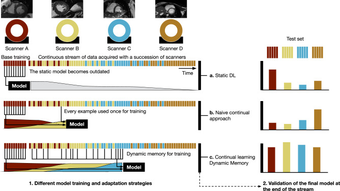
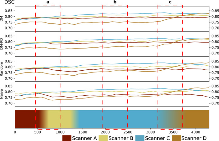
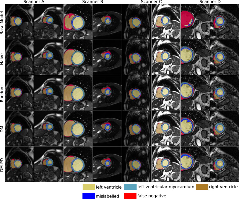
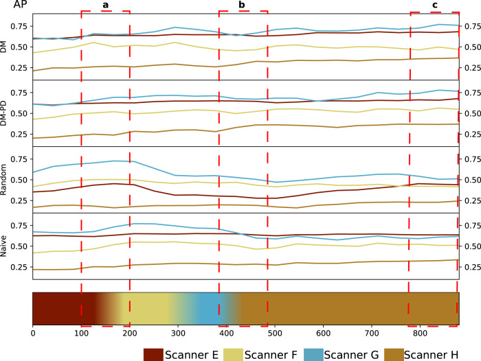
Medical imaging is a central part of clinical diagnosis and treatment guidance. Machine learning has increasingly gained relevance because it captures features of disease and treatment response that are relevant for therapeutic decision-making. In clinical practice, the continuous progress of image acquisition technology or diagnostic procedures, the diversity of scanners, and evolving imaging protocols hamper the utility of machine learning, as prediction accuracy on new data deteriorates, or models become outdated due to these domain shifts. We propose a continual learning approach to deal with such domain shifts occurring at unknown time points. We adapt models to emerging variations in a continuous data stream while counteracting catastrophic forgetting. A dynamic memory enables rehearsal on a subset of diverse training data to mitigate forgetting while enabling models to expand to new domains. The technique balances memory by detecting pseudo-domains, representing different style clusters within the data stream. Evaluation of two different tasks, cardiac segmentation in magnetic resonance imaging and lung nodule detection in computed tomography, demonstrate a consistent advantage of the method.
Overview
The claims and statistics of this paper haven’t been checked yet.
Run the check — free, no account needed. It reads the open-access text and checks the sources it cites.
Integrity signals
Registry
Cites
Statistics
Figures
Verification
Claims the paper makes
The claims and statistics of this paper haven’t been checked yet.
Run the check — free, no account needed. It reads the open-access text and checks the sources it cites.
Claims other papers make about this one
Each is checked against this paper’s own text: does it say what the citing paper says it does?
In the citing paper
“Continuous monitoring systems detect when models encounter data outside their training distribution or when performance begins to degrade [147,148,149,150,151].”
From Artificial Intelligence in Clinical Oncology: From Productivity Enhancement to Creative Discovery (2025) · cites this paper as [149]
Self-check
Percentages, sums and reported differences re-computed from the paper’s own numbers.
The self-check needs the paper’s full text.
Citations
Cites
Deep Residual Learning for Image Recognition
He, K., Zhang, X., Ren, S., & Sun, J. (2016) · 2016 IEEE Conference on Computer Vision and Pattern Recognition (CVPR) · doi:10.1109/cvpr.2016.90 · Cited by 235,474
Faster R-CNN: Towards Real-Time Object Detection with Region Proposal Networks
Ren, S., He, K., Girshick, R., & Sun, J. (2017) · IEEE Transactions on Pattern Analysis and Machine Intelligence · doi:10.1109/tpami.2016.2577031 · Cited by 73,472
(2008) · Journal of Machine Learning Research · Cited by 33,051
PyTorch: An Imperative Style, High-Performance Deep Learning Library
Paszke, A., Gross, S., Massa, F., Lerer, A., Bradbury, J., Chanan, G., Killeen, T., Lin, Z., Gimelshein, N., Antiga, L., et al. (2019) · arXiv · doi:10.48550/arxiv.1912.01703 · Cited by 31,473
The pascal visual object classes (VOC) challenge
Everingham, M., Van Gool, L., Williams, C. K. I., Winn, J., & Zisserman, A. (2010) · International Journal of Computer Vision · doi:10.1007/s11263-009-0275-4 · Cited by 19,599
Fully convolutional networks for semantic segmentation
Shelhamer, E., Long, J., & Darrell, T. (2017) · IEEE Transactions on Pattern Analysis and Machine Intelligence · doi:10.1109/tpami.2016.2572683 · Cited by 11,898
Overcoming catastrophic forgetting in neural networks
Kirkpatrick, J., Pascanu, R., Rabinowitz, N., Veness, J., Desjardins, G., Rusu, A. A., Milan, K., Quan, J., Ramalho, T., Grabska-Barwinska, A., et al. (2017) · Proceedings of the National Academy of Sciences · doi:10.1073/pnas.1611835114 · Cited by 7,899
U1 snRNP regulates cancer cell migration and invasion in vitro
Oh, J.-M., Venters, C. C., Di, C., Pinto, A. M., Wan, L., Younis, I., Cai, Z., Arai, C., So, B. R., Duan, J., et al. (2020) · Nature Communications · doi:10.1038/s41467-019-13993-7 · Cited by 7,157
A Comprehensive Survey on Transfer Learning
Zhuang, F., Qi, Z., Duan, K., Xi, D., Zhu, Y., Zhu, H., Xiong, H., & He, Q. (2021) · Proceedings of the IEEE · doi:10.1109/jproc.2020.3004555 · Cited by 6,678
Liu, F. T., Ting, K. M., & Zhou, Z.-H. (2008) · 2008 Eighth IEEE International Conference on Data Mining · doi:10.1109/icdm.2008.17 · Cited by 6,601
Research works printed but not linked: 2
- Lopez-Paz, D. & Ranzato, M. Gradient episodic memory for continual learning. Adv. Neural Inf. Process. Syst. 30, 6467–6476 (2017).
- Glocker, B., Robinson, R., Castro, D. C., Dou, Q. & Konukoglu, E. Machine learning with multi-site imaging data: an empirical study on the impact of scanner effects. Preprint at https://arxiv.org/abs/1910.04597 (2019). · arXiv
Cited by
Sorted by citations
Detecting and Remediating Harmful Data Shifts for the Responsible Deployment of Clinical AI Models
Subasri, V., Krishnan, A., Kore, A., Dhalla, A., Pandya, D., Wang, B., Malkin, D., Razak, F., Verma, A. A., Goldenberg, A., et al. (2025) · JAMA Network Open · doi:10.1001/jamanetworkopen.2025.13685 · Cited by 87
Lambin, P., Woodruff, H. C., Mali, S. A., Zhong, X., Kuang, S., Lavrova, E., Khan, H., Lekadir, K., Zwanenburg, A., Deasy, J. O., et al. (2025) · Nature Reviews Clinical Oncology · doi:10.1038/s41571-025-01067-1 · Cited by 72
Advancements and Applications of Lateral Flow Assays (LFAs): A Comprehensive Review
Kinyua, D. M., Memeu, D. M., Mwenda, C. M., Ventura, B. D., & Velotta, R. (2025) · Sensors · doi:10.3390/s25175414 · Cited by 63
Lifelong nnU-Net: a framework for standardized medical continual learning
González, C., Ranem, A., dos Santos, D. P., Othman, A. E., & Mukhopadhyay, A. (2023) · Scientific Reports · doi:10.1038/s41598-023-34484-2 · Cited by 52
Wang, W., Chen, K., Ma, X., & Guo, J. (2022) · Fundamental Research · doi:10.1016/j.fmre.2022.03.025 · Cited by 39
Su, H., Gao, H., Wang, X., Fang, X., Liu, Q., Huang, G.-B., Li, X., & Cao, Q. P. (2024) · IEEE Transactions on Instrumentation and Measurement · doi:10.1109/tim.2024.3472860 · Cited by 34
Huang, Y., Zhao, W., Wang, S., Fu, Y., Jiang, Y., & Yu, L. (2023) · IEEE/CVF International Conference on Computer Vision (ICCV) · doi:10.1109/iccv51070.2023.01952 · Cited by 32
Xue, W., Cao, H., Ma, J., Bai, T., Wang, T., & Ni, D. (2022) · IEEE Journal of Biomedical and Health Informatics · doi:10.1109/jbhi.2022.3221429 · Cited by 26
Mapping brain endophenotypes associated with idiopathic pulmonary fibrosis genetic risk
Mohammadi‐Nejad, A., Allen, R. J., Kraven, L. M., Leavy, O. C., Jenkins, G. R., Wain, L. V., Auer, D. P., & Sotiropoulos, S. N. (2022) · EBioMedicine · doi:10.1016/j.ebiom.2022.104356 · Cited by 26
Aslam, S., Rasool, A., Li, X., & Wu, H. (2025) · Interdisciplinary Sciences: Computational Life Sciences · doi:10.1007/s12539-024-00675-2 · Cited by 23
Continual learning in medical image analysis: A survey
Wu, X., Xu, Z., & Tong, R. K. (2024) · Computers in Biology and Medicine · doi:10.1016/j.compbiomed.2024.109206 · Cited by 29
Wang, K., Ye, Z., Xie, X., Cui, H., Chen, T., & Liu, B. (2023) · Knowledge-Based Systems · doi:10.1016/j.knosys.2023.111127 · Cited by 22
Muneer, A., Waqas, M. M., Saad, M. B., Showkatian, E., Bandyopadhyay, R., Xu, H. M., Li, W., Chang, J., Liao, Z., Haymaker, C., et al. (2026) · Artificial Intelligence Review · doi:10.1007/s10462-026-11522-9 · Cited by 24
Lin, Y., Li, Z., Gong, C. Y., Lu, C., Wang, X., & Gong, J. (2023) · IEEE Transactions on Intelligent Transportation Systems · doi:10.1109/tits.2023.3324983 · Cited by 19
Zhu, Z., Ma, X., Wang, W., Dong, S., Wang, K., Wu, L., Luo, G., Wang, G., & Li, S. (2024) · Medical Image Analysis · doi:10.1016/j.media.2024.103112 · Cited by 20
The Evolving Role of Artificial Intelligence in Radiotherapy Treatment Planning—A Literature Review
Kalsi, S., French, H., Chhaya, S., Madani, H., Mir, R., Anosova, A., & Dubash, S. (2024) · Clinical Oncology · doi:10.1016/j.clon.2024.06.005 · Cited by 19
Assessing radiomics feature stability with simulated CT acquisitions
Flouris, K., Jiménez–del–Toro, O. A., Aberle, C., Bach, M., Schaer, R., Obmann, M. M., Stieltjes, B., Müller, H., Depeursinge, A., & Konukoğlu, E. (2022) · Scientific Reports · doi:10.1038/s41598-022-08301-1 · Cited by 17
Singh, V., Shrivastava, S., Singh, S. K., Kumar, A., & Saxena, S. (2022) · Expert Systems with Applications · doi:10.1016/j.eswa.2022.119295 · Cited by 16
Towards precision medicine based on a continuous deep learning optimization and ensemble approach
Li, J., Jin, L., Wang, Z., Peng, Q., Wang, Y., Luo, J., Zhou, J., Cao, Y., Zhang, Y., Zhang, M., et al. (2023) · npj Digital Medicine · doi:10.1038/s41746-023-00759-1 · Cited by 15
Li, W., Zhang, Y., Zhou, H., Yang, W., Xie, Z., & He, Y. (2024) · Medical Image Analysis · doi:10.1016/j.media.2024.103404 · Cited by 15
Task-Agnostic Continual Hippocampus Segmentation for Smooth Population Shifts
González, C., Ranem, A., Othman, A. E., & Mukhopadhyay, A. (2022) · Lecture notes in computer science · doi:10.1007/978-3-031-16852-9_11 · Cited by 12
BiasPruner: Debiased Continual Learning for Medical Image Classification
Bayasi, N., Fayyad, J., Bissoto, A. E., Hamarneh, G., & Garbi, R. (2024) · Lecture notes in computer science · doi:10.1007/978-3-031-72117-5_9 · Cited by 11
Experimental demonstration of quantum continual learning with superconducting qubits
Zhang, C., Lu, Z., Zhao, L., Xu, S., Li, W., Wang, K., Chen, J., Wu, Y., Jin, F., Zhu, X., et al. (2026) · npj Quantum Information · doi:10.1038/s41534-025-01174-y · Cited by 11
Cross Domain Lifelong Learning Based on Task Similarity
Yang, S., & Cai, Z. (2023) · IEEE Transactions on Pattern Analysis and Machine Intelligence · doi:10.1109/tpami.2023.3276991 · Cited by 11
Diagnosing and remediating harmful data shifts for the responsible deployment of clinical AI modelsPreprint
Subasri, V., Krishnan, A., Dhalla, A., Pandya, D., Malkin, D. M., Razak, F. A., Verma, A. A., Goldenberg, A., & Dolatabadi, E. (2023) · medRxiv · doi:10.1101/2023.03.26.23286718 · Cited by 14
LLM-Guided Decoupled Probabilistic Prompt for Continual Learning in Medical Image Diagnosis
Luo, Y., Li, W., Chen, C., Li, X., Liu, T., Niu, T., & Yuan, Y. (2025) · IEEE Transactions on Medical Imaging · doi:10.1109/tmi.2025.3566105 · Cited by 10
Huynh, B. N., Groendahl, A. R., Tomić, O., Liland, K. H., Knudtsen, I. S., Hoebers, F. J. P., van Elmpt, W., Dale, E., Malinen, E., & Futsæther, C. M. (2024) · Biomedical Physics & Engineering Express · doi:10.1088/2057-1976/ad6dcd · Cited by 11
Li, H., Whitney, H. M., Ji, Y., Edwards, A. V., Papaioannou, J., Liu, P., & Giger, M. L. (2022) · Journal of medical imaging · doi:10.1117/1.jmi.9.3.034502 · Cited by 9
Replay-Oriented Gradient Projection Memory for Continual Learning in Medical Scenarios
Shu, K., Li, H., Cheng, J., Guo, Q., Leng, L., Liao, J., Hu, Y., & Liu, J. (2022) · IEEE International Conference on Bioinformatics and Biomedicine (BIBM) · doi:10.1109/bibm55620.2022.9995580 · Cited by 9
Multi-scale Multi-task Distillation for Incremental 3D Medical Image Segmentation
Tian, M., Yang, Q., & Gao, Y. (2023) · Lecture notes in computer science · doi:10.1007/978-3-031-25066-8_20 · Cited by 9
Continual learning for seizure prediction via memory projection strategy
Shi, Y., Tang, S., Li, Y., He, Z., Tang, S., Wang, R., Zheng, W., Chen, Z., & Zhou, Y. (2024) · Computers in Biology and Medicine · doi:10.1016/j.compbiomed.2024.109028 · Cited by 9
Development and validation of deep learning for predicting the growth of ovarian cancer organoids
Wu, H., Ma, L., Wang, L., Zhu, X., Luo, X., Zhang, C., Ha, C., Dang, Y. M., Wang, H., & Zou, D. (2025) · Chinese Medical Journal · doi:10.1097/cm9.0000000000003575 · Cited by 9
Harman, F. (2026) · IEEE Access · doi:10.1109/access.2026.3659025 · Cited by 8
Continual-GEN: Continual Group Ensembling for Domain-agnostic Skin Lesion Classification
Bayasi, N., Du, S., Hamarneh, G., & Garbi, R. (2023) · Lecture notes in computer science · doi:10.1007/978-3-031-47401-9_1 · Cited by 8
Resource-Efficient Continual Learning for Personalized Online Seizure Detection
Shahbazinia, A., Ponzina, F., Miranda, J., Dan, J., Ansaloni, G., & Atienza, D. (2024) · Annual International Conference of the IEEE Engineering in Medicine and Biology Society (EMBC) · doi:10.1109/embc53108.2024.10781699 · Cited by 8
Characterizing Continual Learning Scenarios for Tumor Classification in Histopathology Images
Kaustaban, V., Ba, Q., Bhattacharya, I., Sobh, N., Mukherjee, S., Martin, J. C., Miri, M. S., Guetter, C., & Chaturvedi, A. (2022) · Lecture notes in computer science · doi:10.1007/978-3-031-16961-8_18 · Cited by 8
Relay learning: a physically secure framework for clinical multi-site deep learning
Bo, Z.-H., Guo, Y., Lyu, J., Liang, H., He, J., Deng, S., Xu, F., Lou, X., & Dai, Q. (2023) · npj Digital Medicine · doi:10.1038/s41746-023-00934-4 · Cited by 9
Artificial Intelligence in Clinical Oncology: From Productivity Enhancement to Creative Discovery
Kuno, M., Osumi, H., Udagawa, S., Yoshikawa, K., Ooki, A., Shinozaki, E., Ishikawa, T., Oba, J., Yamaguchi, K., & Sakurada, K. (2025) · Current Oncology · doi:10.3390/curroncol32110588 · Cited by 7
Application of Machine Learning to Breast MR Imaging
Gullo, R. L., van Veldhuizen, V., Roa, T., Kapetas, P., Teuwen, J., & Pinker, K. (2025) · Magnetic Resonance in Medical Sciences · doi:10.2463/mrms.rev.2025-0021 · Cited by 7
Lie, S., & Lysdahlgaard, S. (2022) · Radiography · doi:10.1016/j.radi.2022.01.001 · Cited by 9
Multi-Organ Registration With Continual Learning
Ding, W., Sun, H., Pei, C., Jia, D., & Huang, L. (2024) · IEEE Signal Processing Letters · doi:10.1109/lsp.2024.3388954 · Cited by 6
BiasPruner: Mitigating bias transfer in continual learning for fair medical image analysis
Bayasi, N., Fayyad, J., Bissoto, A. E., Hamarneh, G., & Garbi, R. (2025) · Medical Image Analysis · doi:10.1016/j.media.2025.103764 · Cited by 5
Álvarez, L., Valverde, S., Rovira, À., & Lladó, X. (2025) · NeuroImage Clinical · doi:10.1016/j.nicl.2025.103795 · Cited by 6
Meta-Learning Based Tasks Similarity Representation for Cross Domain Lifelong Learning
Shen, M., Chen, D., & Ren, T. (2023) · IEEE Access · doi:10.1109/access.2023.3264769 · Cited by 5
Closing the loop for AI-ready radiology
Fuchs, M., González, C., Frisch, Y., Hahn, P., Matthies, P., Grüning, M., dos Santos, D. P., Dratsch, T., Kim, M., Nensa, F., et al. (2023) · RöFo - Fortschritte auf dem Gebiet der Röntgenstrahlen und der bildgebenden Verfahren · doi:10.1055/a-2124-1958 · Cited by 6
Class incremental learning of remote sensing images based on class similarity distillation
Shen, M., Chen, D., Hu, S., & Xu, G. (2023) · PeerJ Computer Science · doi:10.7717/peerj-cs.1583 · Cited by 5
Perkonigg, M., Hofmanninger, J., Herold, C., Prosch, H., & Langs, G. (2022) · The Journal of Machine Learning for Biomedical Imaging · doi:10.59275/j.melba.2022-4g6b · Cited by 5
Self-citationDeep Learning for Cancer Detection Based on Genomic and Imaging Data: A Comprehensive Review
Wang, X., & Su, C. (2025) · Cancer Management and Research · doi:10.2147/cmar.s533522 · Cited by 4
Artificial intelligence in medical imaging is a tool for clinical routine and scientific discovery
Langs, G. (2023) · Seminars in Arthritis and Rheumatism · doi:10.1016/j.semarthrit.2023.152321 · Cited by 4
Self-citationMTPret: Improving X-Ray Image Analytics With Multitask Pretraining
Liao, W., Wang, Q., Li, X., Liu, Y., Chen, Z., Huang, S., Dou, D., Xu, Y., & Xiong, H. (2024) · IEEE Transactions on Artificial Intelligence · doi:10.1109/tai.2024.3400750 · Cited by 4
Automated detection and delineation of lymph nodes in haematoxylin & eosin stained digitised slides
Beuque, M. P. L., Magee, D. R., Chatterjee, A., Woodruff, H. C., Langley, R. E., Allum, W., Nankivell, M. G., Cunningham, D., Lambin, P., & Grabsch, H. I. (2023) · Journal of Pathology Informatics · doi:10.1016/j.jpi.2023.100192 · Cited by 4
Embracing Massive Medical Data
Chou, Y.-C., Zhou, Z., & Yuille, A. (2024) · Lecture notes in computer science · doi:10.1007/978-3-031-72378-0_3 · Cited by 4
Wei, S., Yu, S., Qian, C., Xu, Z., & Jia, Y. (2025) · European journal of medical research · doi:10.1186/s40001-025-03615-y · Cited by 3
HMEN: A hybrid modular network with dynamic expansion for continual learning
Fang, Z., Wan, B., Guo, S., & Liu, J. K. (2025) · Knowledge-Based Systems · doi:10.1016/j.knosys.2025.115182 · Cited by 4
Probabilistic Spiking Neural Network for Robotic Tactile Continual Learning
Fang, S., Liu, Y., Liu, C., Wang, J., Su, Y., Zhang, Y., Kong, H., Yi, Z., & Wu, X. (2024) · Proceedings - IEEE International Conference on Robotics and Automation/Proceedings · doi:10.1109/icra57147.2024.10610553 · Cited by 3
A biologically inspired architecture with switching units can learn to generalize across backgrounds
Voina, D., Shea-Brown, E., & Mihalas, S. (2023) · Neural Networks · doi:10.1016/j.neunet.2023.09.014 · Cited by 2
Bai, F., Wei, R., Bai, X., Jin, D., Ye, X., Lü, L., Yan, K., & Meng, M. Q. ‐. (2025) · IEEE Transactions on Medical Imaging · doi:10.1109/tmi.2025.3558861 · Cited by 2
Anterograde interference in multitask perceptual learning
Yang, J., Yan, F.-F., Wang, T., Wang, Z., Ma, Q., Xiao, J., Yang, X., Lu, Z., & Huang, C. (2025) · npj Science of Learning · doi:10.1038/s41539-025-00312-7 · Cited by 2
Wen, Z., Huang, J., Yu, K., Li, Y., Wang, Z., Liao, X., Li, B., Tao, Z., & He, H. (2025) · Scientific Reports · doi:10.1038/s41598-025-30050-0 · Cited by 2
Chen, E., Saenz, A. D., Banerjee, O., Marklund, H., Zhang, X., Johri, S., Zhou, H.-Y., Luo, L., Adithan, S., Wu, K., et al. (2025) · NEJM AI · doi:10.1056/aioa2500522 · Cited by 2
Ding, Z., Huang, J., Zhao, Y., & Cai, Z. (2026) · ACM Transactions on Autonomous and Adaptive Systems · doi:10.1145/3820769 · Cited by 3
Deep Convolutional Neural Network based Fabric Color Difference Detection
Ho, C.-C., & Yu, C.-W. (2022) · International Conference on Mechatronics Technology (ICMT) · doi:10.1109/icmt56556.2022.9997628 · Cited by 2
An Analysis of Current Continual Learning Algorithms in an Image Classification Context
Wakelin, J., & Mohammedali, N. A. (2022) · International Symposium on Computer Science and Intelligent Control (ISCSIC) · doi:10.1109/iscsic57216.2022.00019 · Cited by 2
Navigating the unknown: out-of-distribution detection for medical imaging
Fuchs, M., Angelopoulos, A. N., Paschali, M., Baumgartner, C. F., & Mukhopadhyay, A. (2025) · Elsevier eBooks · doi:10.1016/b978-0-44-323761-4.00013-4 · Cited by 2
Liu, C., Liu, Z., Liu, J., Cai, C., Clifton, D. A., Wang, H., & Yang, Y. (2025) · Journal of the American Medical Informatics Association · doi:10.1093/jamia/ocaf070 · Cited by 1
Exploring DAC Methods for Carbon Dioxide Removal
Chi, Y. (2025) · MATEC Web of Conferences · doi:10.1051/matecconf/202541002002 · Cited by 1
Dual-Stage Clean-Sample Selection for Incremental Noisy Label Learning
Li, J., Ma, X., & Shi, Y. (2025) · Bioengineering · doi:10.3390/bioengineering12070743 · Cited by 1
Krishnan, C., Onuoha, E. E., Hung, A., Sung, K., & Kim, H. (2026) · Medical & Biological Engineering & Computing · doi:10.1007/s11517-026-03522-2 · Cited by 1
Zheng, W., Huang, A., Yuan, J., Zhao, H., Zhao, Z., Xu, Y., & Géraud, T. (2025) · IEEE International Conference on Acoustics Speech and Signal Processing · doi:10.1109/icassp49660.2025.10890868 · Cited by 1
Artificial intelligence and transforming cancer care
Reddy, A. M., Kaur, G., Ribaya, V. S. D., Ribaya, E. L. A., Reddy, M. C., Shah, T., Shinde, D. A., & Suri, G. S. (2026) · Discover Oncology · doi:10.1007/s12672-026-05242-3 · Cited by 1
BAYDİLİ, I., Taşçı, B., Taşçı, G., Doğan, Ş., & Tuncer, T. (2026) · Bioengineering · doi:10.3390/bioengineering13101175 · Cited by 1
Chen, D., Song, H., Ma, A., & Zhong, Y. (2025) · IEEE Transactions on Geoscience and Remote Sensing · doi:10.1109/tgrs.2025.3542974 · Cited by 1
Innovations in Deep Learning to Predict Individual Risk and Treatment Outcome
Langs, G. (2023) · Radiology · doi:10.1148/radiol.231116 · Cited by 1
Self-citationWang, R., Erus, G., Chaudhari, P., & Davatzikos, C. A. (2023) · PubMed · doi:10.48550/arxiv.2308.03175 · Cited by 1
Dai, W., Feng, C., Chen, S., Li, W., Yang, J., & Zhao, D. (2023) · IEEE International Conference on Bioinformatics and Biomedicine (BIBM) · doi:10.1109/bibm58861.2023.10385386
Multi-Task Learning Framework for Molecular Property Predictionusing Continual LearningPreprint
Ranjan, S., SHARMA, R., SINGH, V., & Singh, S. K. (2024) · SSRN Electronic Journal · doi:10.2139/ssrn.4873060
Lichode, R., & Karmore, S. P. (2024) · Intelligent Decision Technologies · doi:10.3233/idt-240543
Optimised Continually Evolved Classifier for Few-Shot Learning Overcoming Catastrophic Forgetting
Lichode, R., & Karmore, S. P. (2025) · Journal of Information & Knowledge Management · doi:10.1142/s0219649225500078
Wu, M., & Peng, Y. (2025) · Medical Physics · doi:10.1002/mp.17842
Causality-Adjusted Data Augmentation for Domain Continual Medical Image Segmentation
Zhu, Z., Dong, Q., Luo, G., Wang, W., Dong, S., Wang, K., Tian, Y., Wang, G., & Li, S. (2025) · IEEE Journal of Biomedical and Health Informatics · doi:10.1109/jbhi.2025.3584068
Incrementally Learning to Segment the Lungs: Similarities and Differences Across Institutions
Sousa, J. V., Oliveira, H. P., & Pereira, T. (2025) · IEEE International Conference on Bioinformatics and Bioengineering (BIBE) · doi:10.1109/bibe66822.2025.00039
FOCUS: Feature Replay with Optimized Channel-Consistent Dropout for U-Net Skip-Connections
Joham, S. J., Thaler, F., Hadzic, A., & Urschler, M. (2025) · Lecture notes in computer science · doi:10.1007/978-3-032-05185-1_22
Moratinos, G. L. (2025) · Revista de derecho y genoma humano: genética, biotecnología y medicina avanzada = Law and the human genome review: genetics, biotechnology and advanced medicine · doi:10.1387/rdgh.28061
Zhu, Z., Gu, W., Li, X., Li, Q., Yuan, Y., Wang, W., Wang, K., Dong, S., & Li, S. (2026) · IEEE Journal of Biomedical and Health Informatics · doi:10.1109/jbhi.2026.3656447
Origin of non-zero overlap in Hopfield neural networks beyond storage capacity
Fang, F., Wang, S.-J., & Huang, Z. (2026) · Cognitive Neurodynamics · doi:10.1007/s11571-026-10413-7
Agarwal, N., Chauhan, A. S., & Bours, P. (2026) · Advance Sustainable Science Engineering and Technology · doi:10.26877/asset.v8i2.2787
Wang, R., Liu, S., Zuo, B., Yan, G., Shen, B., Li, P., Chen, Z., & Xu, X. (2026) · Nanoscale · doi:10.1039/d5nr05291b
Chen, K., Wang, H., Tian, P., Huo, J., Zhen, T., & Gao, Y. (2026) · IEEE Transactions on Circuits and Systems for Video Technology · doi:10.1109/tcsvt.2026.3682343
Routing-Guided Multi-Expert LoRA Fine-Tuning for Image Restoration
Li, Z., & Wang, Y. (2026) · IEEE International Conference on Acoustics Speech and Signal Processing · doi:10.1109/icassp55912.2026.11464482
FineCL: Exemplar-Free Continual Learning for Fine-Grained Remote Sensing Panoptic Segmentation
Yuan, B., Zhao, D., Li, T., Li, W., Yang, B., & Shi, Z. (2026) · IEEE Transactions on Geoscience and Remote Sensing · doi:10.1109/tgrs.2026.3685956
Gao, Z., & Morel, P. (2026) · IEEE Winter Conference on Applications of Computer Vision · doi:10.1109/wacv61042.2026.00743
Alves, R. R. D. C., Nascimento, O. T., Tavares, F. C. J. D. A., Seixas, J. M., & Trajman, A. (2026) · IEEE Access · doi:10.1109/access.2026.3697134
Label-free Pathogen Identification with Microscopy Imaging and Deep LearningPreprint
Zhang, X., Zhou, T., Guo, S., Du, W., Tong, Z., Zheng, J., Shen, N., Zhu, J., & Wang, J. M. (2026) · bioRxiv (Cold Spring Harbor Laboratory) · doi:10.64898/2026.06.22.733661
Feng, S., Wu, Y., Mei, X., Zhou, Y., Zhai, T., Li, J., & Shen, C. (2026) · Liver International · doi:10.1111/liv.70758
MMDiffuzzy: Fuzzy memory guided diffusion for uncertainty-aware multimodal fusion in WSI analysis
Zhou, T., Ding, W., Chen, Y., Huang, J., & Luo, H. (2026) · Pattern Recognition · doi:10.1016/j.patcog.2026.114394
Sex and APOE ε4 allele dose relate to longitudinal brain aging by amyloid statusPreprint
Rajabli, R., Soltaninejad, M., Villeneuve, S., Collins, D. L., & Initiative, T. A. D. N. (2026) · medRxiv · doi:10.64898/2026.07.13.26357678
Metadata
- Authors
- Perkonigg, M., Hofmanninger, J., Herold, C. J., Brink, J. A., Pianykh, O., Prosch, H., & Langs, G.
- Affiliations
- Medical University of Vienna (AT)
- Harvard University (US)
- Corresponding author
- Langs, Georg
- ORCID
- Venue
- Nature Communications
- Published
- 2021-09-28
- Volume
- 12
- Issue
- 1
- Pages
- 5678-5678
- ISSN
- 2041-1723 (ISSN-L)
- PMC
- PMC8479083
- OpenAlex
- W3202253200
- Cited by
- 106
- References
- 61
- Citations by year
- 2026: 24 · 2025: 28 · 2024: 15 · 2023: 23 · 2022: 14 · 2021: 2
- FWCI
- 4.75
- Type
- journal article
- Language
- en
- Open access
- gold
- doi.org
- Locations
- Nature Communications (publishedVersion, cc-by)
- PubMed Central (publishedVersion, cc-by)
- PubMed (publishedVersion)
- RePEc: Research Papers in Economics (submittedVersion)
- DOAJ (DOAJ: Directory of Open Access Journals) (submittedVersion, cc-by-sa)
- Funders
- Boehringer Ingelheim
- European Commission (780495, 765148)
- Austrian Science Fund (P34198, P35189)
- Oesterreichische Nationalbank (18207)
- Novartis Pharmaceuticals Corporation
- Topics
- Physical Sciences › Computer Science › Artificial Intelligence › Domain Adaptation and Few-Shot Learning
- Physical Sciences › Computer Science › Artificial Intelligence › Data Stream Mining Techniques
- Physical Sciences › Computer Science › Computer Vision and Pattern Recognition › Medical Image Segmentation Techniques
- Keywords
- catastrophic forgetting, continual learning, dynamic memory, domain shift, diagnostic imaging, cardiac image segmentation, lung nodule detection, magnetic resonance imaging, computed tomography, model updating
- MeSH terms
- Machine Learning, Diagnostic Imaging, Humans, Learning, Lung, Memory, Tomography, X-Ray Computed, Neural Networks, Computer
About these checks
Registry, retraction, Cites and statistics results are computed, not judged, with one exception: a printed reference that no index lists is read once by a model before it counts as a citation error. Claims are extracted by a model and kept only when their quote appears verbatim in the text; each cited source is then checked, with a model’s help, for whether it says what the claim relies on. Those verdicts can be wrong — read the claim’s quote and the cited source before relying on one.
Statistics and arithmetic are recomputed, not judged.
Confidence bands are shown only for fields calibrated on our annotated test set. None is calibrated yet, so no band is shown.
What counts as a claim ›How we measure confidence ›How we check this ›
Are you an author, or do you see a problem with this page? Request a review
