Cited by 156

The DOI matches its Crossref record; 5 of the 5 references it cites were found in a registry or an index.

Microvascular stabilization via blood-brain barrier regulation prevents seizure activity

Greene, Chris, Hanley, Nicole, Reschke, Cristina R., Reddy, Avril, Mäe, Maarja A., Connolly, Ruairi, Behan, Claire, O’Keeffe, Eoin, Bolger, Isobel, Hudson, Natalie, Delaney, Conor, Farrell, Michael A., O’Brien, Donncha F., Cryan, Jane, Brett, Francesca M., Beausang, Alan, Betsholtz, Christer, Henshall, David C., Doherty, Colin P., Campbell, Matthew·2022·Nature Communications

Abstract

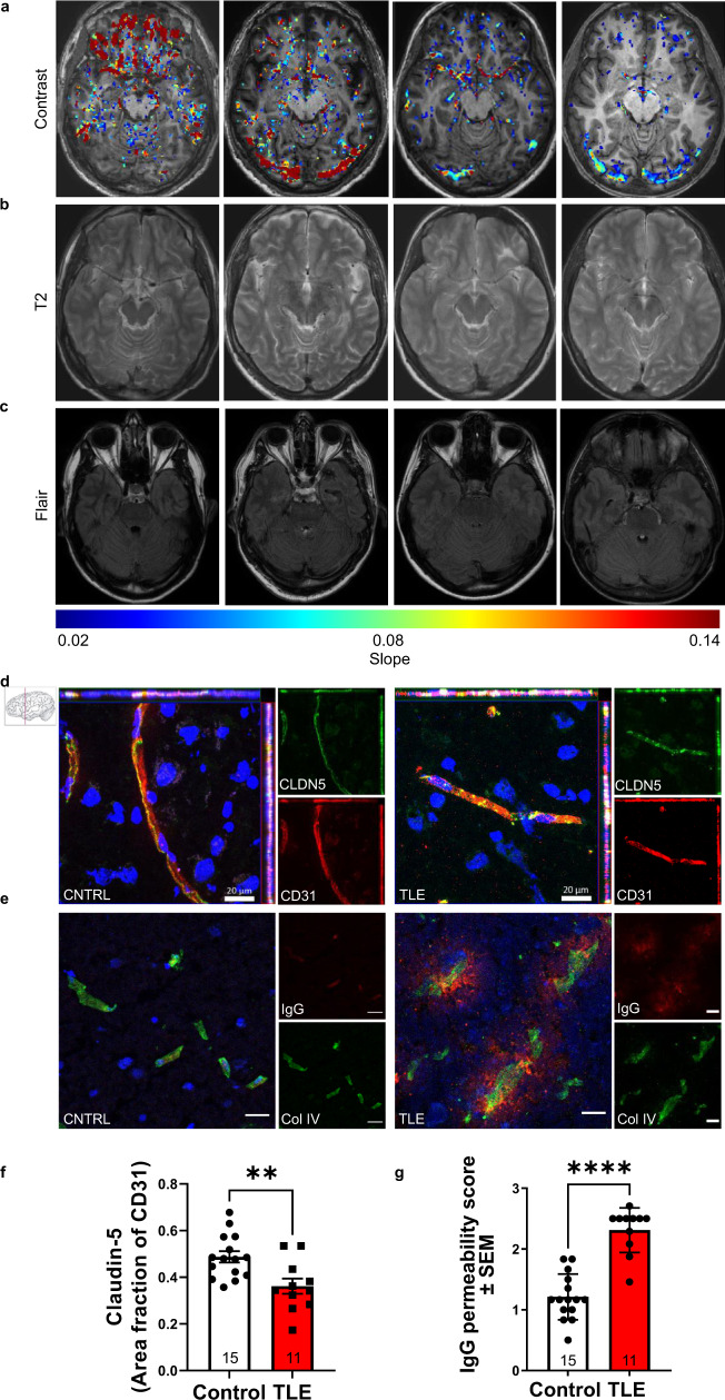
Blood-brain barrier (BBB) dysfunction is associated with worse epilepsy outcomes however the underlying molecular mechanisms of BBB dysfunction remain to be elucidated. Tight junction proteins are important regulators of BBB integrity and in particular, the tight junction protein claudin-5 is the most enriched in brain endothelial cells and regulates size-selectivity at the BBB. Additionally, disruption of claudin-5 expression has been implicated in numerous disorders including schizophrenia, depression and traumatic brain injury, yet its role in epilepsy has not been fully deciphered. Here we report that claudin-5 protein levels are significantly diminished in surgically resected brain tissue from patients with treatment-resistant epilepsy. Concomitantly, dynamic contrast-enhanced MRI in these patients showed widespread BBB disruption. We show that targeted disruption of claudin-5 in the hippocampus or genetic heterozygosity of claudin-5 in mice exacerbates kainic acid-induced seizures and BBB disruption. Additionally, inducible knockdown of claudin-5 in mice leads to spontaneous recurrent seizures, severe neuroinflammation, and mortality. Finally, we identify that RepSox, a regulator of claudin-5 expression, can prevent seizure activity in experimental epilepsy. Altogether, we propose that BBB stabilizing drugs could represent a new generation of agents to prevent seizure activity in epilepsy patients.

Overview

The claims and statistics of this paper haven’t been checked yet.

Run the check — free, no account needed. It reads the open-access text and checks the sources it cites.

A few free checks a day · papers already verified open instantly · results are public

Integrity signals

Registry

Crossref · 10.1038/s41467-022-29657-y

Cites

5 of 5 references verified0 citation errors · 0 not checkable

Statistics

Figures

Fig. 1 Drug resistant temporal lobe epilepsy induces blood-brain barrier leakage and Cldn5 downregulation in the hippocampus. a Representative dynamic contrast-enhanced MRI (DCE-MRI) images from 4 patients with temporal lobe epilepsy (TLE, top panel). Colour bar represents slope of contrast agent. b T2 weighted images (middle panel) and (c) fluid attenuated inversion recovery (FLAIR) images (bottom panel). d Claudin-5 (CLDN5; green) and CD31 (red) staining in non-diseased autopsy control (CNTRL) and TLE cases. Scale bars, 20 μm. e Collagen-IV (ColIV; green) and IgG (red) staining in CNTRL and TLE cases. Scale bars, 50 μm. f Levels of CLDN5 were significantly decreased (**p = 0.0042) and g IgG was significantly increased (****p < 0.0001) in the brains of TLE cases vs CNTRL. Data represent means ± s.e.m.; each datapoint represents one patient. Number of subjects (n) is indicated on graphs. Two-sided Mann–Whitney test for immunohistochemical and western blot analysis. **p < 0.01; ****p < 0.0001. Source data are provided as a Source Data file.
Fig. 1 Drug resistant temporal lobe epilepsy induces blood-brain barrier leakage and Cldn5 downregulation in the hippocampus. a Representative dynamic contrast-enhanced MRI (DCE-MRI) images from 4 patients with temporal lobe epilepsy (TLE, top panel). Colour bar represents slope of contrast agent. b T2 weighted images (middle panel) and (c) fluid attenuated inversion recovery (FLAIR) images (bottom panel). d Claudin-5 (CLDN5; green) and CD31 (red) staining in non-diseased autopsy control (CNTRL) and TLE cases. Scale bars, 20 μm. e Collagen-IV (ColIV; green) and IgG (red) staining in CNTRL and TLE cases. Scale bars, 50 μm. f Levels of CLDN5 were significantly decreased (**p = 0.0042) and g IgG was significantly increased (****p < 0.0001) in the brains of TLE cases vs CNTRL. Data represent means ± s.e.m.; each datapoint represents one patient. Number of subjects (n) is indicated on graphs. Two-sided Mann–Whitney test for immunohistochemical and western blot analysis. **p < 0.01; ****p < 0.0001. Source data are provided as a Source Data file.CC BY · PubMed Central
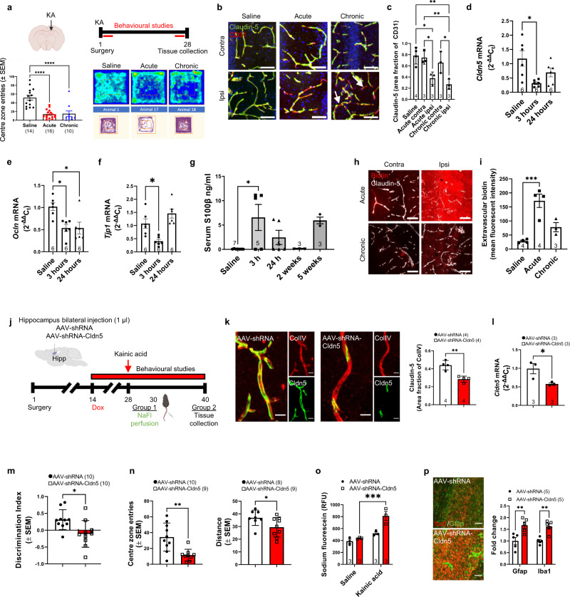
Fig. 2 Conditional knockdown of Cldn5 expression in the hippocampus exacerbates kainic acid-induced epilepsy. a Kainic acid (KA) injection site and timeline of experiment (top). Figure Created with BioRender. Open field testing of mice post KA injection with significant decreases in the number of entries in the centre zone (bottom) at both timepoints compared to saline (****p < 0.0001 for acute and chronic). b Claudin-5 (green) and CD31 (red) staining in the hippocampus in saline injected animals and the acute and chronic stages post KA injection. Scale bars, 50 μm. c Levels of claudin-5 expression were significantly decreased following KA injection at the acute (*p = 0.0187) and chronic stage (**p = 0.0036). d Cldn5 (*p = 0.0102), e Ocln (*p = 0.0124) and f Tjp1 (*p = 0.0129) mRNA levels were decreased at 3 h and 24 h post KA injection. g S100B levels were significantly increased in serum of mice 3 h post KA injection (*p = 0.011). h Immunohistochemistry for Sulfo-NHS-Biotin and claudin-5 following intrahippocampal injection of KA. Scale bars, 50 μm. i Biotin extravasated into the brain at the acute stage post injection of KA (***p = 0.0007). Figure Created with BioRender. j AAV expressing claudin-5 shRNA injected bilaterally into the hippocampus and timeline of experiment. k Claudin-5 protein (**p = 0.0024) (green) and l mRNA (*p = 0.043) was significantly decreased in brain endothelial cells (red) following AAV-shRNA-Cldn5 injection in the hippocampus of mice compared to that in AAV-shRNA-injected mice, following doxycycline (Dox) treatment. Scale bar, 20 μm. m Reduced performance in the novel object recognition (*p = 0.0146) and n open field test (**p = 0.0023) post claudin-5 suppression in KA injected mice. o Increased sodium fluorescein (NaFl) extravasation post claudin-5 suppression in KA injected mice (***p = 0.0006). p Increased IBA1 positive microglia (**p = 0.0044) and GFAP positive astrocytes (**p = 0.0024) post claudin-5 suppression in kainic acid injected mice. Scale bar, 20 μm. Data represent means ± s.e.m.; each datapoint represents one animal. Number of animals (n) is indicated on graphs. Two-way ANOVA followed by Bonferroni’s multiple comparison test for sodium fluorescein permeability; two-sided unpaired t-test for AAV experiments; one-way ANOVA followed by Tukey’s multiple comparison test for all other graphs. *p < 0.05; **p < 0.01; ***p < 0.001; ****p < 0.0001. Source data are provided as a Source Data file.
Fig. 2 Conditional knockdown of Cldn5 expression in the hippocampus exacerbates kainic acid-induced epilepsy. a Kainic acid (KA) injection site and timeline of experiment (top). Figure Created with BioRender. Open field testing of mice post KA injection with significant decreases in the number of entries in the centre zone (bottom) at both timepoints compared to saline (****p < 0.0001 for acute and chronic). b Claudin-5 (green) and CD31 (red) staining in the hippocampus in saline injected animals and the acute and chronic stages post KA injection. Scale bars, 50 μm. c Levels of claudin-5 expression were significantly decreased following KA injection at the acute (*p = 0.0187) and chronic stage (**p = 0.0036). d Cldn5 (*p = 0.0102), e Ocln (*p = 0.0124) and f Tjp1 (*p = 0.0129) mRNA levels were decreased at 3 h and 24 h post KA injection. g S100B levels were significantly increased in serum of mice 3 h post KA injection (*p = 0.011). h Immunohistochemistry for Sulfo-NHS-Biotin and claudin-5 following intrahippocampal injection of KA. Scale bars, 50 μm. i Biotin extravasated into the brain at the acute stage post injection of KA (***p = 0.0007). Figure Created with BioRender. j AAV expressing claudin-5 shRNA injected bilaterally into the hippocampus and timeline of experiment. k Claudin-5 protein (**p = 0.0024) (green) and l mRNA (*p = 0.043) was significantly decreased in brain endothelial cells (red) following AAV-shRNA-Cldn5 injection in the hippocampus of mice compared to that in AAV-shRNA-injected mice, following doxycycline (Dox) treatment. Scale bar, 20 μm. m Reduced performance in the novel object recognition (*p = 0.0146) and n open field test (**p = 0.0023) post claudin-5 suppression in KA injected mice. o Increased sodium fluorescein (NaFl) extravasation post claudin-5 suppression in KA injected mice (***p = 0.0006). p Increased IBA1 positive microglia (**p = 0.0044) and GFAP positive astrocytes (**p = 0.0024) post claudin-5 suppression in kainic acid injected mice. Scale bar, 20 μm. Data represent means ± s.e.m.; each datapoint represents one animal. Number of animals (n) is indicated on graphs. Two-way ANOVA followed by Bonferroni’s multiple comparison test for sodium fluorescein permeability; two-sided unpaired t-test for AAV experiments; one-way ANOVA followed by Tukey’s multiple comparison test for all other graphs. *p < 0.05; **p < 0.01; ***p < 0.001; ****p < 0.0001. Source data are provided as a Source Data file.CC BY · PubMed Central
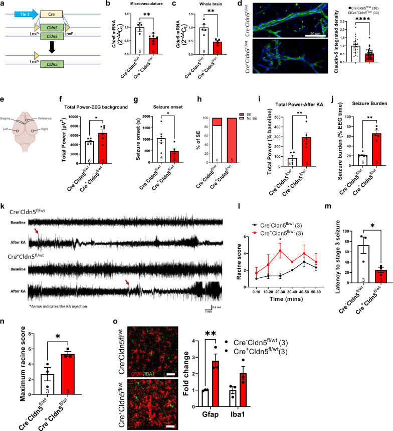
Fig. 3 Cldn5 heterozygous mice have reduced threshold for kainic acid-evoked seizures. a Schematic overview of Cldn5fl/wt mouse. b Cldn5 transcript levels were significantly decreased Cre+Cldn5fl/wt mice in isolated microvasculature (**p = 0.0023) and c whole brain (**p = 0.0014). d Claudin-5 protein expression significantly decreased in isolated microvessels of Cre+Cldn5fl/wt mice (****p < 0.0001). Scale bar, 50 µm. e Schematic representation of electrode placement for electroencephalography (EEG) studies (top). f Increased total power in Cre+Cldn5fl/wt mice prior to kainic acid (KA) injection (*p = 0.0311). g Reduced latency to the first electrographic seizure (*p = 0.0432) in Cre+Cldn5fl/wt mice. h Percentage of mice entering status epilepticus (SE) following kainic acid injection. i Increased total power in Cre+Cldn5fl/wt mice following mildly convulsant KA injection (**p = 0.0015). j Increased seizure burden in Cre+Cldn5fl/wt mice following KA injection (**p = 0.0043). k Representative EEG recording post mildly convulsive KA injection. l Increased Racine score over time in Cre+Cldn5fl/wt mice (*p = 0.0148 at 20–30 min post KA injection). m Decreased latency to stage 3 seizures in Cre+Cldn5fl/wt mice (*=0.0459). n Increased racine score in Cre+Cldn5fl/wt mice post injection of KA (*p = 0.0474). o Increased GFAP (red) expression in Cre+Cldn5fl/wt mice (**p = 0.0072). IBA1 (green) expression did not change (p = 0.0894). Scale bars, 50 μm. Data represent means ± s.e.m.; each data point represents one animal or vessel. Number of animals (n) is indicated on graphs. Two-way ANOVA followed by Bonferroni’s multiple comparison test for Racine score over time; two-sided Mann–Whitney test for seizure burden; two-sided unpaired t-test for all other graphs. *p < 0.05; **p < 0.01; ****p < 0.0001. Source data are provided as a Source Data file.
Fig. 3 Cldn5 heterozygous mice have reduced threshold for kainic acid-evoked seizures. a Schematic overview of Cldn5fl/wt mouse. b Cldn5 transcript levels were significantly decreased Cre+Cldn5fl/wt mice in isolated microvasculature (**p = 0.0023) and c whole brain (**p = 0.0014). d Claudin-5 protein expression significantly decreased in isolated microvessels of Cre+Cldn5fl/wt mice (****p < 0.0001). Scale bar, 50 µm. e Schematic representation of electrode placement for electroencephalography (EEG) studies (top). f Increased total power in Cre+Cldn5fl/wt mice prior to kainic acid (KA) injection (*p = 0.0311). g Reduced latency to the first electrographic seizure (*p = 0.0432) in Cre+Cldn5fl/wt mice. h Percentage of mice entering status epilepticus (SE) following kainic acid injection. i Increased total power in Cre+Cldn5fl/wt mice following mildly convulsant KA injection (**p = 0.0015). j Increased seizure burden in Cre+Cldn5fl/wt mice following KA injection (**p = 0.0043). k Representative EEG recording post mildly convulsive KA injection. l Increased Racine score over time in Cre+Cldn5fl/wt mice (*p = 0.0148 at 20–30 min post KA injection). m Decreased latency to stage 3 seizures in Cre+Cldn5fl/wt mice (*=0.0459). n Increased racine score in Cre+Cldn5fl/wt mice post injection of KA (*p = 0.0474). o Increased GFAP (red) expression in Cre+Cldn5fl/wt mice (**p = 0.0072). IBA1 (green) expression did not change (p = 0.0894). Scale bars, 50 μm. Data represent means ± s.e.m.; each data point represents one animal or vessel. Number of animals (n) is indicated on graphs. Two-way ANOVA followed by Bonferroni’s multiple comparison test for Racine score over time; two-sided Mann–Whitney test for seizure burden; two-sided unpaired t-test for all other graphs. *p < 0.05; **p < 0.01; ****p < 0.0001. Source data are provided as a Source Data file.CC BY · PubMed Central
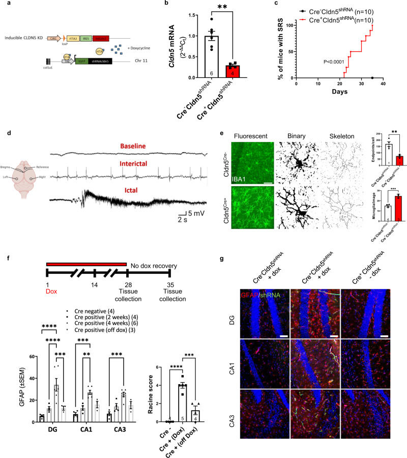
Fig. 4 Inducible Cldn5 knockdown mice develop spontaneous seizures and neuroinflammation. a Schematic outline of inducible knockdown mouse model. b Cldn5 significantly suppressed following 4 weeks of doxycycline (Dox) treatment (**p = 0.001). c Percentage of mice with spontaneous recurrent seizures (SRS) following 4 weeks of Dox treatment (****p < 0.0001). d Representative EEG traces following 4 weeks of Dox treatment. e IBA1 (green) quantification post claudin-5 suppression with decreased cellular endpoints (**p = 0.0088), and increased IBA1 positive cells (***p = 0.0005). Scale bar, 50 µm. f, g Experimental outline for recovery experiment. Increased GFAP levels in the dentate gyrus (DG ****p < 0.0001), CA1 (***p = 0.0001) and CA3 (***p = 0.0007) of mice following 4 weeks of doxycycline treatment are reversed following doxycycline withdrawal in the DG (***p = 0.0002 vs Cre positive 4 weeks). Scale bar, 100 µm. Increased seizure behaviour as scored according to Racine scale (****p < 0.0001 for Cre− vs Cre+ (Dox)) decreased following Dox removal (***p = 0.0004 for Cre+ (Dox) vs Cre+ (off Dox). Data represent means ± s.e.m.; each datapoint represents one animal. Number of animals (n) is indicated on graphs. Mantel–Cox test for SRS analysis; two-sided unpaired t-test for qPCR and microglia morphology; two-way ANOVA followed by Bonferroni’s multiple comparison test for reversibility study. **p < 0.01; ***p < 0.001; ****p < 0.0001. Source data are provided as a Source Data file.
Fig. 4 Inducible Cldn5 knockdown mice develop spontaneous seizures and neuroinflammation. a Schematic outline of inducible knockdown mouse model. b Cldn5 significantly suppressed following 4 weeks of doxycycline (Dox) treatment (**p = 0.001). c Percentage of mice with spontaneous recurrent seizures (SRS) following 4 weeks of Dox treatment (****p < 0.0001). d Representative EEG traces following 4 weeks of Dox treatment. e IBA1 (green) quantification post claudin-5 suppression with decreased cellular endpoints (**p = 0.0088), and increased IBA1 positive cells (***p = 0.0005). Scale bar, 50 µm. f, g Experimental outline for recovery experiment. Increased GFAP levels in the dentate gyrus (DG ****p < 0.0001), CA1 (***p = 0.0001) and CA3 (***p = 0.0007) of mice following 4 weeks of doxycycline treatment are reversed following doxycycline withdrawal in the DG (***p = 0.0002 vs Cre positive 4 weeks). Scale bar, 100 µm. Increased seizure behaviour as scored according to Racine scale (****p < 0.0001 for Cre− vs Cre+ (Dox)) decreased following Dox removal (***p = 0.0004 for Cre+ (Dox) vs Cre+ (off Dox). Data represent means ± s.e.m.; each datapoint represents one animal. Number of animals (n) is indicated on graphs. Mantel–Cox test for SRS analysis; two-sided unpaired t-test for qPCR and microglia morphology; two-way ANOVA followed by Bonferroni’s multiple comparison test for reversibility study. **p < 0.01; ***p < 0.001; ****p < 0.0001. Source data are provided as a Source Data file.CC BY · PubMed Central
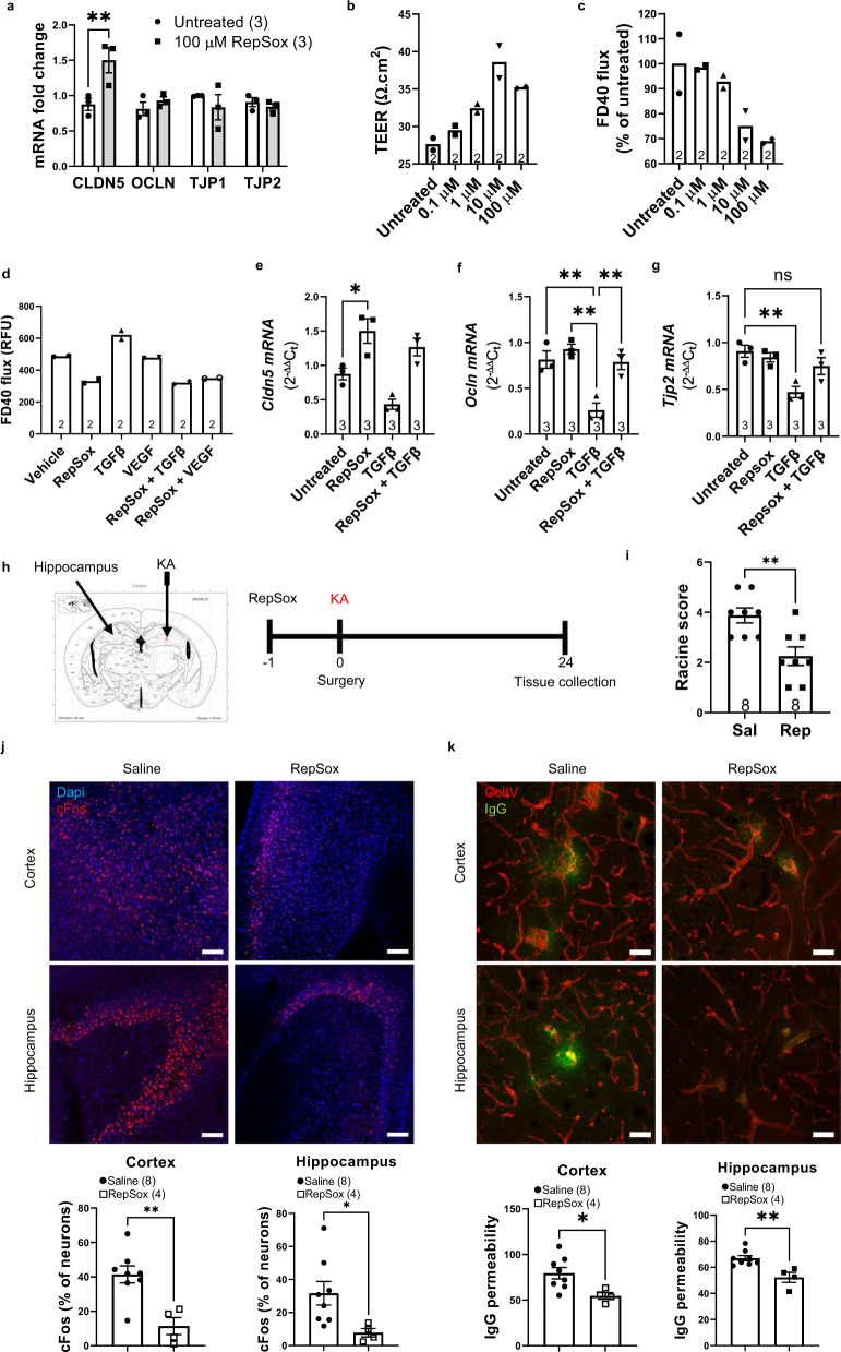
Fig. 5 ALK5 inhibition induces Cldn5 expression and attenuates kainic acid-induced seizures and BBB leakiness. a Increased Cldn5 expression 24 h post treatment with RepSox (100 μM) (**p = 0.0029). b Increased trans-endothelial electrical resistance (TEER) with increasing doses of RepSox. c Decreased flux of FITC-Dextran-40 (FD40) with increasing doses of RepSox. d Decreased flux of FD40 following exposure of endothelial cells to transforming growth factor-β1 (TGFβ) or vascular endothelial growth factor (VEGF) in combination with 100 µM RepSox. e RepSox significantly increased Cldn5 mRNA (*p = 0.0182) without changing (f) Ocln and (g) Tjp2 expression. h Experimental outline of RepSox treatment in the intrahippocampal kainic acid (KA) induced seizure model. i Reduced Racine score in RepSox treated mice (**p = 0.0039) compared to saline (Sal). j Decreased cFos expression in the cortex (**p = 0.0036) and hippocampus (*p = 0.0461) post treatment with 10 mg/kg RepSox. Scale bar, 100 μm. k Decreased IgG (green) extravasation in the cortex (*p = 0.0243) and hippocampus (**p = 0.0038) post treatment with 10 mg/kg RepSox. Scale bar, 100 μm. Data represent means ± s.e.m.; number of independent experiments or animals (n) is indicated on graphs. Two-way ANOVA followed by Bonferroni’s multiple comparison test for tight junction qPCRs; one-way ANOVA followed by Tukey’s multiple comparison test for cell culture experiments; two-sided unpaired t-test for in vivo studies. *p < 0.05; **p < 0.01; ****p < 0.0001. Source data are provided as a Source Data file.
Fig. 5 ALK5 inhibition induces Cldn5 expression and attenuates kainic acid-induced seizures and BBB leakiness. a Increased Cldn5 expression 24 h post treatment with RepSox (100 μM) (**p = 0.0029). b Increased trans-endothelial electrical resistance (TEER) with increasing doses of RepSox. c Decreased flux of FITC-Dextran-40 (FD40) with increasing doses of RepSox. d Decreased flux of FD40 following exposure of endothelial cells to transforming growth factor-β1 (TGFβ) or vascular endothelial growth factor (VEGF) in combination with 100 µM RepSox. e RepSox significantly increased Cldn5 mRNA (*p = 0.0182) without changing (f) Ocln and (g) Tjp2 expression. h Experimental outline of RepSox treatment in the intrahippocampal kainic acid (KA) induced seizure model. i Reduced Racine score in RepSox treated mice (**p = 0.0039) compared to saline (Sal). j Decreased cFos expression in the cortex (**p = 0.0036) and hippocampus (*p = 0.0461) post treatment with 10 mg/kg RepSox. Scale bar, 100 μm. k Decreased IgG (green) extravasation in the cortex (*p = 0.0243) and hippocampus (**p = 0.0038) post treatment with 10 mg/kg RepSox. Scale bar, 100 μm. Data represent means ± s.e.m.; number of independent experiments or animals (n) is indicated on graphs. Two-way ANOVA followed by Bonferroni’s multiple comparison test for tight junction qPCRs; one-way ANOVA followed by Tukey’s multiple comparison test for cell culture experiments; two-sided unpaired t-test for in vivo studies. *p < 0.05; **p < 0.01; ****p < 0.0001. Source data are provided as a Source Data file.CC BY · PubMed Central

Verification

Claims the paper makes

The claims and statistics of this paper haven’t been checked yet.

Run the check — free, no account needed. It reads the open-access text and checks the sources it cites.

A few free checks a day · papers already verified open instantly · results are public

Claims other papers make about this one

Each is checked against this paper’s own text: does it say what the citing paper says it does?

Self-check

Percentages, sums and reported differences re-computed from the paper’s own numbers.

The self-check needs the paper’s full text.

Citations

Cites

Show all 5 references

Research works printed but not linked: 45

  1. Kwan, P., Schachter, S. C. & Brodie, M. J. Drug-resistant epilepsy. N. Engl. J. Med. 365, 919–926 (2011). · DOI
  2. Thijs, R. D., Surges, R., O’Brien, T. J. & Sander, J. W. Epilepsy in adults. Lancet 393, 689–701 (2019). · DOI
  3. Tomkins, O. et al. Blood-brain barrier breakdown following traumatic brain injury: a possible role in posttraumatic epilepsy. Cardiovasc. Psychiatry Neurol. 2011, 765923 (2011). · DOI
  4. Herman, S. T. Epilepsy after brain insult: targeting epileptogenesis. Neurology 59, S21–S26 (2002). · DOI
  5. van Vliet, E. A., Aronica, E. & Gorter, J. A. Blood-brain barrier dysfunction, seizures and epilepsy. Semin. Cell Dev. Biol. 38, 26–34 (2015). · DOI
  6. van Vliet, E. A. et al. Blood-brain barrier leakage may lead to progression of temporal lobe epilepsy. Brain J. Neurol. 130, 521–534 (2007). · DOI · Paper page
  7. Marchi, N. et al. Seizure-promoting effect of blood-brain barrier disruption. Epilepsia 48, 732–742 (2007). · DOI
  8. Rüber, T. et al. Evidence for peri-ictal blood-brain barrier dysfunction in patients with epilepsy. Brain J. Neurol. 141, 2952–2965 (2018). · DOI
  9. Abbott, N. J., Patabendige, A. A., Dolman, D. E., Yusof, S. R. & Begley, D. J. Structure and function of the blood-brain barrier. Neurobiol. Dis. 37, 13–25 (2010). · DOI · Paper page
  10. Williams, S. et al. Neurovascular drug biotransformation machinery in focal human epilepsies: brain CYP3A4 correlates with seizure frequency and antiepileptic drug therapy. Mol. Neurobiol. 56, 8392–8407 (2019). · DOI
  11. Yan, B. C. et al. Changes in the blood-brain barrier function are associated with hippocampal neuron death in a kainic acid mouse model of epilepsy. Front. Neurol. 9, 775–775 (2018). · DOI
  12. Rempe, R. G. et al. Matrix metalloproteinase-mediated blood-brain barrier dysfunction in epilepsy. J. Neurosci. Off. J. Soc. Neurosci. 38, 4301–4315 (2018). · DOI · Paper page
  13. Weissberg, I. et al. Albumin induces excitatory synaptogenesis through astrocytic TGF-beta/ALK5 signaling in a model of acquired epilepsy following blood-brain barrier dysfunction. Neurobiol. Dis. 78, 115–125 (2015). · DOI
  14. Morin-Brureau, M. et al. Epileptiform activity induces vascular remodeling and zonula occludens 1 downregulation in organotypic hippocampal cultures: role of VEGF signaling pathways. J. Neurosci.: Off. J. Soc. Neurosci. 31, 10677–10688 (2011). · DOI · Paper page
  15. Munji, R. N. et al. Profiling the mouse brain endothelial transcriptome in health and disease models reveals a core blood-brain barrier dysfunction module. Nat. Neurosci. 22, 1892–1902 (2019). · DOI
  16. Castro Dias, M. et al. Claudin-12 is not required for blood-brain barrier tight junction function. Fluids Barriers CNS 16, 30 (2019). · DOI
  17. Castro Dias, M. et al. Claudin-3-deficient C57BL/6J mice display intact brain barriers. Sci. Rep. 9, 203 (2019). · DOI
  18. Greene, C. et al. Dose-dependent expression of claudin-5 is a modifying factor in schizophrenia. Mol. Psychiatry 23, 2156–2166 (2018). · DOI · Paper page
  19. Blyth, B. J. et al. Validation of serum markers for blood-brain barrier disruption in traumatic brain injury. J. Neurotrauma 26, 1497–1507 (2009). · DOI
  20. Kevil, C. G. et al. Intercellular adhesion molecule-1 (ICAM-1) regulates endothelial cell motility through a nitric oxide-dependent pathway. J. Biol. Chem. 279, 19230–19238 (2004). · DOI
  21. Roudnicky, F. et al. Inducers of the endothelial cell barrier identified through chemogenomic screening in genome-edited hPSC-endothelial cells. Proc. Natl Acad. Sci. USA 117, 19854–19865 (2020). · DOI
  22. Liu, J. Y. et al. Neuropathology of the blood-brain barrier and pharmaco-resistance in human epilepsy. Brain J. Neurol. 135, 3115–3133 (2012). · DOI
  23. Seiffert, E. et al. Lasting blood-brain barrier disruption induces epileptic focus in the rat somatosensory cortex. J. Neurosci. Off. J. Soc. Neurosci. 24, 7829–7836 (2004). · DOI
  24. Marchi, N. et al. Blood-brain barrier damage and brain penetration of antiepileptic drugs: role of serum proteins and brain edema. Epilepsia 50, 664–677 (2009). · DOI
  25. Hüls, A. et al. Brain DNA methylation patterns in CLDN5 associated with cognitive decline. Biol. Psychiatry, https://doi.org/10.1016/j.biopsych.2021.01.015 (2021). · DOI
  26. Xu, D. et al. Peripherally derived T regulatory and γδ T cells have opposing roles in the pathogenesis of intractable pediatric epilepsy. J. Exp. Med. 215, 1169–1186 (2018). · DOI
  27. Fabene, P. F. et al. A role for leukocyte-endothelial adhesion mechanisms in epilepsy. Nat. Med. 14, 1377–1383 (2008). · DOI
  28. Löscher, W. & Friedman, A. Structural, molecular, and functional alterations of the blood-brain barrier during epileptogenesis and epilepsy: a cause, consequence, or both? Int J. Mol. Sci. 21, 591 (2020). · DOI
  29. Volk, H. A. & Löscher, W. Multidrug resistance in epilepsy: rats with drug-resistant seizures exhibit enhanced brain expression of P-glycoprotein compared with rats with drug-responsive seizures. Brain J. Neurol. 128, 1358–1368 (2005). · DOI
  30. Potschka, H., Volk, H. A. & Löscher, W. Pharmacoresistance and expression of multidrug transporter P-glycoprotein in kindled rats. Neuroreport 15, 1657–1661 (2004). · DOI
  31. Rizzi, M. et al. Limbic seizures induce P-glycoprotein in rodent brain: functional implications for pharmacoresistance. J. Neurosci. Off. J. Soc. Neurosci. 22, 5833–5839 (2002). · DOI
  32. Brandt, C., Bethmann, K., Gastens, A. M. & Löscher, W. The multidrug transporter hypothesis of drug resistance in epilepsy: Proof-of-principle in a rat model of temporal lobe epilepsy. Neurobiol. Dis. 24, 202–211 (2006). · DOI
  33. Feldmann, M. et al. P-glycoprotein expression and function in patients with temporal lobe epilepsy: a case-control study. Lancet Neurol. 12, 777–785 (2013). · DOI
  34. Greene, C., Hanley, N. & Campbell, M. Claudin-5: gatekeeper of neurological function. Fluids barriers CNS 16, 3 (2019). · DOI · Paper page
  35. Ma, S.-C. et al. Claudin-5 regulates blood-brain barrier permeability by modifying brain microvascular endothelial cell proliferation, migration, and adhesion to prevent lung cancer metastasis. CNS Neurosci. Ther. 23, 947–960 (2017). · DOI
  36. Yang, Z. et al. The tight junction protein Claudin-5 limits endothelial cell motility. J. Cell Sci. 134, https://doi.org/10.1242/jcs.248237 (2021). · DOI
  37. Nagatake, T. et al. Selective expression of claudin-5 in thymic endothelial cells regulates the blood-thymus barrier and T-cell export. Int. Immunol., https://doi.org/10.1093/intimm/dxaa069 (2020). · DOI
  38. Paul, D. et al. Appearance of claudin-5(+) leukocytes in the central nervous system during neuroinflammation: a novel role for endothelial-derived extracellular vesicles. J. Neuroinflammation 13, 292–292 (2016). · DOI
  39. Pulido, R. S. et al. Neuronal activity regulates blood-brain barrier efflux transport through endothelial circadian genes. Neuron 108, 937–952 (2020). e937. · DOI
  40. Honkura, N. et al. Intravital imaging-based analysis tools for vessel identification and assessment of concurrent dynamic vascular events. Nat. Commun. 9, 2746–2746 (2018). · DOI
  41. Lévesque, M. & Avoli, M. The kainic acid model of temporal lobe epilepsy. Neurosci. Biobehav. Rev. 37, 2887–2899 (2013). · DOI
  42. Campbell, M. et al. Systemic low-molecular weight drug delivery to pre-selected neuronal regions. EMBO Mol. Med. 3, 235–245 (2011). · DOI · Paper page
  43. Jimenez-Mateos, E. M. et al. Silencing microRNA-134 produces neuroprotective and prolonged seizure-suppressive effects. Nat. Med. 18, 1087–1094 (2012). · DOI
  44. Young, K. & Morrison, H. Quantifying microglia morphology from photomicrographs of immunohistochemistry prepared tissue using ImageJ. J. Vis. Exp. 57648, https://doi.org/10.3791/57648 (2018). · DOI
  45. O’Keeffe, E. et al. Dynamic blood-brain barrier regulation in mild traumatic brain injury. J. Neurotrauma 37, 347–356 (2020). · DOI

Cited by

Sorted by citations

Metadata

Authors
Greene, C., Hanley, N., Reschke, C. R., Reddy, A., Mäe, M. A., Connolly, R., Behan, C., O’Keeffe, E., Bolger, I., Hudson, N., Delaney, C., Farrell, M. A., O’Brien, D. F., Cryan, J., Brett, F. M., Beausang, A., Betsholtz, C., Henshall, D. C., Doherty, C. P., & Campbell, M.
Venue
Nature Communications
Published
2022-04-14
Volume
13
Issue
1
ISSN
2041-1723
Cited by
156
References
5
Language
en
Funders
Science Foundation Ireland (16/RC/3948)

About these checks

Registry, retraction, Cites and statistics results are computed, not judged, with one exception: a printed reference that no index lists is read once by a model before it counts as a citation error. Claims are extracted by a model and kept only when their quote appears verbatim in the text; each cited source is then checked, with a model’s help, for whether it says what the claim relies on. Those verdicts can be wrong — read the claim’s quote and the cited source before relying on one.

Statistics and arithmetic are recomputed, not judged.

Confidence bands are shown only for fields calibrated on our annotated test set. None is calibrated yet, so no band is shown.

What counts as a claim ›How we measure confidence ›How we check this ›

Are you an author, or do you see a problem with this page? Request a review