

The DOI matches its Crossref record; 3 of the 3 references it cites were found in a registry or an index.
Robust total X-ray scattering workflow to study correlated motion of proteins in crystals
Open accessPublished version of a preprint
Abstract
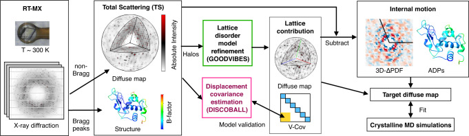
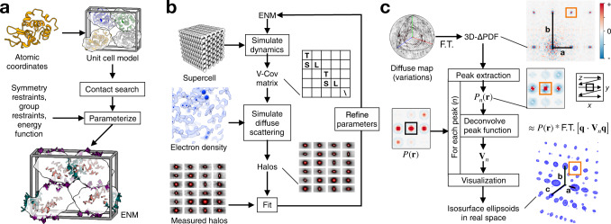
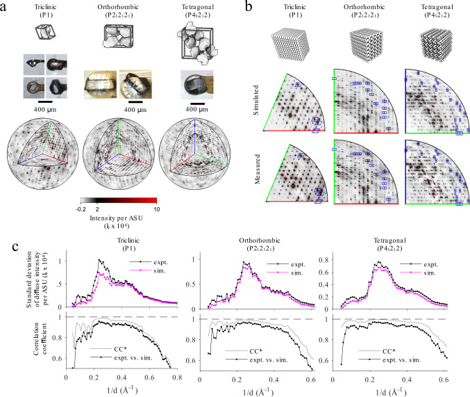
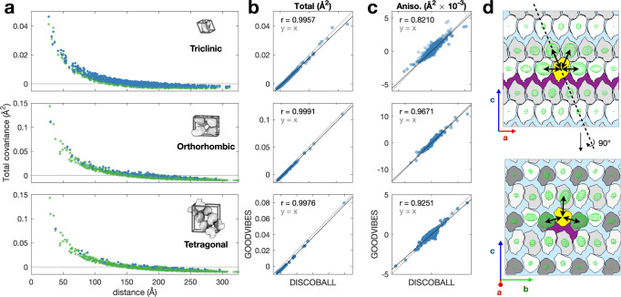
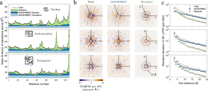
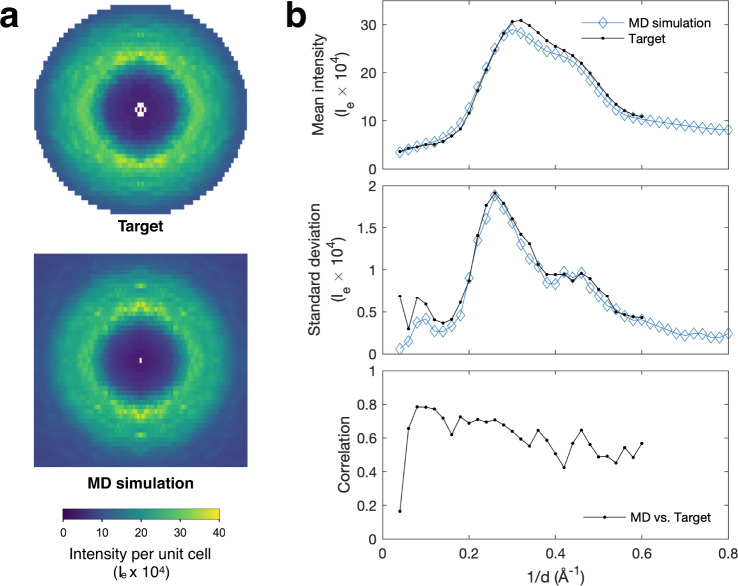
The breathing motions of proteins are thought to play a critical role in function. However, current techniques to study key collective motions are limited to spectroscopy and computation. We present a high-resolution experimental approach based on the total scattering from protein crystals at room temperature (TS/RT-MX) that captures both structure and collective motions. To reveal the scattering signal from protein motions, we present a general workflow that enables robust subtraction of lattice disorder. The workflow introduces two methods: GOODVIBES, a detailed and refinable lattice disorder model based on the rigid-body vibrations of a crystalline elastic network; and DISCOBALL, an independent method of validation that estimates the displacement covariance between proteins in the lattice in real space. Here, we demonstrate the robustness of this workflow and further demonstrate how it can be interfaced with MD simulations towards obtaining high-resolution insight into functionally important protein motions.
Overview
The claims and statistics of this paper haven’t been checked yet.
Run the check — free, no account needed. It reads the open-access text and checks the sources it cites.
Integrity signals
Registry
Cites
Statistics
Figures
Verification
Claims the paper makes
The claims and statistics of this paper haven’t been checked yet.
Run the check — free, no account needed. It reads the open-access text and checks the sources it cites.
Self-check
Percentages, sums and reported differences re-computed from the paper’s own numbers.
The self-check needs the paper’s full text.
Citations
Cites
Highly accurate protein structure prediction with AlphaFold
Jumper, J., Evans, R., Pritzel, A., Green, T., Figurnov, M., Ronneberger, O., Tunyasuvunakool, K., Bates, R., Žídek, A., Potapenko, A., et al. (2021) · Nature · doi:10.1038/s41586-021-03819-2 · Cited by 43,764
Accurate prediction of protein structures and interactions using a 3-track network
Baek, M., DiMaio, F., Anishchenko, I., Dauparas, J., Ovchinnikov, S., Lee, G. R., Wang, J., Cong, Q., Kinch, L. N., Schaeffer, R. D., et al. (2021) · doi:10.1101/2021.06.14.448402 · Cited by 5,549
Linking Crystallographic Model and Data Quality
Karplus, P. A., & Diederichs, K. (2012) · Science · doi:10.1126/science.1218231 · Cited by 1,781
Research works printed but not linked: 51
- Nogales, E. The development of cryo-EM into a mainstream structural biology technique. Nat. Methods 13, 24–27 (2016). · DOI
- Shoemaker, S. C. & Ando, N. X-rays in the cryo-electron microscopy era: structural biology’s dynamic future. Biochemistry 57, 277–285 (2018). · DOI
- Xu, D., Meisburger, S. P. & Ando, N. Correlated motions in structural biology. Biochemistry 60, 2331–2340 (2021). · DOI
- Welberry, T. R. & Weber, T. One hundred years of diffuse scattering. Crystallogr. Rev. 22, 2–78 (2016). · DOI
- Meisburger, S. P., Thomas, W. C., Watkins, M. B. & Ando, N. X-ray scattering studies of protein structural dynamics. Chem. Rev. 117, 7615–7672 (2017). · DOI
- Wall, M. E., Wolff, A. M. & Fraser, J. S. Bringing diffuse x-ray scattering into focus. Curr. Opin. Struct. Biol. 50, 109–116 (2018). · DOI
- Woldeyes, R. A., Sivak, D. A. & Fraser, J. S. E pluribus unum, no more: From one crystal, many conformations. Curr. Opin. Struct. Biol. 28, 56–62 (2014). · DOI
- Helliwell, J. R. Macromolecular crystallography with synchrotron radiation. (Cambridge University Press, 2005).
- Fischer, M. Macromolecular room temperature crystallography. Q. Rev. Biophys. 54, e1 (2021). · DOI
- Meisburger, S. P. & Ando, N. Correlated motions from crystallography beyond diffraction. Acc. Chem. Res. 50, 580–583 (2017). · DOI
- Meisburger, S. P., Case, D. A. & Ando, N. Diffuse X-ray scattering from correlated motions in a protein crystal. Nat. Commun. 11, 1271 (2020). · DOI
- Holton, J. M., Classen, S., Frankel, K. A. & Tainer, J. A. The R-factor gap in macromolecular crystallography: An untapped potential for insights on accurate structures. FEBS J. 281, 4046–4060 (2014). · DOI
- Janowski, P. A., Liu, C., Deckman, J. & Case, D. A. Molecular dynamics simulation of triclinic lysozyme in a crystal lattice. Protein Sci. 25, 87–102 (2016). · DOI
- Wall, M. E. Internal protein motions in molecular-dynamics simulations of Bragg and diffuse X-ray scattering. IUCrJ 5, 172–181 (2018). · DOI
- Press, W. H., Teukolsky, S. A., Vetterling, W. T. & Flannery, B. P. Numerical Recipes 3rd Edition: The Art of Scientific Computing. (Cambridge University Press, 2007).
- Meisburger, S. & Ando, N. X-ray diffraction data for: Hen lysozyme. PDB code 6O2H. https://doi.org/10.15785/SBGRID/747 (2020). · DOI
- Meisburger, S. P., Case, D. A. & Ando, N. Diffuse x-ray scattering from correlated motions in a protein crystal (CXIDB ID 128) https://doi.org/10.11577/1601281 (2020). · DOI
- Wall, M. E. et al. Conformational dynamics of a crystalline protein from microsecond-scale molecular dynamics simulations and diffuse X-ray scattering. Proc. Natl Acad. Sci. USA 111, 17887–17892 (2014). · DOI
- Agarwal, P. K., Billeter, S. R., Rajagopalan, P. R., Benkovic, S. J. & Hammes-Schiffer, S. Network of coupled promoting motions in enzyme catalysis. Proc. Natl Acad. Sci. USA 99, 2794–2799 (2002). · DOI
- Wang, L., Goodey, N. M., Benkovic, S. J. & Kohen, A. Coordinated effects of distal mutations on environmentally coupled tunneling in dihydrofolate reductase. Proc. Natl Acad. Sci. USA 103, 15753–15758 (2006). · DOI
- Boehr, D. D., McElheny, D., Dyson, H. J. & Wright, P. E. The dynamic energy landscape of dihydrofolate reductase catalysis. Science 313, 1638–1642 (2006). · DOI
- Bhabha, G. et al. A dynamic knockout reveals that conformational fluctuations influence the chemical step of enzyme catalysis. Science 332, 234–238 (2011). · DOI
- Stojković, V., Perissinotti, L. L., Willmer, D., Benkovic, S. J. & Kohen, A. Effects of the donor–acceptor distance and dynamics on hydride tunneling in the dihydrofolate reductase catalyzed reaction. J. Am. Chem. Soc. 134, 1738–1745 (2012). · DOI
- Caspar, D., Clarage, J., Salunke, D. & Clarage, M. Liquid-like movements in crystalline insulin. Nature 332, 659–662 (1988). · DOI
- Rose, Y. et al. RCSB protein data bank: architectural advances towards integrated searching and efficient access to macromolecular structure data from the PDB archive. J. Mol. Biol. 433, 166704 (2021). · DOI
- De Klijn, T., Schreurs, A. & Kroon-Batenburg, L. Rigid-body motion is the main source of diffuse scattering in protein crystallography. IUCrJ 6, 277–289 (2019). · DOI
- Peck, A., Poitevin, F. & Lane, T. J. Intermolecular correlations are necessary to explain diffuse scattering from protein crystals. IUCrJ 5, 211–222 (2018). · DOI
- Wych, D. C. et al. Molecular-dynamics simulation methods for macromolecular crystallography. Acta Crystallogr. D. Struct. Biol. 79, 50–65 (2023). · DOI
- Hura, G. et al. Water structure as a function of temperature from x-ray scattering experiments and ab initio molecular dynamics. Phys. Chem. Chem. Phys. 5, 1981–1991 (2003). · DOI
- Schanda, P. & Ernst, M. Studying dynamics by magic-angle spinning solid-state NMR spectroscopy: Principles and applications to biomolecules. Prog. Nucl. Magn. Reson. Spectrosc. 96, 1–46 (2016). · DOI
- Kabsch, W. XDS. Acta Crystallogr., Sect. D: Biol. Crystallogr. 66, 125–132 (2010). · DOI · Paper page
- Potterton, L. et al. CCP4i2: The new graphical user interface to the CCP4 program suite. Acta Crystallogr., Sect. D: Biol. Crystallogr. 74, 68–84 (2018). · DOI
- Winn, M. D. et al. Overview of the CCP4 suite and current developments. Acta Crystallogr., Sect. D: Biol. Crystallogr. 67, 235–242 (2011). · DOI · Paper page
- Evans, P. R. & Murshudov, G. N. How good are my data and what is the resolution? Acta Crystallogr., Sect. D: Biol. Crystallogr. 69, 1204–1214 (2013). · DOI
- McCoy, A. J. et al. Phaser crystallographic software. J. Appl. Crystallogr 40, 658–674 (2007). · DOI · Paper page
- Emsley, P., Lohkamp, B., Scott, W. G. & Cowtan, K. Features and development of coot. Acta Crystallogr., Sect. D: Biol. Crystallogr. 66, 486–501 (2010). · DOI · Paper page
- Murshudov, G. N. et al. REFMAC5 for the refinement of macromolecular crystal structures. Acta Crystallogr., Sect. D: Biol. Crystallogr. 67, 355–367 (2011). · DOI
- Chen, V. B. et al. MolProbity: all-atom structure validation for macromolecular crystallography. Acta Crystallogr., Sect. D: Biol. Crystallogr. 66, 12–21 (2010). · DOI · Paper page
- Schomaker, V. & Trueblood, K. N. On the rigid-body motion of molecules in crystals. Acta Crystallogr., Sect. B: Struct. Crystallogr. Cryst. Chem. 24, 63–76 (1968). · DOI
- Willis, B. T. M. Thermal diffuse scattering of X-rays and neutrons. in International Tables for Crystallography Volume B: Reciprocal space (ed. Shmueli, U.) 400–406 (Springer Netherlands, 2001). https://doi.org/10.1107/97809553602060000563 (2001). · DOI
- Born, M. & Huang, K. Dynamical theory of crystal lattices. (Clarendon Press, 1954).
- Wall, M. E., Clarage, J. B. & Phillips, G. N. Motions of calmodulin characterized using both bragg and diffuse x-ray scattering. Structure 5, 1599–1612 (1997). · DOI
- Maier, J. A. et al. ff14SB: improving the accuracy of protein side chain and backbone parameters from ff99SB. J. Chem. Theory Comput. 11, 3696–3713 (2015). · DOI
- Hornak, V. et al. Comparison of multiple Amber force fields and development of improved protein backbone parameters. Proteins: Struct., Funct., Bioinf 65, 712–725 (2006). · DOI
- Izadi, S., Anandakrishnan, R. & Onufriev, A. V. Building water models: a different approach. J. Phys. Chem. Lett. 5, 3863–3871 (2014). · DOI
- Crosas, E. et al. Uridine as a new scavenger for synchrotron-based structural biology techniques. J. Synchrotron Radiat. 24, 53–62 (2017). · DOI
- Hopkins, C. W., Le Grand, S., Walker, R. C. & Roitberg, A. E. Long-time-step molecular dynamics through hydrogen mass repartitioning. J. Chem. Theory Comput. 11, 1864–1874 (2015). · DOI
- Croarkin, C. 2.6.5.2.2. Bisquare weighting. NIST/SEMATECH e-Handbook of Statistical Methods https://doi.org/10.18434/M32189 (2012). · DOI
- Meisburger, S. & Ando, N. X-ray diffraction data for: Hen lysozyme. PDB code 8DYZ. https://doi.org/10.15785/SBGRID/957 (2022). · DOI
- Meisburger, S., Imran, SM. S. & Ando, N. X-ray diffraction data for: Hen lysozyme. PDB code 8DZ7. https://doi.org/10.15785/SBGRID/958 (2022). · DOI
- Morin, A. et al. Cutting edge: Collaboration gets the most out of software. eLife 2, e01456 (2013). · DOI
Metadata
- Authors
- Meisburger, S. P., Case, D. A., & Ando, N.
- Affiliations
- Cornell University (US)
- Rutgers, The State University of New Jersey (US)
- Corresponding author
- Ando, Nozomi
- Venue
- Nature Communications
- Published
- 2023-03-03
- Volume
- 14
- Issue
- 1
- Pages
- 1228-1228
- ISSN
- 2041-1723 (ISSN-L)
- PMC
- PMC9984388
- OpenAlex
- W4323048702
- Cited by
- 20
- References
- 3
- Citations by year
- 2026: 3 · 2025: 2 · 2024: 6 · 2023: 9
- FWCI
- 1.47
- Type
- journal article
- Language
- en
- Open access
- gold
- doi.org
- Locations
- Nature Communications (publishedVersion, cc-by)
- bioRxiv (Cold Spring Harbor Laboratory) (acceptedVersion, cc-by-nc-nd)
- PubMed Central (publishedVersion, cc-by)
- PubMed (publishedVersion)
- RePEc: Research Papers in Economics (submittedVersion)
- DOAJ (DOAJ: Directory of Open Access Journals) (submittedVersion, cc-by-sa)
- Funders
- National Science Foundation (DMR-1829070)
- U.S. Department of Health & Human Services | NIH | National Institute of General Medical Sciences (1-P30-GM124166-01A1)
- U.S. Department of Health & Human Services | National Institutes of Health (F32GM117757, GM122086, GM124847)
- Startup funds from Cornell University
- Topics
- Life Sciences › Biochemistry, Genetics and Molecular Biology › Structural Biology › Enzyme Structure and Function
- Life Sciences › Biochemistry, Genetics and Molecular Biology › Molecular Biology › Protein Structure and Dynamics
- Physical Sciences › Physics and Astronomy › Radiation › Advanced X-ray Imaging Techniques
- Keywords
- collective protein motions, protein crystals, rigid body vibration, elastic network model, protein function
- MeSH terms
- Motion, Radiography, Vibration, X-Rays, Workflow
About these checks
Registry, retraction, Cites and statistics results are computed, not judged, with one exception: a printed reference that no index lists is read once by a model before it counts as a citation error. Claims are extracted by a model and kept only when their quote appears verbatim in the text; each cited source is then checked, with a model’s help, for whether it says what the claim relies on. Those verdicts can be wrong — read the claim’s quote and the cited source before relying on one.
Statistics and arithmetic are recomputed, not judged.
Confidence bands are shown only for fields calibrated on our annotated test set. None is calibrated yet, so no band is shown.
What counts as a claim ›How we measure confidence ›How we check this ›
Are you an author, or do you see a problem with this page? Request a review
