

The DOI matches its Crossref record; 5 of the 5 references it cites were found in a registry or an index.
HLA3DB: comprehensive annotation of peptide/HLA complexes enables blind structure prediction of T cell epitopes
Open accessPublished version of a preprint
Abstract
The class I proteins of the major histocompatibility complex (MHC-I) display epitopic peptides derived from endogenous proteins on the cell surface for immune surveillance. Accurate modeling of peptides bound to the human MHC, HLA, has been mired by conformational diversity of the central peptide residues, which are critical for recognition by T cell receptors. Here, analysis of X-ray crystal structures within our curated database (HLA3DB) shows that pHLA complexes encompassing multiple HLA allotypes present a discrete set of peptide backbone conformations. Leveraging these backbones, we employ a regression model trained on terms of a physically relevant energy function to develop a comparative modeling approach for nonamer pHLA structures named RepPred. Our method outperforms the top pHLA modeling approach by up to 19% in structural accuracy, and consistently predicts blind targets not included in our training set. Insights from our work may be applied towards predicting antigen immunogenicity, and receptor cross-reactivity.
Overview
The claims and statistics of this paper haven’t been checked yet.
Run the check — free, no account needed. It reads the open-access text and checks the sources it cites.
Integrity signals
Registry
Cites
Statistics
Figures
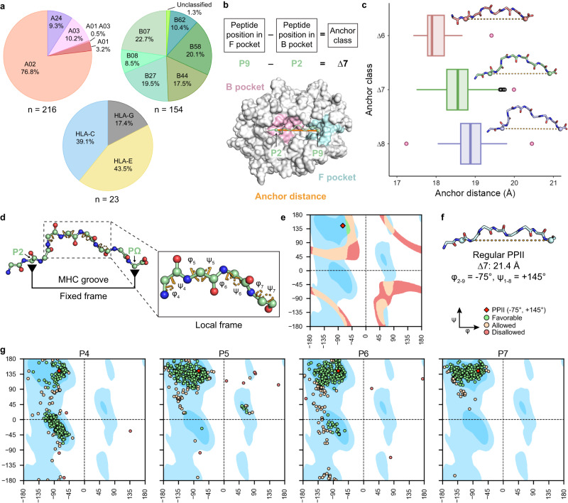
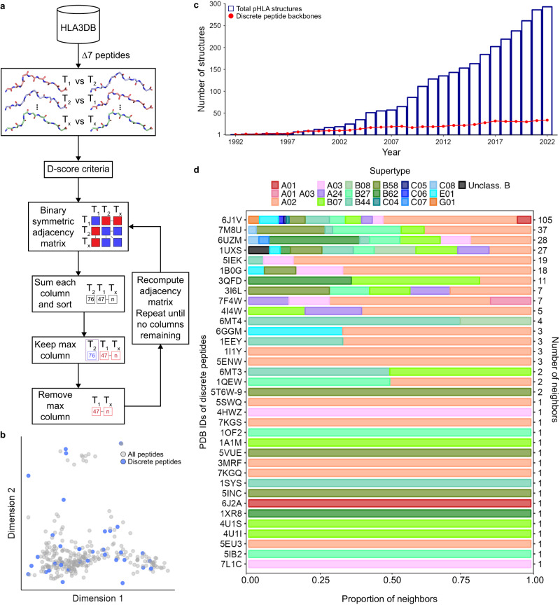
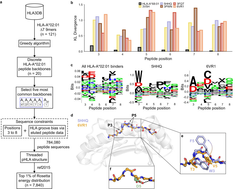
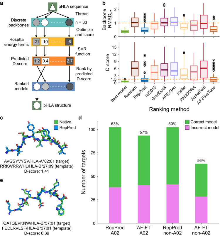
Verification
Claims the paper makes
The claims and statistics of this paper haven’t been checked yet.
Run the check — free, no account needed. It reads the open-access text and checks the sources it cites.
Self-check
Percentages, sums and reported differences re-computed from the paper’s own numbers.
The self-check needs the paper’s full text.
Citations
Cites
Highly accurate protein structure prediction with AlphaFold
Jumper, J., Evans, R., Pritzel, A., Green, T., Figurnov, M., Ronneberger, O., Tunyasuvunakool, K., Bates, R., Žídek, A., Potapenko, A., et al. (2021) · Nature · doi:10.1038/s41586-021-03819-2 · Cited by 43,764
Burley, S. K., Bhikadiya, C., Bi, C., Bittrich, S., Chen, L., Crichlow, G. V., Christie, C. H., Dalenberg, K., Di Costanzo, L., Duarte, J. M., et al. (2021) · Nucleic Acids Research · doi:10.1093/nar/gkaa1038 · Cited by 1,581
The Rosetta All-Atom Energy Function for Macromolecular Modeling and Design
Alford, R. F., Leaver-Fay, A., Jeliazkov, J. R., O’Meara, M. J., DiMaio, F. P., Park, H., Shapovalov, M. V., Renfrew, P. D., Mulligan, V. K., Kappel, K., et al. (2017) · Journal of Chemical Theory and Computation · doi:10.1021/acs.jctc.7b00125 · Cited by 1,532
A new clustering of antibody CDR loop conformations
North, B., Lehmann, A., & Dunbrack, R. L. (2011) · Journal of Molecular Biology · doi:10.1016/j.jmb.2010.10.030 · Cited by 380
Defining a new nomenclature for the structures of active and inactive kinases
Modi, V., & Dunbrack, R. L. (2019) · Proceedings of the National Academy of Sciences · doi:10.1073/pnas.1814279116 · Cited by 367
Research works printed but not linked: 83
- Rock, K. L., Reits, E. & Neefjes, J. Present yourself! By MHC class I and MHC class II molecules. Trends Immunol. 37, 724–737 (2016). · DOI · Paper page
- Mungall, A. J. et al. The DNA sequence and analysis of human chromosome 6. Nature 425, 805–811 (2003). · DOI
- Wieczorek, M. et al. Major histocompatibility complex (MHC) class I and MHC class II proteins: conformational plasticity in antigen presentation. Front. Immunol. 8, 292 (2017). · DOI
- Barker, D. J. et al. The IPD-IMGT/HLA database. Nucleic Acids Res. 51, D1053–D1060 (2023). · DOI
- Gfeller, D. & Bassani-Sternberg, M. Predicting antigen presentation—what could we learn from a million peptides? Front. Immunol. 9. https://doi.org/10.3389/fimmu.2018.01716 (2018). · DOI
- Petersdorf, E. W. HLA matching in allogeneic stem cell transplantation. Curr. Opin. Hematol. 11, 386–391 (2004). · DOI
- Nguyen, A. T., Szeto, C. & Gras, S. The pockets guide to HLA class I molecules. Biochem. Soc. Trans. 49, 2319–2331 (2021). · DOI
- Josephs, T. M., Grant, E. J. & Gras, S. Molecular challenges imposed by MHC-I restricted long epitopes on T cell immunity. Biol. Chem. 398, 1027–1036 (2017). · DOI
- Jurtz, V. et al. NetMHCpan-4.0: improved peptide-MHC class I interaction predictions integrating eluted ligand and peptide binding affinity data. J. Immunol. 199, 3360–3368 (2017). · DOI
- Riley, T. P. et al. Structure based prediction of neoantigen immunogenicity. Front. Immunol. 10. https://doi.org/10.3389/fimmu.2019.02047 (2019). · DOI
- Nerli, S. & Sgourakis, N. G. Structure-based modeling of SARS-CoV-2 Peptide/HLA-A02 antigens. Front. Med. Technol. 2. https://doi.org/10.3389/fmedt.2020.553478 (2020). · DOI
- Toor, J. S. et al. A recurrent mutation in anaplastic lymphoma kinase with distinct neoepitope conformations. Front. Immunol. 9. https://doi.org/10.3389/fimmu.2018.00099 (2018). · DOI
- Bjorkman, P. J., Strominger, J. L. & Wiley, D. C. Structure of the human class I histocompatibility antigen, HLA-A2. 7. Nature 329, 506–512 (1987). · DOI
- Berman, H. M. et al. The protein data bank. Nucleic Acids Res. 28, 235–242 (2000). · DOI · Paper page
- Antunes, D. A., Abella, J. R., Devaurs, D., Rigo, M. M. & Kavraki, L. E. Structure-based methods for binding mode and binding affinity prediction for peptide-MHC complexes. Curr. Top. Med. Chem. 18, 2239–2255 (2018). · DOI
- Antunes, D. A. et al. Structural allele-specific patterns adopted by epitopes in the MHC-I cleft and reconstruction of MHC:peptide complexes to cross-reactivity assessment. PLoS One 5, e10353 (2010). · DOI
- Khan, J. M. & Ranganathan, S. pDOCK: a new technique for rapid and accurate docking of peptide ligands to Major Histocompatibility Complexes. Immunome Res. 6, S2 (2010).
- Liu, T. et al. Subangstrom accuracy in pHLA-I modeling by Rosetta FlexPepDock refinement protocol. J. Chem. Inf. Model. 54, 2233–2242 (2014). · DOI
- Menegatti Rigo, M. et al. DockTope: a Web-based tool for automated pMHC-I modelling. Sci. Rep. 5, 18413 (2015). · DOI
- Todman, S. J. et al. Toward the atomistic simulation of T cell epitopes automated construction of MHC: peptide structures for free energy calculations. J. Mol. Graph Model 26, 957–961 (2008). · DOI
- Yanover, C. & Bradley, P. Large-scale characterization of peptide-MHC binding landscapes with structural simulations. Proc. Natl Acad. Sci. USA 108, 6981–6986 (2011). · DOI
- Bordner, A. J. & Abagyan, R. Ab initio prediction of peptide-MHC binding geometry for diverse class I MHC allotypes. Proteins 63, 512–526 (2006). · DOI
- Bui, H.-H., Schiewe, A. J., von Grafenstein, H. & Haworth, I. S. Structural prediction of peptides binding to MHC class I molecules. Proteins 63, 43–52 (2006). · DOI
- Kyeong, H.-H., Choi, Y. & Kim, H.-S. GradDock: rapid simulation and tailored ranking functions for peptide-MHC Class I docking. Bioinformatics 34, 469–476 (2018). · DOI
- Marzella, D. F. et al. PANDORA: a fast, anchor-restrained modelling protocol for peptide: MHC complexes. Front. Immunol. 13, 878762 (2022). · DOI
- Rognan, D., Lauemoller, S. L., Holm, A., Buus, S. & Tschinke, V. Predicting binding affinities of protein ligands from three-dimensional models: application to peptide binding to class I major histocompatibility proteins. J. Med. Chem. 42, 4650–4658 (1999). · DOI
- Tong, J. C., Tan, T. W. & Ranganathan, S. Modeling the structure of bound peptide ligands to major histocompatibility complex. Protein Sci. 13, 2523–2532 (2004). · DOI
- Antes, I., Siu, S. W. I. & Lengauer, T. DynaPred: a structure and sequence based method for the prediction of MHC class I binding peptide sequences and conformations. Bioinformatics 22, e16–e24 (2006). · DOI
- Keller, G. L. J., Weiss, L. I. & Baker, B. M. Physicochemical heuristics for identifying high fidelity, near-native structural models of Peptide/MHC complexes. Front. Immunol. 13, 887759 (2022). · DOI
- Rajeshwar, R. & Smith, J. C. Structural patterns in class 1 major histocompatibility complex-restricted nonamer peptide binding to T-cell receptors. Proteins https://doi.org/10.1002/prot.26343 (2022). · DOI
- Rudolph, M. G., Stanfield, R. L. & Wilson, I. A. How TCRs bind MHCs, peptides, and coreceptors. Annu. Rev. Immunol. 24, 419–466 (2006). · DOI
- Cole, D. K. et al. Hotspot autoimmune T cell receptor binding underlies pathogen and insulin peptide cross-reactivity. J. Clin. Invest. 126, 2191–2204 (2016). · DOI
- Abella, J. R., Antunes, D. A., Clementi, C. & Kavraki, L. E. APE-Gen: a fast method for generating ensembles of bound peptide-MHC conformations. Molecules 24, E881 (2019). · DOI
- Motmaen, A. et al. Peptide binding specificity prediction using fine-tuned protein structure prediction networks. Proc. Natl Acad. Sci. USA 120, e2216697120 (2023). · DOI · Paper page
- Antunes, D. A. et al. Interpreting T-cell cross-reactivity through structure: implications for TCR-based cancer immunotherapy. Front. Immunol. 8, 1210 (2017). · DOI
- Hellman, L. M. et al. Improving T cell receptor on-target specificity via structure-guided design. Mol. Ther. 27, 300–313 (2019). · DOI
- Mendes, M. F. et al. MatchTope: a tool to predict the cross reactivity of peptides complexed with Major Histocompatibility Complex I. Front. Immunol. 13, 930590 (2022). · DOI
- Mendes, M. F. A., Antunes, D. A., Rigo, M. M., Sinigaglia, M. & Vieira, G. F. Improved structural method for T-cell cross-reactivity prediction. Mol. Immunol. 67, 303–310 (2015). · DOI
- Schmidt, J. et al. Prediction of neo-epitope immunogenicity reveals TCR recognition determinants and provides insight into immunoediting. Cell Rep. Med. 2, 100194 (2021). · DOI
- Tarabini, R. F. et al. Large-scale structure-based screening of potential T cell cross-reactivities involving peptide-targets from BCG vaccine and SARS-CoV-2. Front. Immunol. 12, 812176 (2022). · DOI
- Menezes Teles e Oliveira, D. et al. pHLA3D: an online database of predicted three-dimensional structures of HLA molecules. Hum. Immunol. 80, 834–841 (2019). · DOI
- Ehrenmann, F., Kaas, Q. & Lefranc, M.-P. IMGT/3Dstructure-DB and IMGT/DomainGapAlign: a database and a tool for immunoglobulins or antibodies, T cell receptors, MHC, IgSF and MhcSF. Nucleic Acids Res. 38, D301–D307 (2010). · DOI
- Ehrenmann, F. & Lefranc, M.-P. IMGT/3Dstructure-DB: querying the IMGT database for 3D structures in immunology and immunoinformatics (IG or Antibodies, TR, MH, RPI, and FPIA). Cold Spring Harb. Protoc. 2011, pdb.prot5637 (2011). · DOI
- Kaas, Q., Ruiz, M. & Lefranc, M.-P. IMGT/3Dstructure-DB and IMGT/StructuralQuery, a database and a tool for immunoglobulin, T cell receptor and MHC structural data. Nucleic Acids Res. 32, D208–D210 (2004). · DOI
- Khan, J. M., Cheruku, H. R., Tong, J. C. & Ranganathan, S. MPID-T2: a database for sequence–structure–function analyses of pMHC and TR/pMHC structures. Bioinformatics 27, 1192–1193 (2011). · DOI
- Tong, J. C., Kong, L., Tan, T. W. & Ranganathan, S. MPID-T: database for sequence-structure-function information on T-cell receptor/peptide/MHC interactions. Appl Bioinforma. 5, 111–114 (2006). · DOI
- Teles e Oliveira, D. M. et al. pHLA3D: updating the database of predicted three-dimensional structures of HLA with HLA-DR, HLA-DQ and HLA-DP molecules. Hum. Immunol. 82, 8–10 (2021). · DOI
- Sinigaglia, M., Antunes, D. A., Rigo, M. M., Chies, J. A. B. & Vieira, G. F. CrossTope: a curate repository of 3D structures of immunogenic peptide: MHC complexes. Database (Oxf.) 2013, bat002 (2013).
- Li, X. et al. HLA3D: an integrated structure-based computational toolkit for immunotherapy. Brief Bioinform 23, bbac076 (2022). · DOI
- Sidney, J., Peters, B., Frahm, N., Brander, C. & Sette, A. HLA class I supertypes: a revised and updated classification. BMC Immunol. 9, 1 (2008). · DOI · Paper page
- Trolle, T. et al. The length distribution of class I restricted T cell epitopes is determined by both peptide supply and MHC allele specific binding preference. J. Immunol. 196, 1480–1487 (2016). · DOI
- Adzhubei, A. A., Sternberg, M. J. E. & Makarov, A. A. Polyproline-II Helix in Proteins: Structure and Function. J. Mol. Biol. 425, 2100–2132 (2013). · DOI
- Kufareva, I. & Abagyan, R. Methods of protein structure comparison. Methods Mol. Biol. 857, 231–257 (2012). · DOI
- Huang, P.-S. et al. RosettaRemodel: a generalized framework for flexible backbone protein design. PLoS One 6, e24109 (2011). · DOI
- Liu, Y. & Kuhlman, B. RosettaDesign server for protein design. Nucleic Acids Res. 34, W235–W238 (2006). · DOI
- Kim, Y., Sidney, J., Pinilla, C., Sette, A. & Peters, B. Derivation of an amino acid similarity matrix for peptide: MHC binding and its application as a Bayesian prior. BMC Bioinforma. 10, 394 (2009).
- Das, R. Four small puzzles that Rosetta doesn’t solve. PLoS One 6, e20044 (2011). · DOI
- Vita, R. et al. The immune epitope database (IEDB): 2018 update. Nucleic Acids Res. 47, D339–D343 (2019). · DOI
- Chersi, A., di Modugno, F. & Rosano, L. Flexibility of amino acid residues at position four of nonapeptides enhances their binding to human leucocyte antigen (HLA) molecules. Z. Naturforsch. C. J. Biosci. 55, 109–114 (2000). · DOI
- Strønen, E. et al. Targeting of cancer neoantigens with donor-derived T cell receptor repertoires. Science 352, 1337–1341 (2016). · DOI
- Allen, T. M. et al. Selective escape from CD8+ T-cell responses represents a major driving force of human immunodeficiency virus type 1 (HIV-1) sequence diversity and reveals constraints on HIV-1 evolution. J. Virol. 79, 13239–13249 (2005). · DOI
- Kelow, S. P., Adolf-Bryfogle, J. & Dunbrack, R. L. Hiding in plain sight: structure and sequence analysis reveals the importance of the antibody DE loop for antibody-antigen binding. mAbs 12, 1840005 (2020). · DOI
- Kelow, S. et al. A penultimate classification of canonical antibody CDR conformations. 2022.10.12.511988 Preprint at https://doi.org/10.1101/2022.10.12.511988 (2022). · DOI
- Shapovalov, M., Vucetic, S. & Dunbrack, R. L. Jr. A new clustering and nomenclature for beta turns derived from high-resolution protein structures. PLOS Comput. Biol. 15, e1006844 (2019). · DOI
- Parker, M. I., Meyer, J. E., Golemis, E. A. & Dunbrack, R. L. Jr. Delineating the RAS conformational landscape. Cancer Res. 82, 2485–2498 (2022). · DOI · Paper page
- Fodor, J., Riley, B. T., Borg, N. A. & Buckle, A. M. Previously hidden dynamics at the TCR–peptide–MHC interface revealed. J. Immunol. 200, 4134–4145 (2018). · DOI
- Ayres, C. M., Corcelli, S. A. & Baker, B. M. Peptide and peptide-dependent motions in MHC proteins: immunological implications and biophysical underpinnings. Front. Immunol. 8, 935 (2017). · DOI
- Riley, T. P. et al. T cell receptor cross-reactivity expanded by dramatic peptide–MHC adaptability. Nat. Chem. Biol. 14, 934–942 (2018). · DOI
- Chandran, S. S. et al. Immunogenicity and therapeutic targeting of a public neoantigen derived from mutated PIK3CA. Nat. Med. 28, 946–957 (2022). · DOI
- Borrman, T., Pierce, B. G., Vreven, T., Baker, B. M. & Weng, Z. High-throughput modeling and scoring of TCR-pMHC complexes to predict cross-reactive peptides. Bioinformatics 36, 5377–5385 (2020). · DOI
- Bradley, P. Structure-based prediction of T cell receptor:peptide-MHC interactions. Elife 12, e82813 (2023). · DOI · Paper page
- Jensen, K. K. et al. TCRpMHCmodels: structural modelling of TCR-pMHC class I complexes. Sci. Rep. 9, 14530 (2019).
- Montemurro, A. et al. NetTCR-2.0 enables accurate prediction of TCR-peptide binding by using paired TCRα and β sequence data. Commun. Biol. 4, 1–13 (2021). · DOI
- Papadaki, G. F. et al. Decoupling peptide binding from T cell receptor recognition with engineered chimeric MHC-I molecules. Front. Immunol. 14, 1116906 (2023). · DOI
- Yarmarkovich, M. et al. Cross-HLA targeting of intracellular oncoproteins with peptide-centric CARs. Nature 599, 477–484 (2021). · DOI
- Cock, P. J. A. et al. Biopython: freely available Python tools for computational molecular biology and bioinformatics. Bioinformatics 25, 1422–1423 (2009). · DOI · Paper page
- Robinson, J. et al. IPD-IMGT/HLA database. Nucleic Acids Res 48, D948–D955 (2020).
- Chaudhury, S., Lyskov, S. & Gray, J. J. PyRosetta: a script-based interface for implementing molecular modeling algorithms using Rosetta. Bioinformatics 26, 689–691 (2010). · DOI
- Leaver-Fay, A., Snoeyink, J. & Kuhlman, B. On-the-fly rotamer pair energy evaluation in protein design. in Bioinformatics Research and Applications (eds Măndoiu, I., Sunderraman, R. & Zelikovsky, A.) 343–354 (Springer, 2008). https://doi.org/10.1007/978-3-540-79450-9_32. · DOI
- Nivón, L. G., Moretti, R. & Baker, D. A Pareto-optimal refinement method for protein design scaffolds. PLoS One 8, e59004 (2013). · DOI
- Virtanen, P. et al. SciPy 1.0: fundamental algorithms for scientific computing in Python. Nat. Methods 17, 261–272 (2020). · DOI · Paper page
- Pedregosa, F. et al. Scikit-learn: machine learning in Python. J. Mach. Learn. Res. 12, 2825–2830 (2011).
- Thomsen, M. C. F. & Nielsen, M. Seq2Logo: a method for construction and visualization of amino acid binding motifs and sequence profiles including sequence weighting, pseudo counts and two-sided representation of amino acid enrichment and depletion. Nucleic Acids Res. 40, W281–W287 (2012). · DOI
Metadata
- Authors
- Gupta, S., Nerli, S., Kutti Kandy, S., Mersky, G. L., & Sgourakis, N. G.
- Affiliations
- Children's Hospital of Philadelphia (US)
- Corresponding author
- Sgourakis, Nikolaos G.
- Venue
- Nature Communications
- Published
- 2023-10-10
- Volume
- 14
- Issue
- 1
- Pages
- 6349-6349
- ISSN
- 2041-1723 (ISSN-L)
- PMC
- PMC10564892
- OpenAlex
- W4387491415
- Cited by
- 26
- References
- 5
- Citations by year
- 2026: 5 · 2025: 6 · 2024: 10 · 2023: 5
- FWCI
- 2.31
- Type
- journal article
- Language
- en
- Open access
- gold
- doi.org
- Locations
- Nature Communications (publishedVersion, cc-by)
- PubMed Central (publishedVersion, cc-by)
- PubMed (publishedVersion)
- DOAJ (DOAJ: Directory of Open Access Journals) (submittedVersion, cc-by-sa)
- Funders
- U.S. Department of Health & Human Services | NIH | National Institute of General Medical Sciences (5R35GM125034)
- Division of Intramural Research, National Institute of Allergy and Infectious Diseases (5R01AI143997)
- Topics
- Life Sciences › Biochemistry, Genetics and Molecular Biology › Molecular Biology › vaccines and immunoinformatics approaches
- Life Sciences › Biochemistry, Genetics and Molecular Biology › Molecular Biology › Protein Structure and Dynamics
- Physical Sciences › Chemistry › Organic Chemistry › Chemical Synthesis and Analysis
- Keywords
- peptide-HLA complex, T cell epitopes, MHC class I, homology modeling, peptide backbone conformation, HLA allotypes, X-ray crystal structure, antigen immunogenicity, receptor cross-reactivity
- MeSH terms
- Histocompatibility Antigens, Humans, Peptides, Receptors, Antigen, T-Cell, Histocompatibility Antigens Class I, Epitopes, T-Lymphocyte
About these checks
Registry, retraction, Cites and statistics results are computed, not judged, with one exception: a printed reference that no index lists is read once by a model before it counts as a citation error. Claims are extracted by a model and kept only when their quote appears verbatim in the text; each cited source is then checked, with a model’s help, for whether it says what the claim relies on. Those verdicts can be wrong — read the claim’s quote and the cited source before relying on one.
Statistics and arithmetic are recomputed, not judged.
Confidence bands are shown only for fields calibrated on our annotated test set. None is calibrated yet, so no band is shown.
What counts as a claim ›How we measure confidence ›How we check this ›
Are you an author, or do you see a problem with this page? Request a review
