

The DOI matches its Crossref record; 1 of the 1 references it cites were found in a registry or an index.
Cryo-EM structures of human magnesium channel MRS2 reveal gating and regulatory mechanisms
Open accessPublished version of a preprint
Abstract
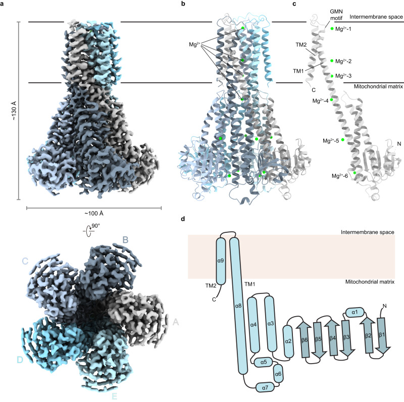
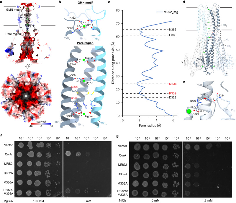
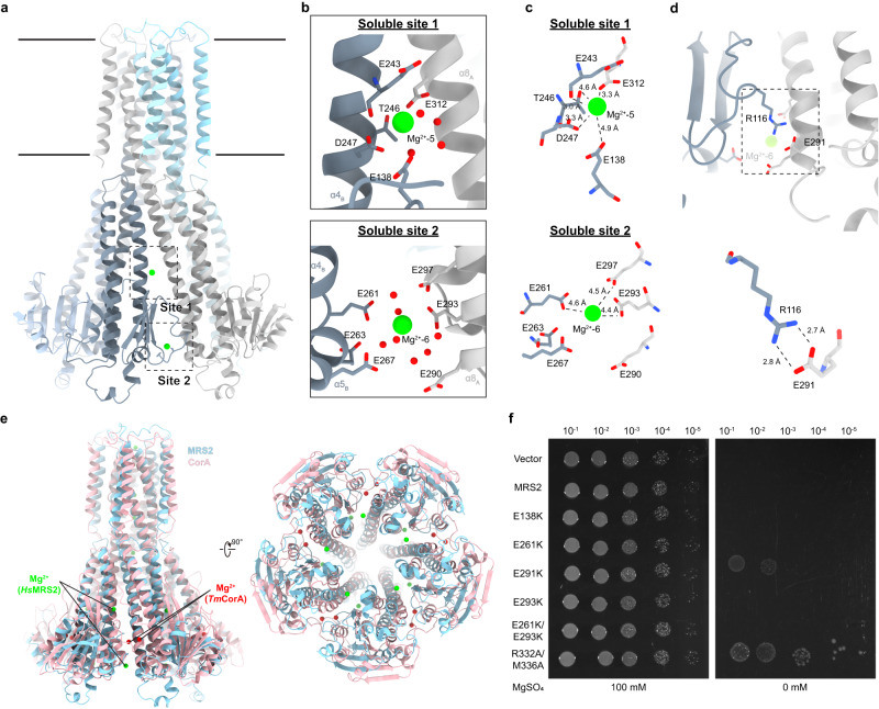
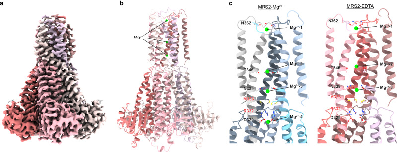
Magnesium ions (Mg 2+ ) play an essential role in cellular physiology. In mitochondria, protein and ATP synthesis and various metabolic pathways are directly regulated by Mg 2+ . MRS2, a magnesium channel located in the inner mitochondrial membrane, mediates the influx of Mg 2+ into the mitochondrial matrix and regulates Mg 2+ homeostasis. Knockdown of MRS2 in human cells leads to reduced uptake of Mg 2+ into mitochondria and disruption of the mitochondrial metabolism. Despite the importance of MRS2, the Mg 2+ translocation and regulation mechanisms of MRS2 are still unclear. Here, using cryo-EM we report the structures of human MRS2 in the presence and absence of Mg 2+ at 2.8 Å and 3.3 Å, respectively. From the homo-pentameric structures, we identify R332 and M336 as major gating residues, which are then tested using mutagenesis and two cellular divalent ion uptake assays. A network of hydrogen bonds is found connecting the gating residue R332 to the soluble domain, potentially regulating the gate. Two Mg 2+ -binding sites are identified in the MRS2 soluble domain, distinct from the two sites previously reported in CorA, a homolog of MRS2 in prokaryotes. Altogether, this study provides the molecular basis for understanding the Mg 2+ translocation and regulatory mechanisms of MRS2.
Overview
The claims and statistics of this paper haven’t been checked yet.
Run the check — free, no account needed. It reads the open-access text and checks the sources it cites.
Integrity signals
Registry
Cites
Statistics
Figures
Verification
Claims the paper makes
The claims and statistics of this paper haven’t been checked yet.
Run the check — free, no account needed. It reads the open-access text and checks the sources it cites.
Self-check
Percentages, sums and reported differences re-computed from the paper’s own numbers.
The self-check needs the paper’s full text.
Citations
Cites
Highly accurate protein structure prediction with AlphaFold
Jumper, J., Evans, R., Pritzel, A., Green, T., Figurnov, M., Ronneberger, O., Tunyasuvunakool, K., Bates, R., Žídek, A., Potapenko, A., et al. (2021) · Nature · doi:10.1038/s41586-021-03819-2 · Cited by 43,764
Research works printed but not linked: 55
- Jahnen-Dechent, W. & Ketteler, M. Magnesium basics. Clin. Kidney J. 5, i3–i14 (2012). · DOI · Paper page
- de Baaij, J. H., Hoenderop, J. G. & Bindels, R. J. Magnesium in man: implications for health and disease. Physiol. Rev. 95, 1–46 (2015). · DOI · Paper page
- Mathew, A. A. & Panonnummal, R. ‘Magnesium’-the master cation-as a drug-possibilities and evidences. Biometals 34, 955–986 (2021). · DOI
- DiNicolantonio, J. J., Liu, J. & O’Keefe, J. H. Magnesium for the prevention and treatment of cardiovascular disease. Open Heart 5, e000775 (2018). · DOI
- Saris, N. E., Mervaala, E., Karppanen, H., Khawaja, J. A. & Lewenstam, A. Magnesium. An update on physiological, clinical and analytical aspects. Clin. Chim. Acta 294, 1–26 (2000). · DOI
- Yamanaka, R., Shindo, Y. & Oka, K. Magnesium is a key player in neuronal maturation and neuropathology. Int. J. Mol. Sci. 20, https://doi.org/10.3390/ijms20143439 (2019). · DOI
- Romani, A. M. Cellular magnesium homeostasis. Arch. Biochem. Biophys. 512, 1–23 (2011). · DOI
- Rodriguez-Zavala, J. S. & Moreno-Sanchez, R. Modulation of oxidative phosphorylation by Mg2+ in rat heart mitochondria. J. Biol. Chem. 273, 7850–7855 (1998). · DOI
- Pilchova, I., Klacanova, K., Tatarkova, Z., Kaplan, P. & Racay, P. The involvement of Mg(2+) in regulation of cellular and mitochondrial functions. Oxid. Med. Cell Longev. 2017, 6797460 (2017). · DOI · Paper page
- Marchi, S. & Pinton, P. The mitochondrial calcium uniporter complex: molecular components, structure and physiopathological implications. J. Physiol. 592, 829–839 (2014). · DOI
- Bui, D. M., Gregan, J., Jarosch, E., Ragnini, A. & Schweyen, R. J. The bacterial magnesium transporter CorA can functionally substitute for its putative homologue Mrs2p in the yeast inner mitochondrial membrane. J. Biol. Chem. 274, 20438–20443 (1999). · DOI
- Zsurka, G., Gregan, J. & Schweyen, R. J. The human mitochondrial Mrs2 protein functionally substitutes for its yeast homologue, a candidate magnesium transporter. Genomics 72, 158–168 (2001). · DOI
- Kolisek, M. et al. Mrs2p is an essential component of the major electrophoretic Mg2+ influx system in mitochondria. EMBO J. 22, 1235–1244 (2003). · DOI
- Schindl, R., Weghuber, J., Romanin, C. & Schweyen, R. J. Mrs2p forms a high conductance Mg2+ selective channel in mitochondria. Biophys. J. 93, 3872–3883 (2007). · DOI
- Piskacek, M., Zotova, L., Zsurka, G. & Schweyen, R. J. Conditional knockdown of hMRS2 results in loss of mitochondrial Mg(2+) uptake and cell death. J. Cell Mol. Med. 13, 693–700 (2009). · DOI
- Yamanaka, R. et al. Mitochondrial Mg(2+) homeostasis decides cellular energy metabolism and vulnerability to stress. Sci. Rep. 6, 30027 (2016). · DOI · Paper page
- Kuramoto, T. et al. A mutation in the gene encoding mitochondrial Mg(2)+ channel MRS2 results in demyelination in the rat. PLoS Genet 7, e1001262 (2011). · DOI
- Daw, C. C. et al. Lactate elicits ER-Mitochondrial Mg(2+) dynamics to integrate cellular metabolism. Cell 183, 474–489 e417 (2020). · DOI
- Madaris, T. R. et al. Limiting Mrs2-dependent mitochondrial Mg(2+) uptake induces metabolic programming in prolonged dietary stress. Cell Rep. 42, 112155 (2023). · DOI
- Dalmas, O., Sompornpisut, P., Bezanilla, F. & Perozo, E. Molecular mechanism of Mg2+-dependent gating in CorA. Nat. Commun. 5, 3590 (2014). · DOI
- Pfoh, R. et al. Structural asymmetry in the magnesium channel CorA points to sequential allosteric regulation. Proc. Natl Acad. Sci. USA 109, 18809–18814 (2012). · DOI
- Khan, M. B. et al. Structural and functional characterization of the N-terminal domain of the yeast Mg2+ channel Mrs2. Acta Crystallogr. D Biol. Crystallogr. 69, 1653–1664 (2013). · DOI
- Uthayabalan, S., Vishnu, N., Madesh, M. & Stathopulos, P. B. The human MRS2 magnesium-binding domain is a regulatory feedback switch for channel activity. Life Sci. Alliance 6, https://doi.org/10.26508/lsa.202201742 (2023). · DOI · Paper page
- Almagro Armenteros, J. J. et al. Detecting sequence signals in targeting peptides using deep learning. Life Sci. Alliance 2, https://doi.org/10.26508/lsa.201900429 (2019). · DOI
- Teufel, F. et al. SignalP 6.0 predicts all five types of signal peptides using protein language models. Nat. Biotechnol. 40, 1023–1025 (2022). · DOI
- Fukasawa, Y. et al. MitoFates: improved prediction of mitochondrial targeting sequences and their cleavage sites. Mol. Cell Proteom. 14, 1113–1126 (2015). · DOI
- Matthies, D. et al. Cryo-EM structures of the magnesium channel CorA reveal symmetry break upon gating. Cell 164, 747–756 (2016). · DOI
- Payandeh, J. & Pai, E. F. A structural basis for Mg2+ homeostasis and the CorA translocation cycle. EMBO J. 25, 3762–3773 (2006). · DOI
- Lunin, V. V. et al. Crystal structure of the CorA Mg2+ transporter. Nature 440, 833–837 (2006). · DOI
- Eshaghi, S. et al. Crystal structure of a divalent metal ion transporter CorA at 2.9 angstrom resolution. Science 313, 354–357 (2006). · DOI
- Lerche, M., Sandhu, H., Flockner, L., Hogbom, M. & Rapp, M. Structure and cooperativity of the cytosolic domain of the CorA Mg(2+) channel from Escherichia coli. Structure 25, 1175–1186 e1174 (2017). · DOI
- Guskov, A. et al. Structural insights into the mechanisms of Mg2+ uptake, transport, and gating by CorA. Proc. Natl Acad. Sci. USA 109, 18459–18464 (2012). · DOI
- Gati, C., Stetsenko, A., Slotboom, D. J., Scheres, S. H. W. & Guskov, A. The structural basis of proton driven zinc transport by ZntB. Nat. Commun. 8, 1313 (2017). · DOI
- Stetsenko, A., Stehantsev, P., Dranenko, N. O., Gelfand, M. S. & Guskov, A. Structural and biochemical characterization of a novel ZntB (CmaX) transporter protein from Pseudomonas aeruginosa. Int. J. Biol. Macromol. 184, 760–767 (2021). · DOI
- Dudev, T. & Lim, C. Importance of metal hydration on the selectivity of Mg2+ versus Ca2+ in magnesium ion channels. J. Am. Chem. Soc. 135, 17200–17208 (2013). · DOI
- Dalmas, O. et al. A repulsion mechanism explains magnesium permeation and selectivity in CorA. Proc. Natl Acad. Sci. USA 111, 3002–3007 (2014). · DOI
- Smart, O. S., Neduvelil, J. G., Wang, X., Wallace, B. A. & Sansom, M. S. HOLE: a program for the analysis of the pore dimensions of ion channel structural models. J. Mol. Graph 14, 354–360 (1996). 376. · DOI
- Payandeh, J. et al. Probing structure-function relationships and gating mechanisms in the CorA Mg2+ transport system. J. Biol. Chem. 283, 11721–11733 (2008). · DOI
- Chakrabarti, N., Neale, C., Payandeh, J., Pai, E. F. & Pomes, R. An iris-like mechanism of pore dilation in the CorA magnesium transport system. Biophys. J. 98, 784–792 (2010). · DOI
- Neale, C., Chakrabarti, N., Pomorski, P., Pai, E. F. & Pomes, R. Hydrophobic gating of ion permeation in magnesium channel CorA. PLoS Comput. Biol. 11, e1004303 (2015). · DOI
- Jin, F. et al. The structure of MgtE in the absence of magnesium provides new insights into channel gating. PLoS Biol. 19, e3001231 (2021). · DOI
- Hattori, M. et al. Mg(2+)-dependent gating of bacterial MgtE channel underlies Mg(2+) homeostasis. EMBO J. 28, 3602–3612 (2009). · DOI · Paper page
- Tomita, A. et al. ATP-dependent modulation of MgtE in Mg(2+) homeostasis. Nat. Commun. 8, 148 (2017). · DOI
- Nemchinova, M., Melcr, J., Wassenaar, T. A., Marrink, S. J. & Guskov, A. Asymmetric CorA gating mechanism as observed by molecular dynamics simulations. J. Chem. Inf. Model 61, 2407–2417 (2021). · DOI
- Li, M. et al. Molecular basis of Mg(2+) permeation through the human mitochondrial Mrs2 channel. Nat. Commun. 14, 4713 (2023). · DOI
- Nordin, N. et al. Exploring the structure and function of Thermotoga maritima CorA reveals the mechanism of gating and ion selectivity in Co2+/Mg2+ transport. Biochem. J. 451, 365–374 (2013). · DOI
- Longo, P. A., Kavran, J. M., Kim, M. S. & Leahy, D. J. Transient mammalian cell transfection with polyethylenimine (PEI). Methods Enzymol. 529, 227–240 (2013). · DOI
- Mastronarde, D. N. Automated electron microscope tomography using robust prediction of specimen movements. J. Struct. Biol. 152, 36–51 (2005). · DOI
- Grant, T., Rohou, A. & Grigorieff, N. cisTEM, user-friendly software for single-particle image processing. eLife 7, https://doi.org/10.7554/eLife.35383 (2018). · DOI
- Punjani, A., Rubinstein, J. L., Fleet, D. J. & Brubaker, M. A. cryoSPARC: algorithms for rapid unsupervised cryo-EM structure determination. Nat. Methods 14, 290–296 (2017). · DOI · Paper page
- Pettersen, E. F. et al. UCSF Chimera-a visualization system for exploratory research and analysis. J. Comput. Chem. 25, 1605–1612 (2004). · DOI
- Emsley, P., Lohkamp, B., Scott, W. G. & Cowtan, K. Features and development of Coot. Acta Crystallogr. D Biol. Crystallogr. 66, 486–501 (2010). · DOI · Paper page
- Afonine, P. V. et al. Real-space refinement in PHENIX for cryo-EM and crystallography. Acta Crystallogr. D Struct. Biol. 74, 531–544 (2018). · DOI
- Williams, C. J. et al. MolProbity: More and better reference data for improved all-atom structure validation. Protein Sci. 27, 293–315 (2018). · DOI · Paper page
- Goddard, T. D. et al. UCSF ChimeraX: Meeting modern challenges in visualization and analysis. Protein Sci. 27, 14–25 (2018). · DOI · Paper page
Metadata
- Authors
- Lai, L. T. F., Balaraman, J., Zhou, F., & Matthies, D.
- Affiliations
- National Institutes of Health (US)
- Corresponding author
- Matthies, Doreen
- Venue
- Nature Communications
- Published
- 2023-11-08
- Volume
- 14
- Issue
- 1
- Pages
- 7207-7207
- ISSN
- 2041-1723 (ISSN-L)
- PMC
- PMC10632456
- OpenAlex
- W4388500134
- Cited by
- 29
- References
- 1
- Citations by year
- 2026: 8 · 2025: 8 · 2024: 13
- FWCI
- 2.57
- Type
- journal article
- Language
- en
- Open access
- gold
- doi.org
- Locations
- Nature Communications (publishedVersion, cc-by)
- PubMed Central (publishedVersion, cc-by)
- PubMed (publishedVersion)
- DOAJ (DOAJ: Directory of Open Access Journals) (submittedVersion, cc-by-sa)
- Funders
- U.S. Department of Health & Human Services | NIH | Eunice Kennedy Shriver National Institute of Child Health and Human Development (ZIA HD008998)
- Topics
- Life Sciences › Biochemistry, Genetics and Molecular Biology › Molecular Biology › Ion channel regulation and function
- Life Sciences › Biochemistry, Genetics and Molecular Biology › Molecular Biology › ATP Synthase and ATPases Research
- Life Sciences › Biochemistry, Genetics and Molecular Biology › Structural Biology › Advanced Electron Microscopy Techniques and Applications
- Keywords
- MRS2, magnesium channel, magnesium homeostasis, channel gating, magnesium binding sites, inner mitochondrial membrane, hydrogen bond network
- MeSH terms
- Agaricales, Humans, Magnesium, Mitochondria, Translocation, Genetic, Cryoelectron Microscopy, Mitochondrial Proteins, Cation Transport Proteins, Mitochondrial Membranes
About these checks
Registry, retraction, Cites and statistics results are computed, not judged, with one exception: a printed reference that no index lists is read once by a model before it counts as a citation error. Claims are extracted by a model and kept only when their quote appears verbatim in the text; each cited source is then checked, with a model’s help, for whether it says what the claim relies on. Those verdicts can be wrong — read the claim’s quote and the cited source before relying on one.
Statistics and arithmetic are recomputed, not judged.
Confidence bands are shown only for fields calibrated on our annotated test set. None is calibrated yet, so no band is shown.
What counts as a claim ›How we measure confidence ›How we check this ›
Are you an author, or do you see a problem with this page? Request a review
How to cite
Cite CoChat for the checks on this page, and the paper for its own findings.
- This page
- CoChat. (2026). Citation check: Cryo-EM structures of human magnesium channel MRS2 reveal gating and regulatory mechanisms. https://app.cochat.ai/papers/10.1038/s41467-023-42599-3
- The paper
- Lai, L. T. F., Balaraman, J., Zhou, F., & Matthies, D. (2023). Cryo-EM structures of human magnesium channel MRS2 reveal gating and regulatory mechanisms. Nature Communications. https://doi.org/10.1038/s41467-023-42599-3
