

The DOI matches its Crossref record; 3 of the 3 references it cites were found in a registry or an index.
Ton motor conformational switch and peptidoglycan role in bacterial nutrient uptake
Open accessPublished version of a preprint
Abstract
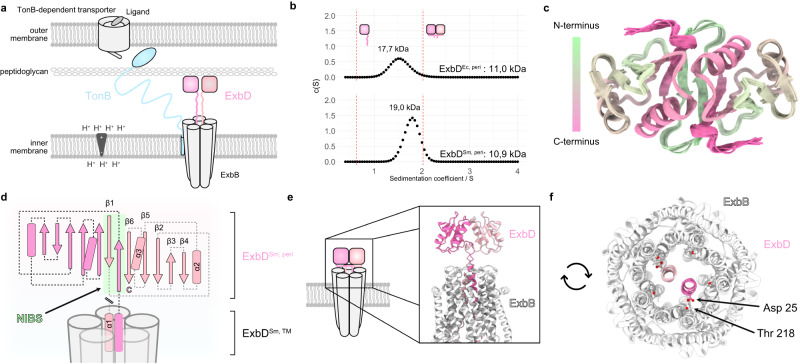
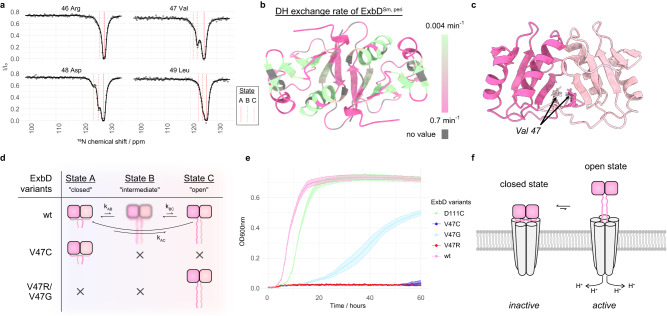
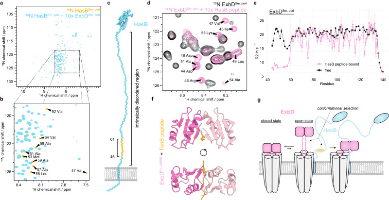
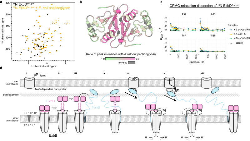
Active nutrient uptake is fundamental for survival and pathogenicity of Gram-negative bacteria, which operate a multi-protein Ton system to transport essential nutrients like metals and vitamins. This system harnesses the proton motive force at the inner membrane to energize the import through the outer membrane, but the mechanism of energy transfer remains enigmatic. Here, we study the periplasmic domain of ExbD, a crucial component of the proton channel of the Ton system. We show that this domain is a dynamic dimer switching between two conformations representing the proton channel’s open and closed states. By in vivo phenotypic assays we demonstrate that this conformational switch is essential for the nutrient uptake by bacteria. The open state of ExbD triggers a disorder to order transition of TonB, enabling TonB to supply energy to the nutrient transporter. We also reveal the anchoring role of the peptidoglycan layer in this mechanism. Herein, we propose a mechanistic model for the Ton system, emphasizing ExbD duality and the pivotal catalytic role of peptidoglycan. Sequence analysis suggests that this mechanism is conserved in other systems energizing gliding motility and membrane integrity. Our study fills important gaps in understanding bacterial motor mechanism and proposes novel antibacterial strategies.
Overview
The claims and statistics of this paper haven’t been checked yet.
Run the check — free, no account needed. It reads the open-access text and checks the sources it cites.
Integrity signals
Registry
Cites
Statistics
Figures
Verification
Claims the paper makes
The claims and statistics of this paper haven’t been checked yet.
Run the check — free, no account needed. It reads the open-access text and checks the sources it cites.
Self-check
Percentages, sums and reported differences re-computed from the paper’s own numbers.
The self-check needs the paper’s full text.
Citations
Cites
Highly accurate protein structure prediction with AlphaFold
Jumper, J., Evans, R., Pritzel, A., Green, T., Figurnov, M., Ronneberger, O., Tunyasuvunakool, K., Bates, R., Žídek, A., Potapenko, A., et al. (2021) · Nature · doi:10.1038/s41586-021-03819-2 · Cited by 43,764
Protein complex prediction with AlphaFold-Multimer
Evans, R., O’Neill, M., Pritzel, A., Antropova, N., Senior, A., Green, T., Žídek, A., Bates, R., Blackwell, S., Yim, J., et al. (2021) · doi:10.1101/2021.10.04.463034 · Cited by 3,808
New substrates for TonB-dependent transport: do we only see the ‘tip of the iceberg’?
Schauer, K., Rodionov, D. A., & de Reuse, H. (2008) · Trends in Biochemical Sciences · doi:10.1016/j.tibs.2008.04.012 · Cited by 360
Research works printed but not linked: 54
- Blair, J. M. A., Webber, M. A., Baylay, A. J., Ogbolu, D. O. & Piddock, L. J. V. Molecular mechanisms of antibiotic resistance. Nat. Rev. Microbiol. 13, 42–51 (2015). · DOI · Paper page
- Noinaj, N., Guillier, M., Barnard, T. J. & Buchanan, S. K. TonB-dependent transporters: regulation, structure, and function. Annu. Rev. Microbiol. 64, 43–60 (2010). · DOI · Paper page
- Paquelin, A., Ghigo, J. M., Bertin, S. & Wandersman, C. Characterization of HasB, a Serratia marcescens TonB-like protein specifically involved in the haemophore-dependent haem acquisition system. Mol. Microbiol. 42, 995–1005 (2001). · DOI
- Biou, V. et al. Structural and molecular determinants for the interaction of ExbB from Serratia marcescens and HasB, a TonB paralog. Commun. Biol. 5, 1–15 (2022). · DOI
- de Amorim, G. C. et al. The structure of hasB reveals a new class of tonb protein fold. PLOS ONE 8, e58964 (2013). · DOI
- Celia, H. et al. Cryo-EM structure of the bacterial Ton motor subcomplex ExbB–ExbD provides information on structure and stoichiometry. Commun. Biol. 2, 1–6 (2019). · DOI
- Celia, H. et al. Structural insight into the role of the Ton complex in energy transduction. Nature 538, 60–65 (2016). · DOI
- Maki-Yonekura, S. et al. Hexameric and pentameric complexes of the ExbBD energizer in the Ton system. eLife 7, e35419 (2018). · DOI
- Garcia-Herrero, A., Peacock, R. S., Howard, S. P. & Vogel, H. J. The solution structure of the periplasmic domain of the TonB system ExbD protein reveals an unexpected structural homology with siderophore-binding proteins. Mol. Microbiol. 66, 872–889 (2007). · DOI
- Rieu, M., Krutyholowa, R., Taylor, N. M. I. & Berry, R. M. A new class of biological ion-driven rotary molecular motors with 5:2 symmetry. Front. Microbiol. 13, 948383 (2022). · DOI
- Deme, J. C. et al. Structures of the stator complex that drives rotation of the bacterial flagellum. Nat. Microbiol. 5, 1553–1564 (2020). · DOI
- Shultis, D. D., Purdy, M. D., Banchs, C. N. & Wiener, M. C. Outer membrane active transport: structure of the btub:tonb complex. Science 312, 1396–1399 (2006). · DOI
- Josts, I., Veith, K. & Tidow, H. Ternary structure of the outer membrane transporter FoxA with resolved signalling domain provides insights into TonB-mediated siderophore uptake. eLife 8, e48528 (2019). · DOI
- Domingo Köhler, S., Weber, A., Howard, S. P., Welte, W. & Drescher, M. The proline-rich domain of TonB possesses an extended polyproline II-like conformation of sufficient length to span the periplasm of Gram-negative bacteria. Protein Sci. 19, 625–630 (2010). · DOI
- Evans, J. S., Levine, B. A., Trayer, I. P., Dorman, C. J. & Higgins, C. F. Sequence-imposed structural constraints in the TonB protein of E. coli. FEBS Lett. 208, 211–216 (1986). · DOI
- Virtanen, S. I., Kiirikki, A. M., Mikula, K. M., Iwaï, H. & Ollila, O. H. S. Heterogeneous dynamics in partially disordered proteins. Phys. Chem. Chem. Phys. 22, 21185–21196 (2020). · DOI
- Ratliff, A. C., Buchanan, S. K. & Celia, H. The Ton Motor. Front. Microbiol. 13, 852955 (2022). · DOI
- Lefèvre, J., Delepelaire, P., Delepierre, M. & Izadi-Pruneyre, N. Modulation by substrates of the interaction between the hasr outer membrane receptor and its specific tonb-like protein, HasB. J. Mol. Biol. 378, 840–851 (2008). · DOI
- Kaserer, W. A. et al. Insight from TonB hybrid proteins into the mechanism of iron transport through the outer membrane. J. Bacteriol. 190, 4001–4016 (2008). · DOI
- Kojima, S. et al. The helix rearrangement in the periplasmic domain of the flagellar stator b subunit activates peptidoglycan binding and ion influx. Structure 26, 590–598.e5 (2018). · DOI
- Szczepaniak, J. et al. The lipoprotein Pal stabilises the bacterial outer membrane during constriction by a mobilisation-and-capture mechanism. Nat. Commun. 11, 1305 (2020). · DOI
- Parsons, L. M., Lin, F. & Orban, J. Peptidoglycan recognition by Pal, an outer membrane lipoprotein. Biochemistry 45, 2122–2128 (2006). · DOI
- Wojdyla, J. A. et al. Structure and function of the escherichia coli tol-pal stator protEin TolR*. J. Biol. Chem. 290, 26675–26687 (2015). · DOI
- Hantke, K. & Braun, V. Functional interaction of the tonA/tonB receptor system in Escherichia coli. J. Bacteriol. 135, 190–197 (1978). · DOI
- Vallurupalli, P., Bouvignies, G. & Kay, L. E. Studying “invisible” excited protein states in slow exchange with a major state conformation. J. Am. Chem. Soc. 134, 8148–8161 (2012). · DOI
- Kopp, D. R. & Postle, K. The intrinsically disordered region of exbd is required for signal transduction. J. Bacteriol. 202, e00687–19 (2020). · DOI
- Moynihan, P. J., Sychantha, D. & Clarke, A. J. Chemical biology of peptidoglycan acetylation and deacetylation. Bioorg. Chem. 54, 44–50 (2014). · DOI
- Parsons, L. M., Grishaev, A. & Bax, A. The periplasmic domain of tolr from haemophilus influenzae forms a dimer with a large hydrophobic groove: nmr solution structure and comparison to SAXS data. Biochemistry 47, 3131–3142 (2008). · DOI
- Gumbart, J., Wiener, M. C. & Tajkhorshid, E. Mechanics of force propagation in tonb-dependent outer membrane transport. Biophys. J. 93, 496–504 (2007). · DOI
- Grinter, R. & Lithgow, T. The structure of the bacterial iron–catecholate transporter Fiu suggests that it imports substrates via a two-step mechanism. J. Biol. Chem. 294, 19523–19534 (2019). · DOI
- Schuck, P. Size-distribution analysis of macromolecules by sedimentation velocity ultracentrifugation and lamm equation modeling. Biophys. J. 78, 1606–1619 (2000). · DOI
- Ortega, A., Amorós, D. & García de la Torre, J. Prediction of hydrodynamic and other solution properties of rigid proteins from atomic- and residue-level models. Biophys. J. 101, 892–898 (2011). · DOI
- Favier, A. & Brutscher, B. NMRlib: user-friendly pulse sequence tools for Bruker NMR spectrometers. J. Biomol. NMR 73, 199–211 (2019). · DOI
- Schanda, P. Kupče, Ē. & Brutscher, B. SOFAST-HMQC experiments for recording two-dimensional deteronuclear correlation spectra of proteins within a few seconds. J. Biomol. NMR 33, 199–211 (2005). · DOI
- Lescop, E., Schanda, P. & Brutscher, B. A set of BEST triple-resonance experiments for time-optimized protein resonance assignment. J. Magn. Reson. 187, 163–169 (2007). · DOI
- Cordier, F., Nisius, L., Dingley, A. J. & Grzesiek, S. Direct detection of N−H⋯O=C hydrogen bonds in biomolecules by NMR spectroscopy. Nat. Protoc. 3, 235–241 (2008). · DOI
- Delaglio, F. et al. NMRPipe: a multidimensional spectral processing system based on UNIX pipes. J. Biomol. NMR 6, 277–293 (1995). · DOI
- Ying, J., Delaglio, F., Torchia, D. A. & Bax, A. Sparse multidimensional iterative lineshape-enhanced (SMILE) reconstruction of both non-uniformly sampled and conventional NMR data. J. Biomol. NMR 68, 101–118 (2017). · DOI
- Vranken, W. F. et al. The CCPN data model for NMR spectroscopy: development of a software pipeline. Proteins Struct. Funct. Bioinforma. 59, 687–696 (2005). · DOI
- Skinner, S. P. et al. CcpNmr analysisassign: a flexible platform for integrated NMR analysis. J. Biomol. NMR 66, 111–124 (2016). · DOI
- Guenneugues, M., Berthault, P. & Desvaux, H. A method for determiningb1field inhomogeneity. are the biases assumed in heteronuclear relaxation experiments usually underestimated? J. Magn. Reson. 136, 118–126 (1999). · DOI
- Allain, F., Mareuil, F., Ménager, H., Nilges, M. & Bardiaux, B. ARIAweb: a server for automated NMR structure calculation. Nucleic Acids Res 48, W41–W47 (2020). · DOI
- Shen, Y. & Bax, A. Protein backbone and sidechain torsion angles predicted from NMR chemical shifts using artificial neural networks. J. Biomol. NMR 56, 227–241 (2013). · DOI
- Bardiaux, B. et al. Influence of different assignment conditions on the determination of symmetric homodimeric structures with ARIA. Proteins Struct. Funct. Bioinforma. 75, 569–585 (2009). · DOI
- Nilges, M. et al. Accurate NMR structures through minimization of an extended hybrid energy. Structure 16, 1305–1312 (2008). · DOI
- Linge, J. P., Williams, M. A., Spronk, C. A. E. M., Bonvin, A. M. J. J. & Nilges, M. Refinement of protein structures in explicit solvent. Proteins Struct. Funct. Bioinforma. 50, 496–506 (2003). · DOI
- Buchner, L. & Güntert, P. Systematic evaluation of combined automated NOE assignment and structure calculation with CYANA. J. Biomol. NMR 62, 81–95 (2015). · DOI
- Weber, P. et al. High-throughput crystallization pipeline at the crystallography core facility of the institut pasteur. Molecules 24, 4451 (2019). · DOI
- Kabsch, W. XDS. Acta Crystallogr. D. Biol. Crystallogr. 66, 125–132 (2010). · DOI · Paper page
- McCoy, A. J. et al. Phaser crystallographic software. J. Appl. Crystallogr. 40, 658–674 (2007). · DOI · Paper page
- Emsley, P. & Cowtan, K. Coot: model-building tools for molecular graphics. Acta Crystallogr. D. Biol. Crystallogr. 60, 2126–2132 (2004). · DOI · Paper page
- Murshudov, G. N. et al. REFMAC5 for the refinement of macromolecular crystal structures. Acta Crystallogr. D. Biol. Crystallogr. 67, 355–367 (2011). · DOI
- Liebschner, D. et al. Macromolecular structure determination using X-rays, neutrons and electrons: recent developments in Phenix. Acta Crystallogr. Sect. Struct. Biol. 75, 861–877 (2019). · DOI · Paper page
- Goddard, T. D. et al. UCSF chimeraX: meeting modern challenges in visualization and analysis. Protein Sci. 27, 14–25 (2018). · DOI · Paper page
Metadata
- Authors
- Zinke, M., Lejeune, M., Mechaly, A., Bardiaux, B., Boneca, I. G., Delepelaire, P., & Izadi-Pruneyre, N.
- Affiliations
- Centre National de la Recherche Scientifique (FR)
- ORCID
- Venue
- Nature Communications
- Published
- 2024-01-06
- Volume
- 15
- Issue
- 1
- Pages
- 331-331
- ISSN
- 2041-1723 (ISSN-L)
- PMC
- PMC10771500
- OpenAlex
- W4390639340
- Cited by
- 27
- References
- 3
- Citations by year
- 2026: 8 · 2025: 12 · 2024: 7
- FWCI
- 7.17
- Type
- journal article
- Language
- en
- Open access
- gold
- doi.org
- Locations
- Nature Communications (publishedVersion, cc-by)
- PubMed Central (publishedVersion, cc-by)
- PubMed (publishedVersion)
- HAL (submittedVersion, cc-by)
- DOAJ (DOAJ: Directory of Open Access Journals) (submittedVersion, cc-by-sa)
- Funders
- Agence Nationale de la Recherche (nvestissement d’Avenir grant ANR-16-CONV-0005)
- Topics
- Life Sciences › Biochemistry, Genetics and Molecular Biology › Genetics › Bacterial Genetics and Biotechnology
- Health Sciences › Medicine › Oncology › Drug Transport and Resistance Mechanisms
- Life Sciences › Immunology and Microbiology › Microbiology › Escherichia coli research studies
- Keywords
- ExbD, TonB, conformational switching, peptidoglycan, proton channel, disorder-to-order transition, Gram-negative bacteria, proton motive force, energy transfer, antibacterial strategies, gliding motility, membrane integrity
- MeSH terms
- Nutrients, Bacteria, Cell Wall, Peptidoglycan, Protons
About these checks
Registry, retraction, Cites and statistics results are computed, not judged, with one exception: a printed reference that no index lists is read once by a model before it counts as a citation error. Claims are extracted by a model and kept only when their quote appears verbatim in the text; each cited source is then checked, with a model’s help, for whether it says what the claim relies on. Those verdicts can be wrong — read the claim’s quote and the cited source before relying on one.
Statistics and arithmetic are recomputed, not judged.
Confidence bands are shown only for fields calibrated on our annotated test set. None is calibrated yet, so no band is shown.
What counts as a claim ›How we measure confidence ›How we check this ›
Are you an author, or do you see a problem with this page? Request a review
How to cite
Cite CoChat for the checks on this page, and the paper for its own findings.
- This page
- CoChat. (2026). Citation check: Ton motor conformational switch and peptidoglycan role in bacterial nutrient uptake. https://app.cochat.ai/papers/10.1038/s41467-023-44606-z
- The paper
- Zinke, M., Lejeune, M., Mechaly, A., Bardiaux, B., Boneca, I. G., Delepelaire, P., & Izadi-Pruneyre, N. (2024). Ton motor conformational switch and peptidoglycan role in bacterial nutrient uptake. Nature Communications. https://doi.org/10.1038/s41467-023-44606-z
