

The DOI matches its Crossref record; 3 of the 3 references it cites were found in a registry or an index.
The biosynthesis of the odorant 2-methylisoborneol is compartmentalized inside a protein shell
Open accessPublished version of a preprint
Abstract
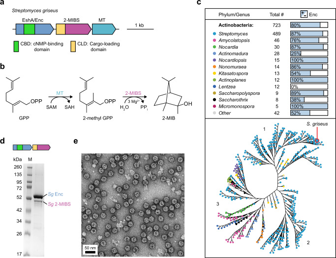
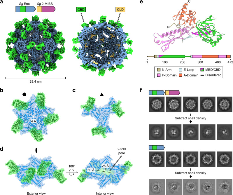
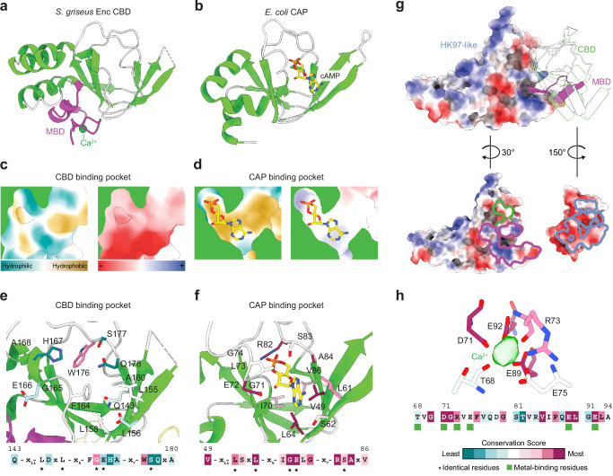
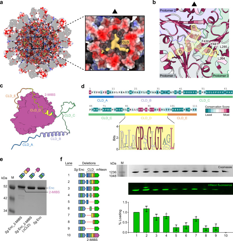
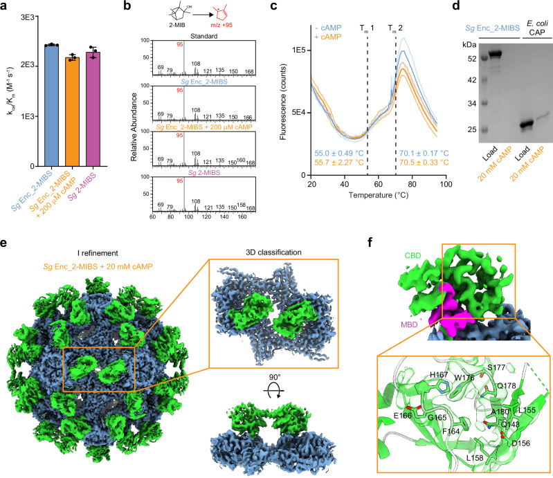
Abstract Terpenoids are the largest class of natural products, found across all domains of life. One of the most abundant bacterial terpenoids is the volatile odorant 2-methylisoborneol (2-MIB), partially responsible for the earthy smell of soil and musty taste of contaminated water. Many bacterial 2-MIB biosynthetic gene clusters were thought to encode a conserved transcription factor, named EshA in the model soil bacterium Streptomyces griseus . Here, we revise the function of EshA, now referred to as Sg Enc, and show that it is a Family 2B encapsulin shell protein. Using cryo-electron microscopy, we find that Sg Enc forms an icosahedral protein shell and encapsulates 2-methylisoborneol synthase (2-MIBS) as a cargo protein. Sg Enc contains a cyclic adenosine monophosphate (cAMP) binding domain (CBD)-fold insertion and a unique metal-binding domain, both displayed on the shell exterior. We show that Sg Enc CBDs do not bind cAMP. We find that 2-MIBS cargo loading is mediated by an N-terminal disordered cargo-loading domain and that 2-MIBS activity and Sg Enc shell structure are not modulated by cAMP. Our work redefines the function of EshA and establishes Family 2B encapsulins as cargo-loaded protein nanocompartments involved in secondary metabolite biosynthesis.
Overview
The claims and statistics of this paper haven’t been checked yet.
Run the check — free, no account needed. It reads the open-access text and checks the sources it cites.
Integrity signals
Registry
Cites
Statistics
Figures
Verification
Claims the paper makes
The claims and statistics of this paper haven’t been checked yet.
Run the check — free, no account needed. It reads the open-access text and checks the sources it cites.
Self-check
Percentages, sums and reported differences re-computed from the paper’s own numbers.
The self-check needs the paper’s full text.
Citations
Cites
Highly accurate protein structure prediction with AlphaFold
Jumper, J., Evans, R., Pritzel, A., Green, T., Figurnov, M., Ronneberger, O., Tunyasuvunakool, K., Bates, R., Žídek, A., Potapenko, A., et al. (2021) · Nature · doi:10.1038/s41586-021-03819-2 · Cited by 43,764
Fast and accurate protein structure search with Foldseek
van Kempen, M., Kim, S. S., Tumescheit, C., Mirdita, M., Lee, J., Gilchrist, C. L. M., Söding, J., & Steinegger, M. (2022) · doi:10.1101/2022.02.07.479398 · Cited by 2,598
Fast and accurate protein structure search with Foldseek
van Kempen, M., Kim, S. S., Tumescheit, C., Mirdita, M., Lee, J., Gilchrist, C. L. M., Söding, J., & Steinegger, M. (2024) · Nature Biotechnology · doi:10.1038/s41587-023-01773-0 · Cited by 2,596
Research works printed but not linked: 110
- Christianson, D. W. Structural and Chemical Biology of Terpenoid Cyclases. Chem. Rev. 117, 11570–11648 (2017). · DOI
- Rudolf, J. D., Alsup, T. A., Xu, B. & Li, Z. Bacterial terpenome. Nat. Prod. Rep. 38, 905–980 (2021). · DOI
- Tholl, D. in Biotechnology of Isoprenoids (eds Jens Schrader & Jörg Bohlmann) 63-106 (Springer International Publishing, 2015). · DOI
- Harvey, A. L. Natural products in drug discovery. Drug Discov. Today 13, 894–901 (2008). · DOI
- Cane, D. E. & Ikeda, H. Exploration and mining of the bacterial terpenome. Acc. Chem. Res. 45, 463–472 (2012). · DOI
- Dickschat, J. S. Bacterial terpene cyclases. Nat. Prod. Rep. 33, 87–110 (2016). · DOI
- Juttner, F. & Watson, S. B. Biochemical and ecological control of geosmin and 2-methylisoborneol in source waters. Appl Environ. Microbiol 73, 4395–4406 (2007). · DOI
- Garbeva, P., Avalos, M., Ulanova, D., van Wezel, G. P. & Dickschat, J. S. Volatile sensation: The chemical ecology of the earthy odorant geosmin. Environ. Microbiol 25, 1565–1574 (2023). · DOI
- Zaroubi, L. et al. The Ubiquitous Soil Terpene Geosmin Acts as a Warning Chemical. Appl Environ. Microbiol 88, e0009322 (2022). · DOI
- Saha, M. & Fink, P. Algal volatiles - the overlooked chemical language of aquatic primary producers. Biol. Rev. Camb. Philos. Soc. 97, 2162–2173 (2022). · DOI
- Wang, C. M. & Cane, D. E. Biochemistry and molecular genetics of the biosynthesis of the earthy odorant methylisoborneol in Streptomyces coelicolor. J. Am. Chem. Soc. 130, 8908–8909 (2008). · DOI
- Giglio, S., Chou, W. K., Ikeda, H., Cane, D. E. & Monis, P. T. Biosynthesis of 2-methylisoborneol in cyanobacteria. Environ. Sci. Technol. 45, 992–998 (2011). · DOI
- Wang, Z., Xu, Y., Shao, J., Wang, J. & Li, R. Genes associated with 2-methylisoborneol biosynthesis in cyanobacteria: isolation, characterization, and expression in response to light. PLoS One 6, e18665 (2011). · DOI
- Dickschat, J. S. et al. Biosynthesis of the off-flavor 2-methylisoborneol by the myxobacterium Nannocystis exedens. Angew. Chem. Int Ed. Engl. 46, 8287–8290 (2007). · DOI
- Avalos, M. et al. Biosynthesis, evolution and ecology of microbial terpenoids. Nat. Prod. Rep. 39, 249–272 (2022). · DOI
- Scholler, C. E., Gurtler, H., Pedersen, R., Molin, S. & Wilkins, K. Volatile metabolites from actinomycetes. J. Agric Food Chem. 50, 2615–2621 (2002). · DOI
- Citron, C. A., Gleitzmann, J., Laurenzano, G., Pukall, R. & Dickschat, J. S. Terpenoids are widespread in actinomycetes: a correlation of secondary metabolism and genome data. Chembiochem 13, 202–214 (2012). · DOI
- Larsen, T. O. & Frisvad, J. C. Characterization of volatile metabolites from 47 Penicillium taxa. Mycological Res. 99, 1153–1166 (1995). · DOI
- Wilkins, K. Volatile Organic Metabolites from Selected Streptomyces Strains. Actinomycetologica 23, 27–33 (2009). · DOI
- Medsker, L. L., Jenkins, D., Thomas, J. F. & Koch, C. Odorous compounds in natural waters. 2-exo-Hydroxy-2-methylbornane, the major odorous compound produced by several actinomycetes. Environ. Sci. Technol. 3, 476–477 (1969). · DOI
- Gerber, N. N. A volatile metabolite of actinomycetes, 2-methylisoborneol. J. Antibiot. (Tokyo) 22, 508–509 (1969). · DOI
- Bentley, R. & Meganathan, R. Geosmin and methylisoborneol biosynthesis in streptomycetes. Evidence for an isoprenoid pathway and its absence in non-differentiating isolates. FEBS Lett. 125, 220–222 (1981). · DOI
- Gu, B., Liang, L. F. & Dickschat, J. S. Functions of enzyme domains in 2-methylisoborneol biosynthesis and enzymatic synthesis of non-natural analogs. Beilstein J. Org. Chem. 19, 1452–1459 (2023). · DOI
- Koksal, M., Chou, W. K., Cane, D. E. & Christianson, D. W. Structure of 2-methylisoborneol synthase from Streptomyces coelicolor and implications for the cyclization of a noncanonical C-methylated monoterpenoid substrate. Biochemistry 51, 3011–3020 (2012). · DOI
- Brock, N. L., Ravella, S. R., Schulz, S. & Dickschat, J. S. A detailed view of 2-methylisoborneol biosynthesis. Angew. Chem. Int Ed. Engl. 52, 2100–2104 (2013). · DOI
- Gu, B., Hou, A. & Dickschat, J. S. The stereochemical course of 2-methylisoborneol biosynthesis. Beilstein J. Org. Chem. 18, 818–824 (2022). · DOI
- Gu, B. & Dickschat, J. S. A non-natural biosynthesis pathway toward 2-methylisoborneol. Chem. Commun. (Camb.) 58, 4316–4319 (2022). · DOI
- Komatsu, M., Tsuda, M., Omura, S., Oikawa, H. & Ikeda, H. Identification and functional analysis of genes controlling biosynthesis of 2-methylisoborneol. Proc. Natl Acad. Sci. USA 105, 7422–7427 (2008). · DOI
- Koksal, M., Chou, W. K., Cane, D. E. & Christianson, D. W. Structure of geranyl diphosphate C-methyltransferase from Streptomyces coelicolor and implications for the mechanism of isoprenoid modification. Biochemistry 51, 3003–3010 (2012). · DOI
- Kawamoto, S. et al. Molecular and functional analyses of the gene (eshA) encoding the 52-kilodalton protein of Streptomyces coelicolor A3(2) required for antibiotic production. J. Bacteriol. 183, 6009–6016 (2001). · DOI
- Passner, J. M., Schultz, S. C. & Steitz, T. A. Modeling the cAMP-induced Allosteric Transition Using the Crystal Structure of CAP-cAMP at 2.1Å Resolution. J. Mol. Biol. 304, 847–859 (2000). · DOI
- Andreas, M. P. & Giessen, T. W. Large-scale computational discovery and analysis of virus-derived microbial nanocompartments. Nat. Commun. 12, 4748 (2021). · DOI
- Nichols, R. J. et al. Discovery and characterization of a novel family of prokaryotic nanocompartments involved in sulfur metabolism. Elife 10. https://doi.org/10.7554/eLife.59288 (2021). · DOI
- Giessen, T. W. Encapsulins. Annu Rev Biochem. https://doi.org/10.1146/annurev-biochem-040320-102858 (2022). · DOI
- Nichols, R. J., Cassidy-Amstutz, C., Chaijarasphong, T. & Savage, D. F. Encapsulins: molecular biology of the shell. Crit. Rev. Biochem Mol. Biol. 52, 583–594 (2017). · DOI
- Sutter, M. et al. Structural basis of enzyme encapsulation into a bacterial nanocompartment. Nat. Struct. Mol. Biol. 15, 939–947 (2008). · DOI
- Giessen, T. W. & Silver, P. A. Widespread distribution of encapsulin nanocompartments reveals functional diversity. Nat. Microbiol 2, 17029 (2017). · DOI
- Heinemann, J. et al. Fossil record of an archaeal HK97-like provirus. Virology 417, 362–368 (2011). · DOI
- Altenburg, W. J., Rollins, N., Silver, P. A. & Giessen, T. W. Exploring targeting peptide-shell interactions in encapsulin nanocompartments. Sci. Rep. 11, 4951 (2021). · DOI
- McHugh, C. A. et al. A virus capsid-like nanocompartment that stores iron and protects bacteria from oxidative stress. EMBO J. 33, 1896–1911 (2014). · DOI
- Giessen, T. W. et al. Large protein organelles form a new iron sequestration system with high storage capacity. Elife 8. https://doi.org/10.7554/eLife.46070 (2019). · DOI
- Akita, F. et al. The crystal structure of a virus-like particle from the hyperthermophilic archaeon Pyrococcus furiosus provides insight into the evolution of viruses. J. Mol. Biol. 368, 1469–1483 (2007). · DOI
- Kwon, S., Andreas, M. P. & Giessen, T. W. Structure and heterogeneity of a highly cargo-loaded encapsulin shell. J. Struct. Biol. 215, 108022 (2023). · DOI
- Eren, E. et al. Structural characterization of the Myxococcus xanthus encapsulin and ferritin-like cargo system gives insight into its iron storage mechanism. Structure 30, 551–563 e554 (2022). · DOI
- Jones, J. A., Andreas, M. P. & Giessen, T. W. Structural basis for peroxidase encapsulation inside the encapsulin from the Gram-negative pathogen Klebsiella pneumoniae. Nat. Commun. 15, 2558 (2024). · DOI
- Sugano, Y. & Yoshida, T. DyP-Type Peroxidases: Recent Advances and Perspectives. Int J Mol Sci. 22. https://doi.org/10.3390/ijms22115556 (2021). · DOI
- Almeida, A. V. et al. Condensation and Protection of DNA by the Myxococcus xanthus Encapsulin: A Novel Function. Int J Mol Sci 23. https://doi.org/10.3390/ijms23147829 (2022). · DOI
- Benisch, R., Andreas, M. P. & Giessen, T. W. A widespread bacterial protein compartment sequesters and stores elemental sulfur. Sci. Adv. 10, eadk9345 (2024). · DOI
- Kwak, J., McCue, L. A., Trczianka, K. & Kendrick, K. E. Identification and characterization of a developmentally regulated protein, EshA, required for sporogenic hyphal branches in Streptomyces griseus. J. Bacteriol. 183, 3004–3015 (2001). · DOI
- Saito, N., Matsubara, K., Watanabe, M., Kato, F. & Ochi, K. Genetic and biochemical characterization of EshA, a protein that forms large multimers and affects developmental processes in Streptomyces griseus. J. Biol. Chem. 278, 5902–5911 (2003). · DOI
- Saito, N. et al. EshA accentuates ppGpp accumulation and is conditionally required for antibiotic production in Streptomyces coelicolor A3(2). J. Bacteriol. 188, 4952–4961 (2006). · DOI
- Jones, J. A., Benisch, R. & Giessen, T. W. Encapsulin cargo loading: progress and potential. J. Mater. Chem. B 11, 4377–4388 (2023). · DOI
- Gu, B. & Dickschat, J. S. Functional Characterisation of Highly Conserved and Structurally Prominent Residues of 2-Methylisoborneol Synthase. Chemistry 29, e202300775 (2023). · DOI
- Berman, H. M. et al. The cAMP binding domain: an ancient signaling module. Proc. Natl Acad. Sci. USA 102, 45–50 (2005). · DOI
- Kannan, N. et al. Evolution of allostery in the cyclic nucleotide binding module. Genome Biol. 8, R264 (2007). · DOI
- Krol, E., Werel, L., Essen, L. O. & Becker, A. Structural and functional diversity of bacterial cyclic nucleotide perception by CRP proteins. microLife 4. https://doi.org/10.1093/femsml/uqad024 (2023). · DOI
- Popovych, N., Tzeng, S.-R., Tonelli, M., Ebright, R. H. & Kalodimos, C. G. Structural basis for cAMP-mediated allosteric control of the catabolite activator protein. Proc. Natl Acad. Sci. 106, 6927–6932 (2009). · DOI
- Belduz, A. O., Lee, E. J. & Harman, J. G. Mutagenesis of the cyclic AMP receptor protein of Escherichia coli: targeting positions 72 and 82 of the cyclic nucleotide binding pocket. Nucleic Acids Res 21, 1827–1835 (1993). · DOI
- Gronenborn, A. M., Sandulache, R., Gärtner, S. & Clore, G. M. Mutations in the cyclic AMP binding site of the cyclic AMP receptor protein of Escherichia coli. Biochem J. 253, 801–807 (1988). · DOI
- Moore, J., Kantorow, M., Vanderzwaag, D. & McKenney, K. Escherichia coli cyclic AMP receptor protein mutants provide evidence for ligand contacts important in activation. J. Bacteriol. 174, 8030–8035 (1992). · DOI
- Lanzilotta, W. N. et al. Structure of the CO sensing transcription activator CooA. Nat. Struct. Biol. 7, 876–880 (2000). · DOI
- Joyce, M. G. et al. CprK Crystal Structures Reveal Mechanism for Transcriptional Control of Halorespiration *. J. Biol. Chem. 281, 28318–28325 (2006). · DOI
- Zhao, M.-X. et al. Structural basis for the allosteric control of the global transcription factor NtcA by the nitrogen starvation signal 2-oxoglutarate. Proc. Natl Acad. Sci. 107, 12487–12492 (2010). · DOI
- Brelidze, T. I., Carlson, A. E., Sankaran, B. & Zagotta, W. N. Structure of the carboxy-terminal region of a KCNH channel. Nature 481, 530–533 (2012). · DOI
- Marques-Carvalho, M. J. et al. Structural, Biochemical, and Functional Characterization of the Cyclic Nucleotide Binding Homology Domain from the Mouse EAG1 Potassium Channel. J. Mol. Biol. 423, 34–46 (2012). · DOI
- Haitin, Y., Carlson, A. E. & Zagotta, W. N. The structural mechanism of KCNH-channel regulation by the eag domain. Nature 501, 444–448 (2013). · DOI
- Holm, L., Laiho, A., Törönen, P. & Salgado, M. DALI shines a light on remote homologs: One hundred discoveries. Protein Sci. 32, e4519 (2023). · DOI
- Gabler, F. et al. Protein Sequence Analysis Using the MPI Bioinformatics Toolkit. Curr. Protoc. Bioinforma. 72, e108 (2020). · DOI · Paper page
- Zimmermann, L. et al. A Completely Reimplemented MPI Bioinformatics Toolkit with a New HHpred Server at its Core. J. Mol. Biol. 430, 2237–2243 (2018). · DOI · Paper page
- Mirdita, M. et al. ColabFold: making protein folding accessible to all. Nat. Methods 19, 679–682 (2022). · DOI · Paper page
- Cassidy-Amstutz, C. et al. Identification of a Minimal Peptide Tag for in Vivo and in Vitro Loading of Encapsulin. Biochemistry 55, 3461–3468 (2016). · DOI
- Jones, J. A., Andreas, M. P. & Giessen, T. W. Exploring the Extreme Acid Tolerance of a Dynamic Protein Nanocage. Biomacromolecules 24, 1388–1399 (2023). · DOI
- Kwon, S. & Giessen, T. W. Engineered Protein Nanocages for Concurrent RNA and Protein Packaging In Vivo. ACS Synth. Biol. 11, 3504–3515 (2022). · DOI
- Faylo, J. L. et al. Structural insight on assembly-line catalysis in terpene biosynthesis. Nat. Commun. 12, 3487 (2021). · DOI
- Ronnebaum, T. A., Eaton, S. A., Brackhahn, E. A. E. & Christianson, D. W. Engineering the Prenyltransferase Domain of a Bifunctional Assembly-Line Terpene Synthase. Biochemistry 60, 3162–3172 (2021). · DOI
- Ronnebaum, T. A., Gupta, K. & Christianson, D. W. Higher-order oligomerization of a chimeric αβγ bifunctional diterpene synthase with prenyltransferase and class II cyclase activities is concentration-dependent. J. Struct. Biol. 210, 107463 (2020). · DOI
- Gao, K., Oerlemans, R. & Groves, M. R. Theory and applications of differential scanning fluorimetry in early-stage drug discovery. Biophys. Rev. 12, 85–104 (2020). · DOI
- Dremier, S., Kopperud, R., Doskeland, S. O., Dumont, J. E. & Maenhaut, C. Search for new cyclic AMP-binding proteins. FEBS Lett. 546, 103–107 (2003). · DOI
- Bertinetti, D. et al. Chemical tools selectively target components of the PKA system. BMC Chem. Biol. 9, 3 (2009). · DOI
- Endoh, T. & Engel Joanne, N. CbpA: a Polarly Localized Novel Cyclic AMP-Binding Protein in Pseudomonas aeruginosa. J. Bacteriol. 191, 7193–7205 (2009). · DOI
- Jäger, A. V. et al. Identification of novel cyclic nucleotide binding proteins in Trypanosoma cruzi. Mol. Biochemical Parasitol. 198, 104–112 (2014). · DOI
- Ross, J. et al. Pore dynamics and asymmetric cargo loading in an encapsulin nanocompartment. Sci. Adv. 8, eabj4461 (2022). · DOI
- Adamson, L. S. R. et al. Pore structure controls stability and molecular flux in engineered protein cages. Sci. Adv. 8, eabl7346 (2022). · DOI
- Joyce, M. G. et al. CprK crystal structures reveal mechanism for transcriptional control of halorespiration. J. Biol. Chem. 281, 28318–28325 (2006). · DOI
- Ober, V. T. et al. Purine nucleosides replace cAMP in allosteric regulation of PKA in trypanosomatid pathogens. eLife 12, RP91040 (2024). · DOI
- Bachmaier, S. et al. Nucleoside analogue activators of cyclic AMP-independent protein kinase A of Trypanosoma. Nat. Commun. 10, 1421 (2019). · DOI
- Zallot, R., Oberg, N. & Gerlt, J. A. The EFI Web Resource for Genomic Enzymology Tools: Leveraging Protein, Genome, and Metagenome Databases to Discover Novel Enzymes and Metabolic Pathways. Biochemistry 58, 4169–4182 (2019). · DOI
- Oberg, N., Zallot, R. & Gerlt, J. A. EFI-EST, EFI-GNT, and EFI-CGFP: Enzyme Function Initiative (EFI) Web Resource for Genomic Enzymology Tools. J. Mol. Biol. https://doi.org/10.1016/j.jmb.2023.168018 (2023). · DOI
- Lemoine, F. et al. NGPhylogeny.fr: new generation phylogenetic services for non-specialists. Nucleic Acids Res. 47, W260–W265 (2019). · DOI
- Katoh, K. & Standley, D. M. MAFFT Multiple Sequence Alignment Software Version 7: Improvements in Performance and Usability. Mol. Biol. Evolution 30, 772–780 (2013). · DOI · Paper page
- Price, M. N., Dehal, P. S. & Arkin, A. P. FastTree 2 – Approximately Maximum-Likelihood Trees for Large Alignments. PLOS ONE 5, e9490 (2010). · DOI · Paper page
- Price, M. N., Dehal, P. S. & Arkin, A. P. FastTree: Computing Large Minimum Evolution Trees with Profiles instead of a Distance Matrix. Mol. Biol. Evolution 26, 1641–1650 (2009). · DOI · Paper page
- Lemoine, F. et al. Renewing Felsenstein’s phylogenetic bootstrap in the era of big data. Nature 556, 452–456 (2018). · DOI
- Letunic, I. & Bork, P. Interactive Tree Of Life (iTOL) v5: an online tool for phylogenetic tree display and annotation. Nucleic Acids Res. 49, W293–W296 (2021). · DOI
- Shannon, P. et al. Cytoscape: a software environment for integrated models of biomolecular interaction networks. Genome Res 13, 2498–2504 (2003). · DOI · Paper page
- Madeira, F. et al. Search and sequence analysis tools services from EMBL-EBI in 2022. Nucleic acids Res. 50, W276–W279 (2022). · DOI
- Andreas, M. P. & Giessen, T. W. Heterologous expression and purification of encapsulins in Streptomyces coelicolor. MethodsX 9, 101787 (2022). · DOI
- Andreas, M. P. & Giessen, T. W. Cyclodipeptide oxidase is an enzyme filament. Nat Commun. 15, 3574 (2024). · DOI
- Schneider, C. A., Rasband, W. S. & Eliceiri, K. W. NIH Image to ImageJ: 25 years of image analysis. Nat. Methods 9, 671–675 (2012). · DOI · Paper page
- Suloway, C. et al. Automated molecular microscopy: the new Leginon system. J. Struct. Biol. 151, 41–60 (2005). · DOI
- Punjani, A., Rubinstein, J. L., Fleet, D. J. & Brubaker, M. A. cryoSPARC: algorithms for rapid unsupervised cryo-EM structure determination. Nat. Methods 14, 290–296 (2017). · DOI · Paper page
- UniProt, C. UniProt: the Universal Protein Knowledgebase in 2023. Nucleic Acids Res 51, D523–D531 (2023). · DOI · Paper page
- Pettersen, E. F. et al. UCSF ChimeraX: Structure visualization for researchers, educators, and developers. Protein Sci. 30, 70–82 (2021). · DOI
- Emsley, P., Lohkamp, B., Scott, W. G. & Cowtan, K. Features and development of Coot. Acta Crystallogr. Sect. D: Biol. Crystallogr. 66, 486–501 (2010). · DOI · Paper page
- Emsley, P. & Cowtan, K. Coot: model-building tools for molecular graphics. Acta Crystallogr D. Biol. Crystallogr 60, 2126–2132 (2004). · DOI · Paper page
- Liebschner, D. et al. Macromolecular structure determination using X-rays, neutrons and electrons: recent developments in Phenix. Acta Crystallogr D. Struct. Biol. 75, 861–877 (2019). · DOI · Paper page
- Adams, P. D. et al. PHENIX: a comprehensive Python-based system for macromolecular structure solution. Acta Crystallogr D. Biol. Crystallogr 66, 213–221 (2010). · DOI · Paper page
- Yariv, B. et al. Using evolutionary data to make sense of macromolecules with a “face-lifted” ConSurf. Protein Sci. 32, e4582 (2023). · DOI
- Ashkenazy, H. et al. ConSurf 2016: an improved methodology to estimate and visualize evolutionary conservation in macromolecules. Nucleic Acids Res. 44, W344–W350 (2016). · DOI · Paper page
- Ashkenazy, H., Erez, E., Martz, E., Pupko, T. & Ben-Tal, N. ConSurf 2010: calculating evolutionary conservation in sequence and structure of proteins and nucleic acids. Nucleic Acids Res. 38, W529–W533 (2010). · DOI
Metadata
- Authors
- Andreas, M. P., & Giessen, T. W.
- Affiliations
- University of Michigan (US)
- Corresponding author
- Giessen, Tobias W.
- Venue
- Nature Communications
- Published
- 2024-11-09
- Volume
- 15
- Issue
- 1
- Pages
- 9715-9715
- ISSN
- 2041-1723 (ISSN-L)
- PMC
- PMC11550324
- OpenAlex
- W4404198673
- Cited by
- 14
- References
- 3
- Citations by year
- 2026: 4 · 2025: 9 · 2024: 1
- FWCI
- 3.98
- Type
- journal article
- Language
- en
- Open access
- gold
- doi.org
- Licence
- Locations
- Nature Communications (publishedVersion, cc-by-nc-nd)
- bioRxiv (Cold Spring Harbor Laboratory) (acceptedVersion, cc-by-nd)
- PubMed Central (publishedVersion, cc-by-nc-nd)
- PubMed (publishedVersion)
- PubMed Central (submittedVersion, cc-by-nd)
- RePEc: Research Papers in Economics (submittedVersion)
- DOAJ (DOAJ: Directory of Open Access Journals) (submittedVersion, cc-by-sa)
- Funders
- U.S. Department of Health & Human Services | NIH | National Institute of General Medical Sciences (R35GM133325)
- Topics
- Life Sciences › Immunology and Microbiology › Applied Microbiology and Biotechnology › Microbial Natural Products and Biosynthesis
- Life Sciences › Biochemistry, Genetics and Molecular Biology › Molecular Biology › Plant biochemistry and biosynthesis
- Life Sciences › Biochemistry, Genetics and Molecular Biology › Structural Biology › Enzyme Structure and Function
- Keywords
- 2-methylisoborneol, encapsulin, Streptomyces griseus, protein shell, cargo loading, cAMP-binding domain, metal-binding domain, secondary metabolite biosynthesis, cryo-electron microscopy
About these checks
Registry, retraction, Cites and statistics results are computed, not judged, with one exception: a printed reference that no index lists is read once by a model before it counts as a citation error. Claims are extracted by a model and kept only when their quote appears verbatim in the text; each cited source is then checked, with a model’s help, for whether it says what the claim relies on. Those verdicts can be wrong — read the claim’s quote and the cited source before relying on one.
Statistics and arithmetic are recomputed, not judged.
Confidence bands are shown only for fields calibrated on our annotated test set. None is calibrated yet, so no band is shown.
What counts as a claim ›How we measure confidence ›How we check this ›
Are you an author, or do you see a problem with this page? Request a review
How to cite
Cite CoChat for the checks on this page, and the paper for its own findings.
- This page
- CoChat. (2026). Citation check: The biosynthesis of the odorant 2-methylisoborneol is compartmentalized inside a protein shell. https://app.cochat.ai/papers/10.1038/s41467-024-54175-4
- The paper
- Andreas, M. P., & Giessen, T. W. (2024). The biosynthesis of the odorant 2-methylisoborneol is compartmentalized inside a protein shell. Nature Communications. https://doi.org/10.1038/s41467-024-54175-4
