

The DOI matches its Crossref record; 76 of the 76 references it cites were found in a registry or an index.
Homeostatic Microglia Initially Seed and Activated Microglia Later Reshape Amyloid Plaques in Alzheimer's Disease
Open accessPublished version of a preprint
Abstract
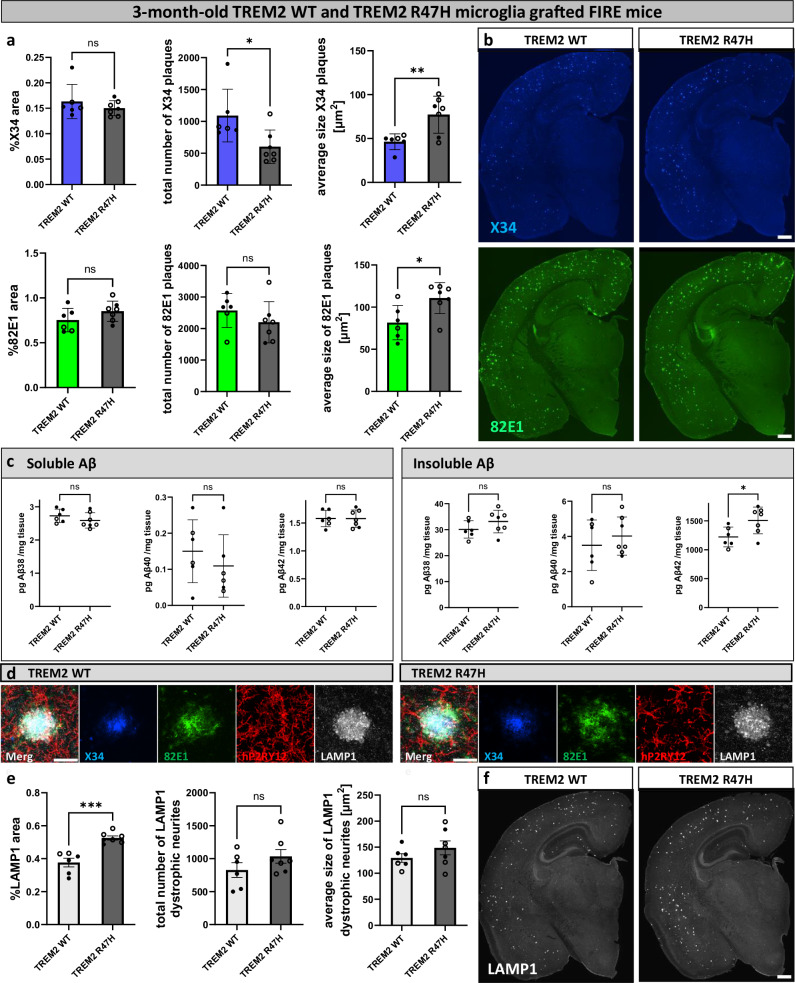
The role of microglia in the amyloid cascade of Alzheimer’s disease (AD) is debated due to conflicting findings. Using a genetic and a pharmacological approach we demonstrate that depletion of microglia before amyloid-β (Aβ) plaque deposition, leads to a reduction in plaque numbers and neuritic dystrophy, confirming their role in plaque initiation. Transplanting human microglia restores Aβ plaque formation. While microglia depletion reduces insoluble Aβ levels, soluble Aβ concentrations stay consistent, challenging the view that microglia clear Aβ. In later stages, microglial depletion decreases plaque compaction and increases neuritic dystrophy, suggesting a protective role. Human microglia with the TREM2 R47H/R47H mutation exacerbate plaque pathology, emphasizing the importance of non-reactive microglia in the initiation of the amyloid cascade. Adaptive immune depletion ( Rag2 -/- ) does not affect microglia’s impact on plaque formation. These findings clarify conflicting reports, identifying microglia as key drivers of amyloid pathology, and raise questions about optimal therapeutic strategies for AD.
Overview
The claims and statistics of this paper haven’t been checked yet.
Run the check — free, no account needed. It reads the open-access text and checks the sources it cites.
Integrity signals
Registry
Cites
Statistics
Figures
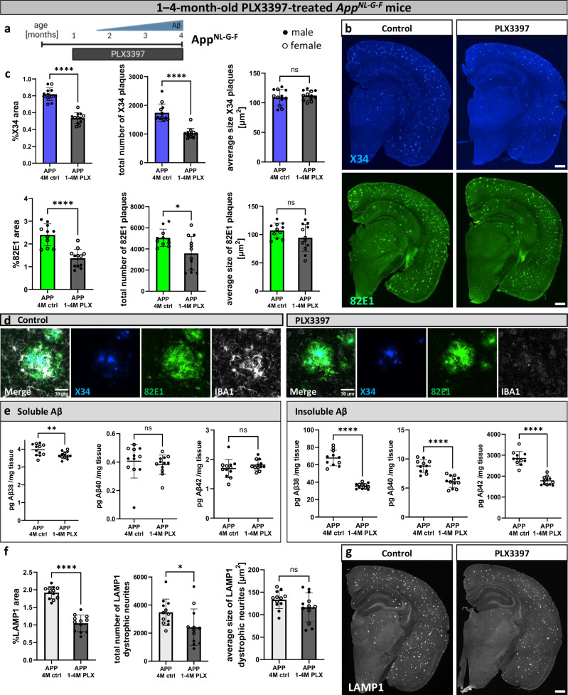
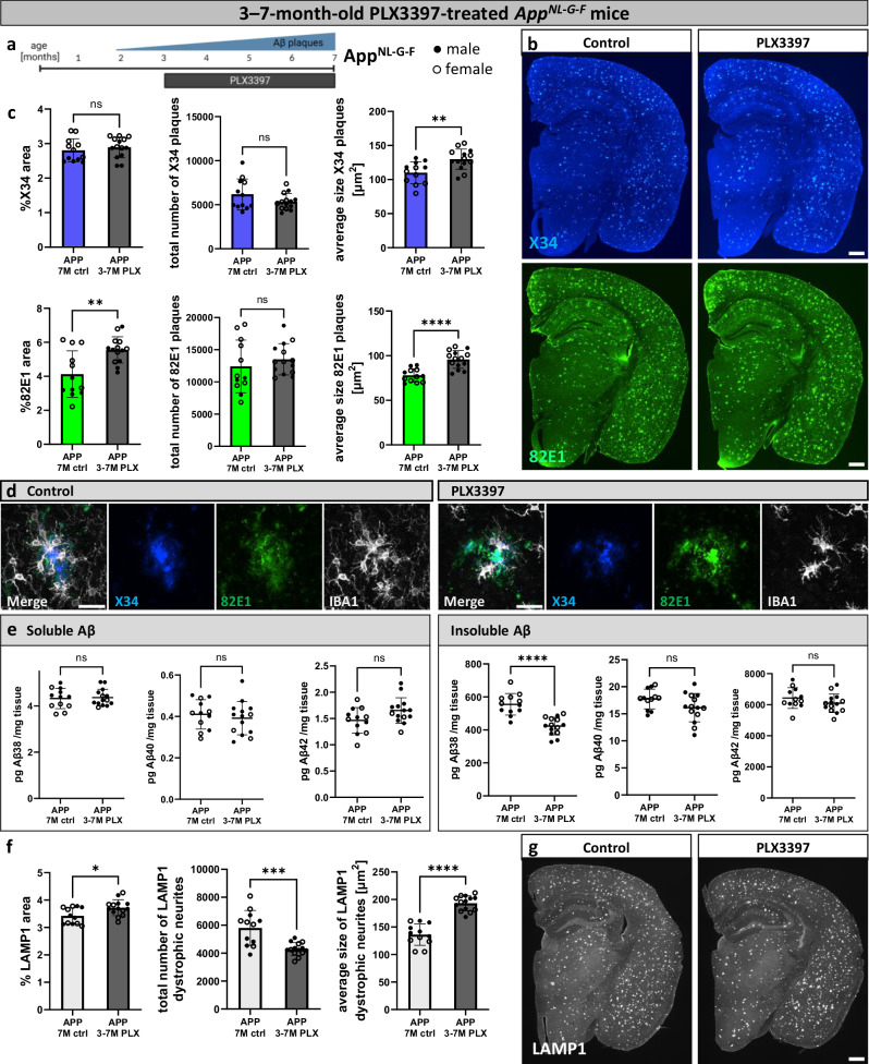
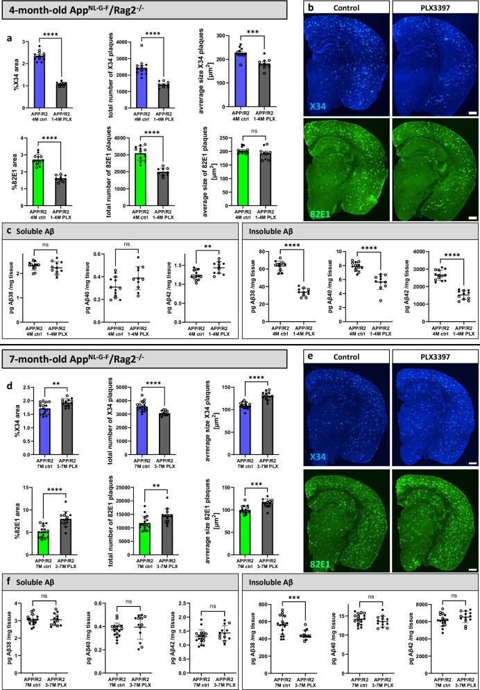
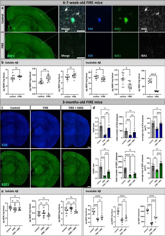
Verification
Claims the paper makes
The claims and statistics of this paper haven’t been checked yet.
Run the check — free, no account needed. It reads the open-access text and checks the sources it cites.
Claims other papers make about this one
Each is checked against this paper’s own text: does it say what the citing paper says it does?
The source backs part of the claim, not all of it — or backs it for a different population, condition or comparison.
In the citing paper
“Plaques were characterized using X34, a dye that strongly binds β‐sheeted amyloid, labelling both dense plaque cores and surrounding amyloid deposits, albeit with lower intensity in the latter”
From Alzheimer's Protection by PLCγ2 Compacts Plaques, Redistributes Microglia, and Protects Synapses in App NL ‐G‐F Mice (2026) · cites this paper as Baligács et al. 2024
Experimental — accuracy not yet measured
Self-check
Percentages, sums and reported differences re-computed from the paper’s own numbers.
The self-check needs the paper’s full text.
Citations
Cites
The Amyloid Hypothesis of Alzheimer’s Disease: Progress and Problems on the Road to Therapeutics
Hardy, J., & Selkoe, D. J. (2002) · Science · doi:10.1126/science.1072994 · Cited by 13,864
Amyloid oligomers exacerbate tau pathology in a mouse model of tauopathy
Selenica, M.-L. B., Brownlow, M., Jimenez, J. P., Lee, D. C., Pena, G., Dickey, C. A., Gordon, M. N., & Morgan, D. (2013) · Neurodegenerative Diseases · doi:10.1159/000337230 · Cited by 10,359
Lecanemab in early Alzheimer's disease
van Dyck, C. H., Swanson, C. J., Aisen, P., Bateman, R. J., Chen, C., Gee, M., Kanekiyo, M., Li, D., Reyderman, L., Cohen, S., et al. (2023) · New England Journal of Medicine · doi:10.1056/nejmoa2212948 · Cited by 5,678
A Unique Microglia Type Associated with Restricting Development of Alzheimer’s Disease
Keren-Shaul, H., Spinrad, A., Weiner, A., Matcovitch-Natan, O., Dvir-Szternfeld, R., Ulland, T. K., David, E., Baruch, K., Lara-Astaiso, D., Toth, B., et al. (2017) · Cell · doi:10.1016/j.cell.2017.05.018 · Cited by 5,493
Microglia-mediated neurotoxicity: uncovering the molecular mechanisms
Block, M. L., Zecca, L., & Hong, J.-S. (2007) · Nature Reviews Neuroscience · doi:10.1038/nrn2038 · Cited by 4,110
Neuroinflammation and microglial activation in Alzheimer disease: where do we go from here?
Leng, F., & Edison, P. (2021) · Nature Reviews Neurology · doi:10.1038/s41582-020-00435-y · Cited by 3,179
TREM2 Variants in Alzheimer's Disease
Guerreiro, R., Wojtas, A., Bras, J., Carrasquillo, M., Rogaeva, E., Majounie, E., Cruchaga, C., Sassi, C., Kauwe, J. S. K., Younkin, S., et al. (2013) · New England Journal of Medicine · doi:10.1056/nejmoa1211851 · Cited by 3,020
Krasemann, S., Madore, C., Cialic, R., Baufeld, C., Calcagno, N., El Fatimy, R., Beckers, L., O’Loughlin, E., Xu, Y., Fanek, Z., et al. (2017) · Immunity · doi:10.1016/j.immuni.2017.08.008 · Cited by 2,979
Donanemab in Early Symptomatic Alzheimer Disease: The TRAILBLAZER-ALZ 2 Randomized Clinical Trial
Sims, J. R., Zimmer, J. A., Evans, C. D., Lu, M., Ardayfio, P., Sparks, J., Wessels, A. M., Shcherbinin, S., Wang, H., Monkul Nery, E. S., et al. (2023) · JAMA · doi:10.1001/jama.2023.13239 · Cited by 2,932
RAG-2-deficient mice lack mature lymphocytes owing to inability to initiate V(D)J rearrangement
Shinkai, Y. (1992) · Cell · doi:10.1016/0092-8674(92)90029-c · Cited by 2,651
Variant of TREM2 Associated with the Risk of Alzheimer's Disease
Jonsson, T., Stefansson, H., Steinberg, S., Jonsdottir, I., Jonsson, P. V., Snaedal, J., Bjornsson, S., Huttenlocher, J., Levey, A. I., Lah, J. J., et al. (2013) · New England Journal of Medicine · doi:10.1056/nejmoa1211103 · Cited by 2,558
SMARCAD1 ATPase activity is required to silence endogenous retroviruses in embryonic stem cells
Sachs, P., Ding, D., Bergmaier, P., Lamp, B., Schlagheck, C., Finkernagel, F., Nist, A., Stiewe, T., & Mermoud, J. E. (2019) · Nature Communications · doi:10.1038/s41467-019-09078-0 · Cited by 2,400
The Cellular Phase of Alzheimer’s Disease
De Strooper, B., & Karran, E. (2016) · Cell · doi:10.1016/j.cell.2015.12.056 · Cited by 1,990
Elmore, M. R. P., Najafi, A. R., Koike, M. A., Dagher, N. N., Spangenberg, E. E., Rice, R. A., Kitazawa, M., Matusow, B., Nguyen, H., West, B. L., et al. (2014) · Neuron · doi:10.1016/j.neuron.2014.02.040 · Cited by 1,968
TREM2 Lipid Sensing Sustains the Microglial Response in an Alzheimer’s Disease Model
Wang, Y., Cella, M., Mallinson, K., Ulrich, J. D., Young, K. L., Robinette, M. L., Gilfillan, S., Krishnan, G. M., Sudhakar, S., Zinselmeyer, B. H., et al. (2015) · Cell · doi:10.1016/j.cell.2015.01.049 · Cited by 1,736
Disease-Associated Microglia: A Universal Immune Sensor of Neurodegeneration
Deczkowska, A., Keren-Shaul, H., Weiner, A., Colonna, M., Schwartz, M., & Amit, I. (2018) · Cell · doi:10.1016/j.cell.2018.05.003 · Cited by 1,474
Single App knock-in mouse models of Alzheimer’s disease
Saito, T., Matsuba, Y., Mihira, N., Takano, J., Nilsson, P., Itohara, S., Iwata, N., & Saido, T. C. (2014) · Nature Neuroscience · doi:10.1038/nn.3697 · Cited by 1,366
Alzheimer’s Disease Risk Genes and Mechanisms of Disease Pathogenesis
Karch, C. M., & Goate, A. M. (2015) · Biological Psychiatry · doi:10.1016/j.biopsych.2014.05.006 · Cited by 1,357
Microglial Dysfunction and Defective β-Amyloid Clearance Pathways in Aging Alzheimer's Disease Mice
Hickman, S. E., Allison, E. K., & El Khoury, J. (2008) · The Journal of Neuroscience · doi:10.1523/jneurosci.0616-08.2008 · Cited by 1,329 · cited 2×
“On one hand, microglia are believed to facilitate amyloid clearance through phagocytosis, potentially limiting plaque pathology 11 – 13 .”
Zhou, Y., Song, W. M., Andhey, P. S., Swain, A., Levy, T., Miller, K. R., Poliani, P. L., Cominelli, M., Grover, S., Gilfillan, S., et al. (2020) · Nature Medicine · doi:10.1038/s41591-019-0695-9 · Cited by 1,150
Clonally expanded CD8 T cells patrol the cerebrospinal fluid in Alzheimer’s disease
Gate, D., Saligrama, N., Leventhal, O., Yang, A. C., Unger, M. S., Middeldorp, J., Chen, K., Lehallier, B., Channappa, D., De Los Santos, M. B., et al. (2020) · Nature · doi:10.1038/s41586-019-1895-7 · Cited by 992
Spangenberg, E., Severson, P. L., Hohsfield, L. A., Crapser, J., Zhang, J., Burton, E. A., Zhang, Y., Spevak, W., Lin, J., Phan, N. Y., et al. (2019) · Nature Communications · doi:10.1038/s41467-019-11674-z · Cited by 939
Sala Frigerio, C., Wolfs, L., Fattorelli, N., Thrupp, N., Voytyuk, I., Schmidt, I., Mancuso, R., Chen, W.-T., Woodbury, M. E., Srivastava, G., et al. (2019) · Cell Reports · doi:10.1016/j.celrep.2019.03.099 · Cited by 926
Spangenberg, E. E., Lee, R. J., Najafi, A. R., Rice, R. A., Elmore, M. R. P., Blurton-Jones, M., West, B. L., & Green, K. N. (2016) · Brain · doi:10.1093/brain/aww016 · Cited by 695
Andreasen, N., Hesse, C., Davidsson, P., Minthon, L., Wallin, A., Winblad, B., Vanderstichele, H., Vanmechelen, E., & Blennow, K. (1999) · Archives of Neurology · doi:10.1001/archneur.56.6.673 · Cited by 660
CSF Aβ 42 levels correlate with amyloid-neuropathology in a population-based autopsy study
Strozyk, D., Blennow, K., White, L. R., & Launer, L. J. (2003) · Neurology · doi:10.1212/01.wnl.0000046581.81650.d0 · Cited by 621
Late onset Alzheimer’s disease genetics implicates microglial pathways in disease risk
Efthymiou, A. G., & Goate, A. M. (2017) · Molecular Neurodegeneration · doi:10.1186/s13024-017-0184-x · Cited by 562
Olmos-Alonso, A., Schetters, S. T. T., Sri, S., Askew, K., Mancuso, R., Vargas-Caballero, M., Holscher, C., Perry, V. H., & Gomez-Nicola, D. (2016) · Brain · doi:10.1093/brain/awv379 · Cited by 532
Kinetic theory of fibrillogenesis of amyloid β-protein
Lomakin, A., Teplow, D. B., Kirschner, D. A., & Benedek, G. B. (1997) · Proceedings of the National Academy of Sciences · doi:10.1073/pnas.94.15.7942 · Cited by 529
Xu, Q., Bernardo, A., Walker, D., Kanegawa, T., Mahley, R. W., & Huang, Y. (2006) · The Journal of Neuroscience · doi:10.1523/jneurosci.5476-05.2006 · Cited by 524
Havis, E., & Duprez, D. (2020) · International Journal of Molecular Sciences · doi:10.3390/ijms21051664 · Cited by 520
Bales, K. R., Verina, T., Cummins, D. J., Du, Y., Dodel, R. C., Saura, J., Fishman, C. E., DeLong, C. A., Piccardo, P., Petegnief, V., et al. (1999) · Proceedings of the National Academy of Sciences · doi:10.1073/pnas.96.26.15233 · Cited by 486
Human microglial state dynamics in Alzheimer’s disease progression
Sun, N., Victor, M. B., Park, Y. P., Xiong, X., Scannail, A. N., Leary, N., Prosper, S., Viswanathan, S., Luna, X., Boix, C. A., et al. (2023) · Cell · doi:10.1016/j.cell.2023.08.037 · Cited by 462
Harper, J. D., Wong, S. S., Lieber, C. M., & Lansbury, P. T. (1999) · Biochemistry · doi:10.1021/bi9904149 · Cited by 454
Microglia drive APOE-dependent neurodegeneration in a tauopathy mouse model
Shi, Y., Manis, M., Long, J., Wang, K., Sullivan, P. M., Remolina Serrano, J., Hoyle, R., & Holtzman, D. M. (2019) · Journal of Experimental Medicine · doi:10.1084/jem.20190980 · Cited by 448
Amyloid seeds formed by cellular uptake, concentration, and aggregation of the amyloid-beta peptide
Hu, X., Crick, S. L., Bu, G., Frieden, C., Pappu, R. V., & Lee, J.-M. (2009) · Proceedings of the National Academy of Sciences · doi:10.1073/pnas.0911281106 · Cited by 426 · cited 1×
“Homeostatic microglia uptake extracellular A β and concentrate it within endosomes and lysosomes at an acidic pH 54,55 , which could generate intracellular high molecular weight species 56,57 .”
Marsh, S. E., Abud, E. M., Lakatos, A., Karimzadeh, A., Yeung, S. T., Davtyan, H., Fote, G. M., Lau, L., Weinger, J. G., Lane, T. E., et al. (2016) · Proceedings of the National Academy of Sciences · doi:10.1073/pnas.1525466113 · Cited by 411
Sosna, J., Philipp, S., Albay, R., Reyes-Ruiz, J. M., Baglietto-Vargas, D., LaFerla, F. M., & Glabe, C. G. (2018) · Molecular Neurodegeneration · doi:10.1186/s13024-018-0244-x · Cited by 406
Rojo, R., Raper, A., Ozdemir, D. D., Lefèvre, L., Grabert, K., Wollscheid-Lengeling, E., Bradford, B. M., Caruso, M., Gažová, I., Sánchez, A., et al. (2019) · Nature Communications · doi:10.1038/s41467-019-11053-8 · Cited by 390
Self-citationInflammation in Alzheimer’s disease: Lessons learned from microglia-depletion models
Spangenberg, E. E., & Green, K. N. (2017) · Brain, Behavior, and Immunity · doi:10.1016/j.bbi.2016.07.003 · Cited by 352
Humanized TREM2 mice reveal microglia-intrinsic and -extrinsic effects of R47H polymorphism
Song, W. M., Joshita, S., Zhou, Y., Ulland, T. K., Gilfillan, S., & Colonna, M. (2018) · Journal of Experimental Medicine · doi:10.1084/jem.20171529 · Cited by 305
Mechanism of amyloid plaque formation suggests an intracellular basis of Aβ pathogenicity
Friedrich, R. P., Tepper, K., Rönicke, R., Soom, M., Westermann, M., Reymann, K., Kaether, C., & Fändrich, M. (2010) · Proceedings of the National Academy of Sciences · doi:10.1073/pnas.0904532106 · Cited by 303
Emerging roles of innate and adaptive immunity in Alzheimer’s disease
Chen, X., & Holtzman, D. M. (2022) · Immunity · doi:10.1016/j.immuni.2022.10.016 · Cited by 297
Stem-cell-derived human microglia transplanted in mouse brain to study human disease
Mancuso, R., Van Den Daele, J., Fattorelli, N., Wolfs, L., Balusu, S., Burton, O., Liston, A., Sierksma, A., Fourne, Y., Poovathingal, S., et al. (2019) · Nature Neuroscience · doi:10.1038/s41593-019-0525-x · Cited by 295
Self-citationFrackowiak, J., Wisniewski, H. M., Wegiel, J., Merz, G. S., Iqbal, K., & Wang, K. C. (1992) · Acta Neuropathologica · doi:10.1007/bf00227813 · Cited by 276
MEG3 activates necroptosis in human neuron xenografts modeling Alzheimer’s disease
Balusu, S., Horré, K., Thrupp, N., Craessaerts, K., Snellinx, A., Serneels, L., T’Syen, D., Chrysidou, I., Arranz, A. M., Sierksma, A. S. R., et al. (2023) · Science · doi:10.1126/science.abp9556 · Cited by 246
Self-citationClayton, K., Delpech, J. C., Herron, S., Iwahara, N., Ericsson, M., Saito, T., Saido, T. C., Ikezu, S., & Ikezu, T. (2021) · Molecular Neurodegeneration · doi:10.1186/s13024-021-00440-9 · Cited by 245
Baik, S. H., Kang, S., Son, S. M., & Mook-Jung, I. (2016) · Glia · doi:10.1002/glia.23074 · Cited by 234
Activated microglia mitigate Aβ-associated tau seeding and spreading
Gratuze, M., Chen, Y., Parhizkar, S., Jain, N., Strickland, M. R., Serrano, J. R., Colonna, M., Ulrich, J. D., & Holtzman, D. M. (2021) · Journal of Experimental Medicine · doi:10.1084/jem.20210542 · Cited by 215
Microglia contribute to the propagation of Aβ into unaffected brain tissue
d’Errico, P., Ziegler-Waldkirch, S., Aires, V., Hoffmann, P., Mezö, C., Erny, D., Monasor, L. S., Liebscher, S., Ravi, V. M., Joseph, K., et al. (2022) · Nature Neuroscience · doi:10.1038/s41593-021-00951-0 · Cited by 214
Chung, H., Brazil, M. I., Soe, T. T., & Maxfield, F. R. (1999) · Journal of Biological Chemistry · doi:10.1074/jbc.274.45.32301 · Cited by 212
Dramatic Shifts in Circulating CD4 but not CD8 T Cell Subsets in Mild Alzheimer's Disease
Larbi, A., Pawelec, G., Witkowski, J. M., Schipper, H. M., Derhovanessian, E., Goldeck, D., & Fülöp, T. (2009) · Journal of Alzheimer s Disease · doi:10.3233/jad-2009-1015 · Cited by 207
Lueg, G., Groß, C. C., Lohmann, H., Johnen, A., Kemmling, A., Deppe, M., Groger, J., Minnerup, J., Wiendl, H., Meuth, S. G., et al. (2014) · Neurobiology of Aging · doi:10.1016/j.neurobiolaging.2014.08.008 · Cited by 195
Casali, B. T., MacPherson, K. P., Reed-Geaghan, E. G., & Landreth, G. E. (2020) · Neurobiology of Disease · doi:10.1016/j.nbd.2020.104956 · Cited by 186
A multi-pronged human microglia response to Alzheimer’s disease Aβ pathology
Mancuso, R., Fattorelli, N., Martinez-Muriana, A., Davis, E., Wolfs, L., Van Den Daele, J., Geric, I., Preman, P., Serneels, L., Poovathingal, S., et al. (2022) · doi:10.1101/2022.07.07.499139 · Cited by 169
Self-citationAbsence of microglia promotes diverse pathologies and early lethality in Alzheimer’s disease mice
Kiani Shabestari, S., Morabito, S., Danhash, E. P., McQuade, A., Sanchez, J. R., Miyoshi, E., Chadarevian, J. P., Claes, C., Coburn, M. A., Hasselmann, J., et al. (2022) · Cell Reports · doi:10.1016/j.celrep.2022.110961 · Cited by 167
Self-citationTranslating genetic risk of Alzheimer’s disease into mechanistic insight and drug targets
Sierksma, A. S. R., Escott‐Price, V., & Strooper, B. D. (2020) · Science · doi:10.1126/science.abb8575 · Cited by 153
Microglia limit the expansion of β-amyloid plaques in a mouse model of Alzheimer’s disease
Zhao, R., Hu, W., Tsai, J., Li, W., & Gan, W.-B. (2017) · Molecular Neurodegeneration · doi:10.1186/s13024-017-0188-6 · Cited by 151
Unger, M. S., Schernthaner, P., Marschallinger, J., Mrowetz, H., & Aigner, L. (2018) · Journal of Neuroinflammation · doi:10.1186/s12974-018-1304-4 · Cited by 115
Stem-cell-derived human microglia transplanted into mouse brain to study human disease
Fattorelli, N., Martínez‐Muriana, A., Wolfs, L., Geric, I., Strooper, B. D., & Mancuso, R. (2021) · Nature Protocols · doi:10.1038/s41596-020-00447-4 · Cited by 114
Kaji, S., Berghoff, S. A., Spieth, L., Schlaphoff, L., Sasmita, A. O., Vitale, S., Büschgens, L., Kedia, S., Zirngibl, M., Nazarenko, T., et al. (2024) · Immunity · doi:10.1016/j.immuni.2024.09.014 · Cited by 104
Measuring heritable contributions to Alzheimer’s disease: polygenic risk score analysis with twins
Karlsson, I., Escott‐Price, V., Gatz, M., Hardy, J., Pedersen, N. L., Shoai, M., & Reynolds, C. A. (2021) · Brain Communications · doi:10.1093/braincomms/fcab308 · Cited by 100
Claes, C., Daele, J. V. D., Boon, R., Schouteden, S., Colombo, A. V., Monasor, L. S., Fiers, M. W. E. J., Ordovás, L., Nami, F., Bohrmann, B., et al. (2018) · Alzheimer s & Dementia · doi:10.1016/j.jalz.2018.09.006 · Cited by 88
Späni, C., Suter, T., Derungs, R., Ferretti, M. T., Welt, T., Wirth, F., Gericke, C., Nitsch, R. M., & Kulic, L. (2015) · Acta Neuropathologica Communications · doi:10.1186/s40478-015-0251-x · Cited by 88
Benítez, D. P., Jiang, S., Wood, J. I., Wang, R., Hall, C. M., Peerboom, C., Wong, N. S. M., Stringer, K. M., Vitanova, K. S., Smith, V. C., et al. (2021) · Molecular Neurodegeneration · doi:10.1186/s13024-021-00457-0 · Cited by 81
Fruhwürth, S., Zetterberg, H., & Paludan, S. R. (2024) · Journal of Neuroimmunology · doi:10.1016/j.jneuroim.2024.578342 · Cited by 77
Delizannis, A. T., Nonneman, A., Tsering, W., Bondt, A. D., den Wyngaert, I. V., Zhang, B., Meymand, E. S., Olufemi, M. F., Koivula, P. P., Maimaiti, S., et al. (2021) · Acta Neuropathologica Communications · doi:10.1186/s40478-021-01251-1 · Cited by 71
Plaque contact and unimpaired Trem2 is required for the microglial response to amyloid pathology
Wood, J. I., Wong, E., Joghee, R., Balbaa, A., Vitanova, K. S., Stringer, K. M., VanSchoiack, A., Phelan, S.-L. J., Launchbury, F., Desai, S., et al. (2022) · Cell Reports · doi:10.1016/j.celrep.2022.111686 · Cited by 65
Aducanumab anti-amyloid immunotherapy induces sustained microglial and immune alterations
Cadiz, M. P., Gibson, K. A., Todd, K. T., Nascari, D. G., Massa, N., Lilley, M. T., Olney, K. C., Al-Amin, M. M., Jiang, H., Holtzman, D. M., et al. (2024) · Journal of Experimental Medicine · doi:10.1084/jem.20231363 · Cited by 55
Cellular mechanism of fibril formation from serum amyloid A1 protein
Claus, S., Meinhardt, K., Aumüller, T., Puşcalău-Gîrţu, I., Linder, J., Haupt, C., Walther, P., Syrovets, T., Simmet, T., & Fändrich, M. (2017) · EMBO Reports · doi:10.15252/embr.201643411 · Cited by 48
Henningfield, C. M., Arreola, M. A., Soni, N., Spangenberg, E. E., & Green, K. N. (2022) · Glia · doi:10.1002/glia.24105 · Cited by 40
De Strooper, B., & Karran, E. (2024) · The EMBO Journal · doi:10.1038/s44318-024-00057-w · Cited by 36
Preman, P., Moechars, D., Fertan, E., Wolfs, L., Serneels, L., Shah, D., Lamote, J., Poovathingal, S., Snellinx, A., Mancuso, R., et al. (2024) · EMBO Molecular Medicine · doi:10.1038/s44321-024-00162-7 · Cited by 32
Self-citationMagy-Bertrand, N., Benson, M. D., Liepnieks, J. J., & Kluve‐Beckerman, B. (2007) · Amyloid · doi:10.1080/13506120601116575 · Cited by 25
Romero‐Molina, C., Navarro, V., Jiménez, S., Muñoz‐Castro, C., Sanchez-Mico, M. V., Gutiérrez, A., Vitórica, J., & Vizuete, M. (2021) · International Journal of Molecular Sciences · doi:10.3390/ijms22189734 · Cited by 14
Microglial APOE4: more is less and less is more
Eskandari‐Sedighi, G., & Blurton-Jones, M. M. (2023) · Molecular Neurodegeneration · doi:10.1186/s13024-023-00693-6 · Cited by 11
Research works printed but not linked: 1
- Mancuso, R. et al. Xenografted human microglia display diverse transcriptomic states in response to Alzheimer’s disease-related amyloid-β pathology. Nat. Neurosci. 27, 886–900 (2024). · DOI · Paper page
Cited by
Sorted by citations
Microglia transcriptional states and their functional significance: Context drives diversity
Depp, C., Doman, J. L., Hingerl, M., Xia, J., & Stevens, B. (2025) · Immunity · doi:10.1016/j.immuni.2025.04.009 · Cited by 84
Albertini, G., Zielonka, M., Cuypers, M., Snellinx, A., Xu, C., Poovathingal, S. K., Wojno, M., Davie, K., van Lieshout, V., Craessaerts, K., et al. (2025) · Nature Neuroscience · doi:10.1038/s41593-025-02125-8 · Cited by 47
Self-citationChen, D., Wang, C., Chen, X., Li, J., Chen, S., Li, Y., Ma, F., Li, T., Zou, M., Li, X., et al. (2025) · Science Translational Medicine · doi:10.1126/scitranslmed.ads6111 · Cited by 42
The Role of Genetic, Environmental, and Dietary Factors in Alzheimer’s Disease: A Narrative Review
Mertaş, B., & Boşgelmez, İ. İ. (2025) · International Journal of Molecular Sciences · doi:10.3390/ijms26031222 · Cited by 35
He, C., Chen, B., Yang, H., & Zhou, X. (2025) · Frontiers in Aging Neuroscience · doi:10.3389/fnagi.2025.1554398 · Cited by 40
Metadata
- Authors
- Baligács, N., Albertini, G., Borrie, S. C., Serneels, L., Pridans, C., Balusu, S., & De Strooper, B.
- Affiliations
- Vlaams Instituut voor Biotechnologie (BE)
- Centre for Inflammation Research (GB)
- ORCID
- Venue
- Nature Communications
- Published
- 2024-12-05
- Volume
- 15
- Issue
- 1
- Pages
- 10634-10634
- ISSN
- 2041-1723 (ISSN-L)
- PMC
- PMC11621353
- OpenAlex
- W4405035908
- Cited by
- 123
- References
- 76
- Citations by year
- 2026: 66 · 2025: 57
- FWCI
- 19.19
- Type
- journal article
- Language
- en
- Open access
- gold
- doi.org
- Locations
- Nature Communications (publishedVersion, cc-by)
- bioRxiv (Cold Spring Harbor Laboratory) (acceptedVersion)
- PubMed Central (publishedVersion, cc-by)
- PubMed (publishedVersion)
- UCL Discovery (University College London) (submittedVersion, cc-by)
- Lirias (KU Leuven) (submittedVersion, cc-by)
- RePEc: Research Papers in Economics (submittedVersion)
- DOAJ (DOAJ: Directory of Open Access Journals) (submittedVersion, cc-by-sa)
- Edinburgh Research Explorer (University of Edinburgh) (submittedVersion, other-oa)
- Funders
- Alzheimer's Association
- Queen Elisabeth Medical Foundation
- European Commission (HORIZON2020, ERC-834682 CELLPHASE_AD, 834682)
- Fonds Wetenschappelijk Onderzoek (11PSA24N)
- KU Leuven
- Vlaams Instituut voor Biotechnologie
- Vlaamse regering
- Fondation pour la Recherche sur Alzheimer
- Fondation Médicale Reine Elisabeth
- Medical Research Council (MR/Y014847/1)
- Topics
- Life Sciences › Neuroscience › Neurology › Neuroinflammation and Neurodegeneration Mechanisms
- Life Sciences › Neuroscience › Cellular and Molecular Neuroscience › Alzheimer's disease research and treatments
- Life Sciences › Neuroscience › Neurology › Neurological Disease Mechanisms and Treatments
- Keywords
- microglia, Alzheimer's disease, amyloid plaques, neuritic dystrophy, microglial depletion, amyloid clearance, Rag2 knockout mice, amyloid hypothesis
- MeSH terms
- Alzheimer Disease, Animals, Disease Models, Animal, Female, Homeostasis, Humans, Male, Membrane Glycoproteins, Mice, Inbred C57BL, Mice, Transgenic, Mutation, Receptors, Immunologic, Amyloid beta-Peptides, Microglia, Mice, Plaque, Amyloid
About these checks
Registry, retraction, Cites and statistics results are computed, not judged, with one exception: a printed reference that no index lists is read once by a model before it counts as a citation error. Claims are extracted by a model and kept only when their quote appears verbatim in the text; each cited source is then checked, with a model’s help, for whether it says what the claim relies on. Those verdicts can be wrong — read the claim’s quote and the cited source before relying on one.
Statistics and arithmetic are recomputed, not judged.
Confidence bands are shown only for fields calibrated on our annotated test set. None is calibrated yet, so no band is shown.
What counts as a claim ›How we measure confidence ›How we check this ›
Are you an author, or do you see a problem with this page? Request a review
