

The DOI matches its Crossref record; 3 of the 3 references it cites were found in a registry or an index.
Structure and function of the human apoptotic scramblase Xkr4
Open accessPublished version of a preprint
Abstract
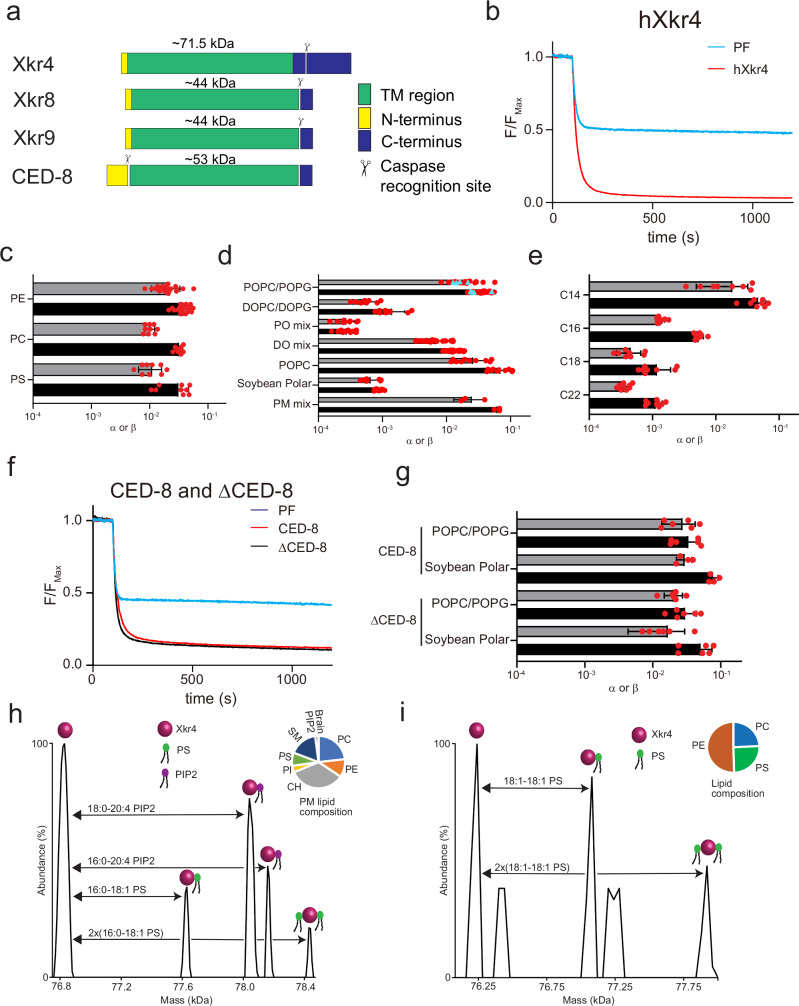
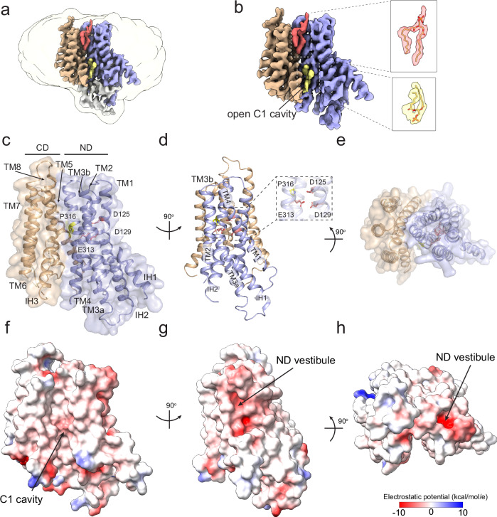
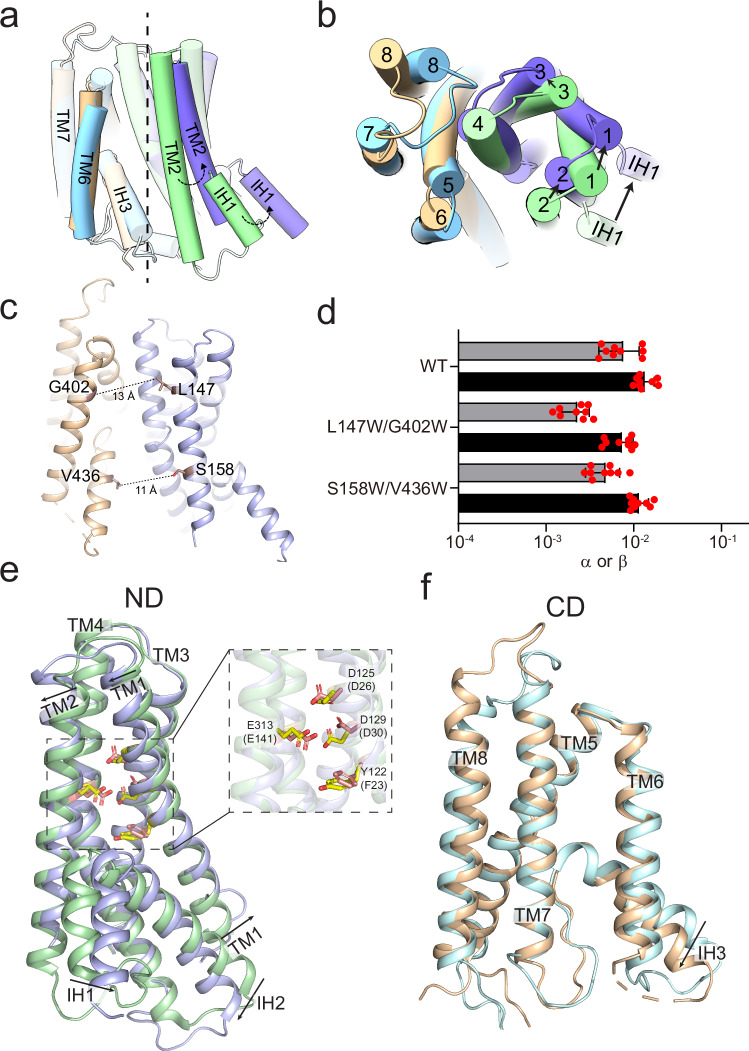
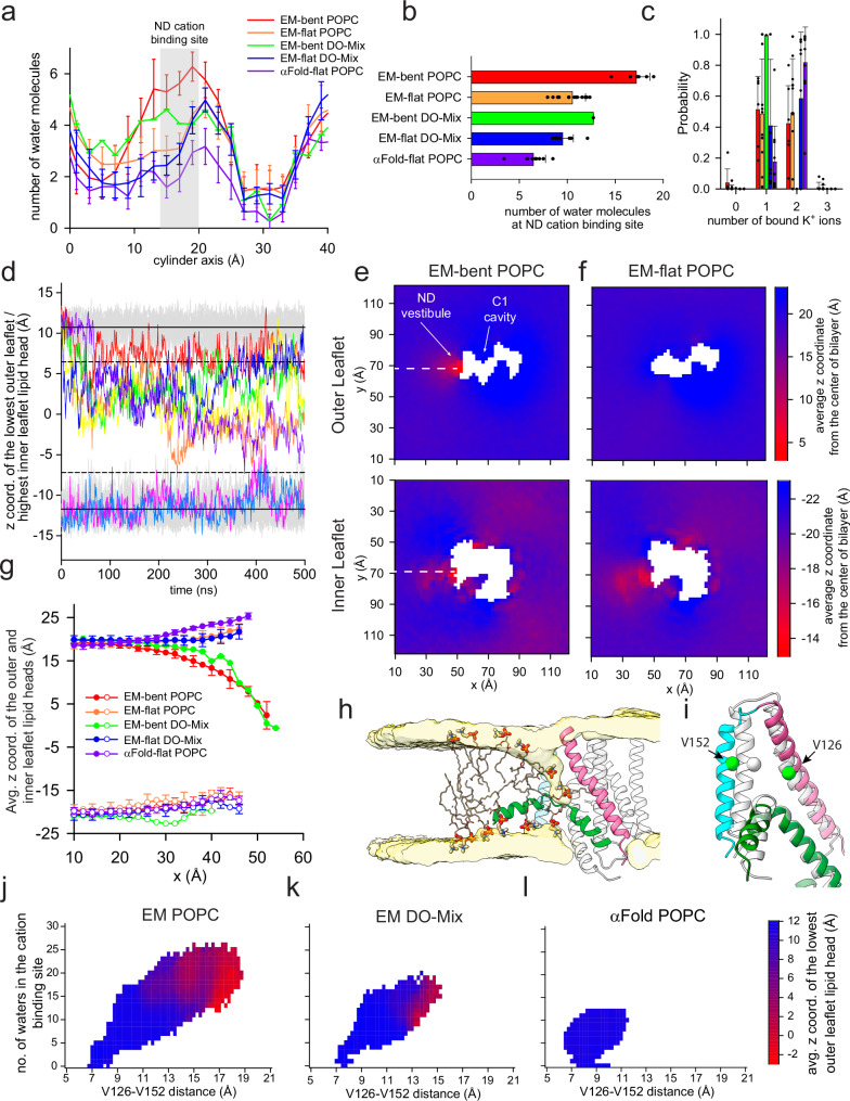
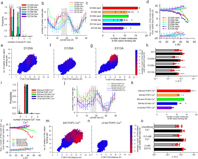
Abstract Phosphatidylserine externalization on the surface of dying cells is a key signal for their recognition and clearance by macrophages and is mediated by members of the X-Kell related (Xkr) protein family. Defective Xkr-mediated scrambling impairs clearance, leading to inflammation. It was proposed that activation of the Xkr4 apoptotic scramblase requires caspase cleavage, followed by dimerization and ligand binding. Here, using a combination of biochemical approaches we show that purified monomeric, full-length human Xkr4 (hXkr4) scrambles lipids. CryoEM imaging shows that hXkr4 adopts a novel conformation, where three conserved acidic residues create an electronegative surface embedded in the membrane. Molecular dynamics simulations show this conformation induces membrane thinning, which could promote scrambling. Thinning is ablated or reduced in conditions where scrambling is abolished or reduced. Our work provides insights into the molecular mechanisms of hXkr4 scrambling and suggests the ability to thin membranes might be a general property of active scramblases.
Overview
The claims and statistics of this paper haven’t been checked yet.
Run the check — free, no account needed. It reads the open-access text and checks the sources it cites.
Integrity signals
Registry
Cites
Statistics
Figures
Verification
Claims the paper makes
The claims and statistics of this paper haven’t been checked yet.
Run the check — free, no account needed. It reads the open-access text and checks the sources it cites.
Self-check
Percentages, sums and reported differences re-computed from the paper’s own numbers.
The self-check needs the paper’s full text.
Citations
Cites
Highly accurate protein structure prediction with AlphaFold
Jumper, J., Evans, R., Pritzel, A., Green, T., Figurnov, M., Ronneberger, O., Tunyasuvunakool, K., Bates, R., Žídek, A., Potapenko, A., et al. (2021) · Nature · doi:10.1038/s41586-021-03819-2 · Cited by 43,764
Accurate structure prediction of biomolecular interactions with AlphaFold 3
Abramson, J., Adler, J., Dunger, J., Evans, R., Green, T., Pritzel, A., Ronneberger, O., Willmore, L., Ballard, A. J., Bambrick, J., et al. (2024) · Nature · doi:10.1038/s41586-024-07487-w · Cited by 15,861
Hodge, S., Hodge, G., Scicchitano, R., Reynolds, P. N., & Holmes, M. (2003) · Immunology & Cell Biology · doi:10.1046/j.1440-1711.2003.t01-1-01170.x · Cited by 440 · cited 1×
“Mutations and/or deletion of Xkr genes contribute to auto-immune disorders, such as systemic lupus erythematosus, favor in fl ammation, asthma, and lung cancer 16 – 21 , further highlighting their importance in human physiology.”
Research works printed but not linked: 80
- Bevers, E. M. & Williamson, P. L. Getting to the Outer Leaflet: Physiology of Phosphatidylserine Exposure at the Plasma Membrane. Physiol. Rev. 96, 605–645 (2016). · DOI
- Sakuragi, T. & Nagata, S. Regulation of phospholipid distribution in the lipid bilayer by flippases and scramblases. Nat. Rev. Mol. Cell Biol. 24, 576–596 (2023). · DOI
- Kobayashi, T. & Menon, A. K. Transbilayer lipid asymmetry. Curr. Biol. 28, R386–R391 (2018). · DOI
- Whitlock, J. M. & Hartzell, H. C. Anoctamins/TMEM16 Proteins: Chloride Channels Flirting with Lipids and Extracellular Vesicles. Annu Rev. Physiol. 10, 119–143 (2017). · DOI
- Singh, N. Apoptosis in health and disease and modulation of apoptosis for therapy: An overview. Indian J. Clin. Biochem 22, 6–16 (2007). · DOI
- Peter, M. E. Programmed cell death: Apoptosis meets necrosis. Nature 471, 310–312 (2011). · DOI
- Shi, Y. Caspase activation, inhibition, and reactivation: a mechanistic view. Protein Sci. 13, 1979–1987 (2004). · DOI
- Arandjelovic, S. & Ravichandran, K. S. Phagocytosis of apoptotic cells in homeostasis. Nat. Immunol. 16, 907–917 (2015). · DOI · Paper page
- Doran, A. C., Yurdagul, A. & Tabas, I. Efferocytosis in health and disease. Nat. Rev. Immunol. 20, 254–267 (2020). · DOI · Paper page
- Silva, M. T., do Vale, A. & dos Santos, N. M. Secondary necrosis in multicellular animals: an outcome of apoptosis with pathogenic implications. Apoptosis 13, 463–482 (2008). · DOI
- Murao, A. et al. Release mechanisms of major DAMPs. Apoptosis 26, 152–162 (2021). · DOI
- Bäck, M. et al. Inflammation and its resolution in atherosclerosis: mediators and therapeutic opportunities. Nat. Rev. Cardiol. 16, 389–406 (2019).
- Calenda, G. et al. Identification of two new members, XPLAC and XTES, of the XK family. Gene 370, 6–16 (2006). · DOI
- Suzuki, J. et al. Xk-Related Protein 8 and CED-8 Promote Phosphatidylserine Exposure in Apoptotic Cells. Science 341, 403–406 (2013). · DOI
- Suzuki, J., Imanishi, E. & Nagata, S. Exposure of phosphatidylserine by Xk-related protein family members during apoptosis. J. Biol. Chem. 289, 30257–30267 (2014). · DOI
- Harley, J. B. et al. Genome-wide association scan in women with systemic lupus erythematosus identifies susceptibility variants in ITGAM, PXK, KIAA1542 and other loci. Nat. Genet 40, 204–210 (2008). · DOI · Paper page
- Kawano, M. & Nagata, S. Lupus-like autoimmune disease caused by a lack of Xkr8, a caspase-dependent phospholipid scramblase. Proc. Natl. Acad. Sci. USA 115, 2132–2137 (2018). · DOI
- Kim, G.-W. et al. Xk-related protein 8 regulates myoblast differentiation and survival. FEBS J. 284, 3575–3588 (2017). · DOI
- Sun, J. et al. XK-related protein 5 (XKR5) is a novel negative regulator of KIT/D816V-mediated transformation. Oncogenesis 7, 48 (2018). · DOI
- Henson, P. M. & Tuder, R. M. Apoptosis in the lung: induction, clearance and detection. Am. J. Physiol. Lung Cell Mol. Physiol. 294, L601–L611 (2008). · DOI
- Maruoka, M. et al. Caspase cleavage releases a nuclear protein fragment that stimulates phospholipid scrambling at the plasma membrane. Mol. Cell 81, 1397–1410 e9 (2021). · DOI
- Shook, D. et al. XKR4 Gene Effects on Cerebellar Development Are Not Specific to ADHD. Front Cell Neurosci. 11, 396 (2017). · DOI
- de Zeeuw, P. et al. Imaging gene and environmental effects on cerebellum in Attention-Deficit/Hyperactivity Disorder and typical development. NeuroImage: Clin. 2, 103–110 (2013). · DOI
- Uhl, G. R. et al. Higher order” addiction molecular genetics: Convergent data from genome-wide association in humans and mice. Biochem. Pharmacol. 75, 98–111 (2008). · DOI
- Chen, Y.-Z. et al. Caspase-mediated activation of Caenorhabditis elegans CED-8 promotes apoptosis and phosphatidylserine externalization. Nat. Commun. 4, 2726 (2013). · DOI
- Sakuragi, T., Kosako, H. & Nagata, S. Phosphorylation-mediated activation of mouse Xkr8 scramblase for phosphatidylserine exposure. Proc. Natl. Acad. Sci. 116, 2907–2912 (2019). · DOI
- Suzuki, J., Imanishi, E. & Nagata, S. Xkr8 phospholipid scrambling complex in apoptotic phosphatidylserine exposure. Proc. Natl. Acad. Sci. USA 113, 9509–9514 (2016). · DOI
- Zhang, P. et al. Extracellular calcium functions as a molecular glue for transmembrane helices to activate the scramblase Xkr4. Nat. Commun. 14, 5592 (2023). · DOI
- Sakuragi, T. et al. The tertiary structure of the human Xkr8-Basigin complex that scrambles phospholipids at plasma membranes. Nat. Struct. Mol. Biol. 28, 825–834 (2021). · DOI
- Straub, M. S. et al. Cryo-EM structures of the caspase-activated protein XKR9 involved in apoptotic lipid scrambling. eLife 10, e69800 (2021). · DOI
- Guillén-Samander, A. et al. A partnership between the lipid scramblase XK and the lipid transfer protein VPS13A at the plasma membrane. Proc. Natl. Acad. Sci. USA 119, e2205425119 (2022). · DOI
- Ryoden, Y. & Nagata, S. The XK plasma membrane scramblase and the VPS13A cytosolic lipid transporter for ATP-induced cell death. Bioessays 44, e2200106 (2022). · DOI
- Ghanbarpour, A. et al. A model for a partnership of lipid transfer proteins and scramblases in membrane expansion and organelle biogenesis. Proc. Natl. Acad. Sci. USA. 118, e2101562118 (2021). · DOI
- Adlakha, J. et al. Structural and biochemical insights into lipid transport by VPS13 proteins. J. Cell Biol. 221, e202202030 (2022). · DOI
- Sakuragi, T. et al. The role of the C-terminal tail region as a plug to regulate XKR8 lipid scramblase. J. Biol. Chem. 300, 105755 (2024). · DOI
- Pomorski, T. & Menon, A. K. Lipid flippases and their biological functions. Cell Mol. Life Sci. 63, 2908–2921 (2006). · DOI
- Falzone, M. E. et al. TMEM16 scramblases thin the membrane to enable lipid scrambling. Nat. Commun. 13, 2604 (2022). · DOI
- Falzone, M. E., et al., Structural basis of Ca(2+)-dependent activation and lipid transport by a TMEM16 scramblase. Elife, 8, (2019). · DOI
- Panda, A. et al. Direct determination of oligomeric organization of integral membrane proteins and lipids from intact customizable bilayer. Nat. Methods 20, 891–897 (2023). · DOI
- Panda, A., Brown, C. & Gupta, K. Studying Membrane Protein-Lipid Specificity through Direct Native Mass Spectrometric Analysis from Tunable Proteoliposomes. J. Am. Soc. Mass Spectrom. 34, 1917–1927 (2023). · DOI
- Hattori, M., Hibbs, R. E. & Gouaux, E. A fluorescence-detection size-exclusion chromatography-based thermostability assay for membrane protein precrystallization screening. Structure 20, 1293–1299 (2012). · DOI
- Goehring, A. et al. Screening and large-scale expression of membrane proteins in mammalian cells for structural studies. Nat. Protoc. 9, 2574–2585 (2014). · DOI
- Malvezzi, M. et al. Out of the groove transport of lipids by TMEM16 and GPCR scramblases. Proc. Natl. Acad. Sci. USA. 115 E7033–E7042 (2018). · DOI
- Falzone, M. E. & Accardi, A. Reconstitution of Proteoliposomes for Phospholipid Scrambling and Nonselective Channel Assays. Methods Mol. Biol. 2127, 207–225 (2020). · DOI
- Feng, Z., Alvarenga, O. E. & Accardi, A. Structural basis of closed groove scrambling by a TMEM16 protein. Nat. Struct. Mol. Biol. 31, 1468–1481 (2024). · DOI
- Malvezzi, M. et al. Ca2+-dependent phospholipid scrambling by a reconstituted TMEM16 ion channel. Nat. Commun. 4, 2367 (2013). · DOI
- Garcia-Calvo, M. et al. Purification and catalytic properties of human caspase family members. Cell Death Differ. 6, 362–369 (1999). · DOI
- Lee, B.-C., Menon, A. nantK. & Accardi, A. The nhTMEM16 Scramblase Is Also a Nonselective Ion Channel. Biophysical J. 111, 1919–1924 (2016). · DOI
- Alvadia, C. et al. Cryo-EM structures and functional characterization of the murine lipid scramblase TMEM16F. eLife 8, e44365 (2019). · DOI
- Brunner, J. D. et al. X-ray structure of a calcium-activated TMEM16 lipid scramblase. Nature 516, 207–212 (2014). · DOI
- Motiwala, Z. et al. Structural basis of GABA reuptake inhibition. Nature 606, 820–826 (2022). · DOI
- Punjani, A. et al. cryoSPARC: algorithms for rapid unsupervised cryo-EM structure determination. Nat. Methods 14, 290 (2017). · DOI · Paper page
- Bushell, S. R. et al. The structural basis of lipid scrambling and inactivation in the endoplasmic reticulum scramblase TMEM16K. Nat. Commun. 10, 3956 (2019). · DOI
- Lee, B. C. et al. Gating mechanism of the extracellular entry to the lipid pathway in a TMEM16 scramblase. Nat. Commun. 9, 3251 (2018). · DOI
- Bethel, N. P. & Grabe, M. Atomistic insight into lipid translocation by a TMEM16 scramblase. Proc. Natl. Acad. Sci. USA 113, 14049–14054 (2016). · DOI
- Jiang, T. et al. Lipids and ions traverse the membrane by the same physical pathway in the nhTMEM16 scramblase. Elife. 6, e28671 (2017). · DOI
- Kostritskii, A. Y. & Machtens, J.-P. Molecular mechanisms of ion conduction and ion selectivity in TMEM16 lipid scramblases. Nat. Commun. 12, 2826 (2021). · DOI
- Arndt, M. et al. Structural basis for the activation of the lipid scramblase TMEM16F. Nat. Commun. 13, 6692 (2022). · DOI
- Kalienkova, V. et al. Stepwise activation mechanism of the scramblase nhTMEM16 revealed by cryo-EM. eLife 8, e44364 (2019). · DOI
- Feng, S. et al. Cryo-EM Studies of TMEM16F Calcium-Activated Ion Channel Suggest Features Important for Lipid Scrambling. Cell Rep. 28, 567–579.e4 (2019). · DOI
- Jarvis, D. L., Chapter 14 Baculovirus–Insect Cell Expression Systems, in Methods in Enzymology, R.R. Burgess and M.P. Deutscher, Editors. 2009, Academic Press. p. 191-222. · DOI
- Berger, I., Fitzgerald, D. J. & Richmond, T. J. Baculovirus expression system for heterologous multiprotein complexes. Nat. Biotechnol. 22, 1583–1587 (2004). · DOI
- Kostelic, M. M. & Marty, M. T. Deconvolving Native and Intact Protein Mass Spectra with UniDec. Methods Mol. Biol. 2500, 159–180 (2022). · DOI
- Rohou, A. & Grigorieff, N. CTFFIND4: Fast and accurate defocus estimation from electron micrographs. J. Struct. Biol. 192, 216–221 (2015). · DOI
- Tegunov, D. & Cramer, P. Real-time cryo-electron microscopy data preprocessing with Warp. Nat. Methods 16, 1146–1152 (2019). · DOI
- Lander, G. C. et al. Appion: an integrated, database-driven pipeline to facilitate EM image processing. J. Struct. Biol. 166, 95–102 (2009). · DOI
- Suloway, C. et al. Automated molecular microscopy: The new Leginon system. J. Struct. Biol. 151, 41–60 (2005). · DOI
- Afonine, P. V. Phenix tool to compute a difference map for cryo-EM. Computational Crystallogr. Newsl. 8, 8–9 (2017).
- Afonine, P. V. et al. PHENIX News. Computational Crystallogr. Newsl. 4, 43–44 (2013).
- Liebschner, D. et al. Macromolecular structure determination using X-rays, neutrons and electrons: recent developments in Phenix. Acta Crystallogr D. Struct. Biol. 75, 861–877 (2019). · DOI · Paper page
- Mirdita, M. et al. ColabFold: making protein folding accessible to all. Nat. Methods 19, 679–682 (2022). · DOI · Paper page
- Webb, B. & Sali, A. Comparative protein structure modeling using MODELLER. Curr Protoc Bioinformatics, 54, 5.6.1–5.6.37 (2016). · DOI
- Humphrey, W., Dalke, A. & Schulten, K. VMD: visual molecular dynamics. J. Mol. Graph. 14, 33–38 (1996). · DOI · Paper page
- Wu, E. L. et al. CHARMM-GUI Membrane Builder toward realistic biological membrane simulations. J. Comp. Chem. 35, 1997–2004 (2014). · DOI
- Best, R. B. et al. Optimization of the additive CHARMM all-atom protein force field targeting improved sampling of the backbone phi, psi and side-chain chi(1) and chi(2) dihedral angles. J. Chem. Theory Comput 8, 3257–3273 (2012). · DOI
- Jorgensen, W. L. et al. Comparison of simple potential functions for simulating liquid water. J. Chem. Phys. 79, 926–935 (1983). · DOI
- Zhang, A. et al. The Ca2+ permeation mechanism of the ryanodine receptor revealed by a multi-site ion model. Nat. Commun. 11, 922 (2020). · DOI
- Abraham, M. J. et al. GROMACS: High performance molecular simulations through multi-level parallelism from laptops to supercomputers. SoftwareX 1-2, 19–25 (2015). · DOI · Paper page
- Fortea, E. et al. Structural basis of pH-dependent activation in a CLC transporter. Nat. Struct. Mol. Biol. 31, 644–656 (2024). · DOI
- Bernhardt, N. & Faraldo-Gómez, J. D. MOSAICS: A software suite for analysis of membrane structure and dynamics in simulated trajectories. Biophysical J. 122, 2023–2040 (2023). · DOI
Metadata
- Authors
- Chakraborty, S., Feng, Z., Lee, S., Alvarenga, O. E., Panda, A., Zhang, S., Bruni, R., Khelashvili, G., Gupta, K., & Accardi, A.
- Affiliations
- Weill Cornell Medicine (US)
- Yale University (US)
- New York Structural Biology Center (US)
- Corresponding author
- Accardi, Alessio
- ORCID
- Venue
- Nature Communications
- Published
- 2025-08-08
- Volume
- 16
- Issue
- 1
- Pages
- 7317-7317
- ISSN
- 2041-1723 (ISSN-L)
- PMC
- PMC12334663
- OpenAlex
- W4413054266
- Cited by
- 5
- References
- 3
- Citations by year
- 2026: 3 · 2025: 1
- FWCI
- 1.20
- Type
- journal article
- Language
- en
- Open access
- gold
- doi.org
- Licence
- Locations
- Nature Communications (publishedVersion, cc-by-nc-nd)
- bioRxiv (Cold Spring Harbor Laboratory) (acceptedVersion, cc-by-nc-nd)
- PubMed Central (publishedVersion, cc-by-nc-nd)
- PubMed (publishedVersion)
- PubMed Central (submittedVersion, cc-by-nc-nd)
- DOAJ (DOAJ: Directory of Open Access Journals) (submittedVersion, cc-by-sa)
- Funders
- Division of Intramural Research, National Institute of Allergy and Infectious Diseases (R01AI178180)
- NSF | Directorate for Biological Sciences (1746886)
- U.S. Department of Health & Human Services | NIH | National Institute of General Medical Sciences (R01GM141192)
- Topics
- Life Sciences › Biochemistry, Genetics and Molecular Biology › Molecular Biology › Lipid Membrane Structure and Behavior
- Life Sciences › Immunology and Microbiology › Immunology › Phagocytosis and Immune Regulation
- Life Sciences › Biochemistry, Genetics and Molecular Biology › Molecular Biology › Cell death mechanisms and regulation
- Keywords
- phosphatidylserine exposure, membrane thinning, protein conformation, caspase cleavage, inflammation, cryo-electron microscopy, molecular dynamics simulation
About these checks
Registry, retraction, Cites and statistics results are computed, not judged, with one exception: a printed reference that no index lists is read once by a model before it counts as a citation error. Claims are extracted by a model and kept only when their quote appears verbatim in the text; each cited source is then checked, with a model’s help, for whether it says what the claim relies on. Those verdicts can be wrong — read the claim’s quote and the cited source before relying on one.
Statistics and arithmetic are recomputed, not judged.
Confidence bands are shown only for fields calibrated on our annotated test set. None is calibrated yet, so no band is shown.
What counts as a claim ›How we measure confidence ›How we check this ›
Are you an author, or do you see a problem with this page? Request a review
How to cite
Cite CoChat for the checks on this page, and the paper for its own findings.
- This page
- CoChat. (2026). Citation check: Structure and function of the human apoptotic scramblase Xkr4. https://app.cochat.ai/papers/10.1038/s41467-025-62739-1
- The paper
- Chakraborty, S., Feng, Z., Lee, S., Alvarenga, O. E., Panda, A., Zhang, S., Bruni, R., Khelashvili, G., Gupta, K., & Accardi, A. (2025). Structure and function of the human apoptotic scramblase Xkr4. Nature Communications. https://doi.org/10.1038/s41467-025-62739-1
