

The DOI matches its Crossref record; 24 of the 24 references it cites were found in a registry or an index.
Organotypic human skin culture models constructed with senescent fibroblasts show hallmarks of skin aging
Open accessAbstract
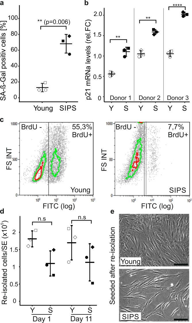
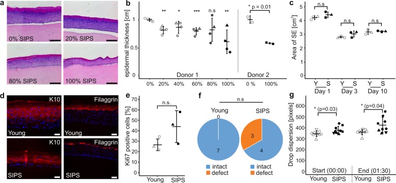
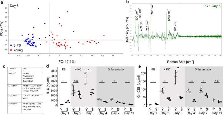
Skin aging is driven by intrinsic and extrinsic factors impacting on skin functionality with progressive age. One factor of this multifaceted process is cellular senescence, as it has recently been identified to contribute to a declining tissue functionality in old age. In the skin, senescent cells have been found to markedly accumulate with age, and thus might impact directly on skin characteristics. Especially the switch from young, extracellular matrix-building fibroblasts to a senescence-associated secretory phenotype (SASP) could alter the microenvironment in the skin drastically and therefore promote skin aging. In order to study the influence of senescence in human skin, 3D organotypic cultures are a well-suited model system. However, only few “aged” skin- equivalent (SE) models are available, requiring complex and long-term experimental setups. Here, we adapted a previously published full-thickness SE model by seeding increasing ratios of stress-induced premature senescent versus normal fibroblasts into the collagen matrix, terming these SE “senoskin”. Immunohistochemistry stainings revealed a shift in the balance between proliferation (Ki67) and differentiation (Keratin 10 and Filaggrin) of keratinocytes within our senoskin equivalents, as well as partial impairment of skin barrier function and changed surface properties. Monitoring of cytokine levels of known SASP factors confirmedly showed an upregulation in 2D cultures of senescent cells and at the time of seeding into the skin equivalent. Surprisingly, we find a blunted response of cytokines in the senoskin equivalent over time during 3D differentiation.
Overview
The claims and statistics of this paper haven’t been checked yet.
Run the check — free, no account needed. It reads the open-access text and checks the sources it cites.
Integrity signals
Registry
Cites
Statistics
Figures
Verification
Claims the paper makes
The claims and statistics of this paper haven’t been checked yet.
Run the check — free, no account needed. It reads the open-access text and checks the sources it cites.
Claims other papers make about this one
Each is checked against this paper’s own text: does it say what the citing paper says it does?
The cited source says what the claim attributes to it.
In the citing paper
“HSEs are used to investigate various aspects of normal and abnormal skin biology, including wound healing [40,41], aging [42,43], and the study of various diseases [18].”
From Diversity of human skin three-dimensional organotypic cultures (2024) · cites this paper as [43]
Self-check
Percentages, sums and reported differences re-computed from the paper’s own numbers.
The self-check needs the paper’s full text.
Citations
Cites
Oliver, W. C., & Pharr, G. M. (2004) · Journal of materials research/Pratt's guide to venture capital sources · doi:10.1557/jmr.2004.19.1.3 · Cited by 8,377
A biomarker that identifies senescent human cells in culture and in aging skin in vivo.
Dimri, G. P., Lee, X., Basile, G., Acosta, M., Scott, G., Roskelley, C., Medrano, E. E., Linskens, M., Rubelj, I., & Pereira-Smith, O. (1995) · Proceedings of the National Academy of Sciences · doi:10.1073/pnas.92.20.9363 · Cited by 7,477
The limited in vitro lifetime of human diploid cell strains
Hayflick, L. (1965) · Experimental Cell Research · doi:10.1016/0014-4827(65)90211-9 · Cited by 5,876
The senescence-associated secretory phenotype: the dark side of tumor suppression
Coppé, J.-P., Desprez, P.-Y., Krtolica, A., & Campisi, J. (2010) · Annual Review of Pathology: Mechanisms of Disease · doi:10.1146/annurev-pathol-121808-102144 · Cited by 5,206
Biomarkers of cellular senescence and skin aging
Wang, A. S., & Dreesen, O. (2018) · Frontiers in Genetics · doi:10.3389/fgene.2018.00247 · Cited by 404
Mine, S., Fortunel, N. O., Pageon, H., & Asselineau, D. (2008) · PLoS ONE · doi:10.1371/journal.pone.0004066 · Cited by 266
Kaplan, G., WALSH, G. P., Guido, L. S., Meyn, P., Burkhardt, R. A., Abalos, R. M., Barker, J., Frindt, P., Fajardo, T. T., & Celona, R. (1992) · The Journal of Experimental Medicine · doi:10.1084/jem.175.6.1717 · Cited by 262
Terlecki‐Ζaniewicz, L., Lämmermann, I., Latreille, J., Bobbili, M. R., Pils, V., Schosserer, M., Weinmüllner, R., Dellago, H., Skalicky, S., Pum, D., et al. (2018) · Aging · doi:10.18632/aging.101452 · Cited by 249
Self-citationSecreted microvesicular miR-31 inhibits osteogenic differentiation of mesenchymal stem cells
Weilner, S., Schraml, E., Wieser, M., Messner, P., Schneider, K. H., Wassermann, K. J., Mičutková, L., Fortschegger, K., Maier, A. B., Westendorp, R. G. J., et al. (2016) · Aging Cell · doi:10.1111/acel.12484 · Cited by 216
Self-citationStress-induced premature senescence and tissue ageing
Toussaint, O., Royer, V., Salmon, M., & Remacle, J. (2002) · Biochemical Pharmacology · doi:10.1016/s0006-2952(02)01170-x · Cited by 141
Cited by
Sorted by citations
Skin Senescence: Mechanisms and Impact on Whole-Body Aging
Franco, A. C., Aveleira, C., & Cavadas, C. (2022) · Trends in Molecular Medicine · doi:10.1016/j.molmed.2021.12.003 · Cited by 321
Connective tissue and fibroblast senescence in skin aging
Wlaschek, M., Maity, P., Makrantonaki, E., & Scharffetter-Kochanek, K. (2021) · Journal of Investigative Dermatology · doi:10.1016/j.jid.2020.11.010 · Cited by 245
The Skin Microbiome: Current Landscape and Future Opportunities
Smythe, P., & Wilkinson, H. N. (2023) · International Journal of Molecular Sciences · doi:10.3390/ijms24043950 · Cited by 218
Faces of cellular senescence in skin aging
Ho, C. Y., & Dreesen, O. (2021) · Mechanisms of Ageing and Development · doi:10.1016/j.mad.2021.111525 · Cited by 172
The role of cellular senescence in skin aging and age-related skin pathologies
Chin, T., Lee, X. E., Ng, P. Y., Lee, Y., & Dreesen, O. (2023) · Frontiers in Physiology · doi:10.3389/fphys.2023.1297637 · Cited by 164
Flavonoids in Skin Senescence Prevention and Treatment
Domaszewska-Szostek, A., Puzianowska‐Kuźnicka, M., & Kuryłowicz, A. (2021) · International Journal of Molecular Sciences · doi:10.3390/ijms22136814 · Cited by 142
How good is the evidence that cellular senescence causes skin ageing?
Low, E., Alimohammadiha, G., Smith, L. A., Costello, L., Przyborski, S. A., von Zglinicki, T., & Miwa, S. (2021) · Ageing Research Reviews · doi:10.1016/j.arr.2021.101456 · Cited by 116
Zhao, X., Liu, Y., Jia, P., Cheng, H., Wang, C., Chen, S., Huang, H., Han, Z., Han, Z., Marycz, K., et al. (2021) · Stem Cell Research & Therapy · doi:10.1186/s13287-021-02262-4 · Cited by 87
Narzt, M.-S., Pils, V., Kremslehner, C., Nagelreiter, I.-M., Schosserer, M., Bessonova, E., Bayer, A., Reifschneider, R., Terlecki-Zaniewicz, L., Waidhofer-Söllner, P., et al. (2021) · Journal of Investigative Dermatology · doi:10.1016/j.jid.2020.11.020 · Cited by 73
Self-citationChoudhury, S., & Das, A. (2020) · Cytotherapy · doi:10.1016/j.jcyt.2020.10.001 · Cited by 68
Mavrogonatou, E., Papadopoulou, A., Pratsinis, H., & Kletsas, D. (2023) · American Journal of Physiology-Cell Physiology · doi:10.1152/ajpcell.00178.2023 · Cited by 72
Cellular Senescence in Human Skin Aging: Leveraging Senotherapeutics
Wyles, S. P., CARRUTHERS, J. D. A., Dashti, P., Yu, G., Yap, J. Q., Gingery, A., Tchkonia, T., & Kirkland, J. L. (2023) · Gerontology · doi:10.1159/000534756 · Cited by 67
tBHP treatment as a model for cellular senescence and pollution-induced skin aging
Wedel, S., Martic, I., Hrapovic, N., Fabre, S., Madreiter‐Sokolowski, C. T., Haller, T., Pierer, G., Ploner, C., Jansen‐Dürr, P., & Cavinato, M. S. (2020) · Mechanisms of Ageing and Development · doi:10.1016/j.mad.2020.111318 · Cited by 62
Senescence as a molecular target in skin aging and disease
Thau, H., Gerjol, B. P., Hahn, K., Gudenberg, R. W. V., Knoedler, L., Stallcup, K., Emmert, M. Y., Buhl, T., Wyles, S. P., Tchkonia, T., et al. (2025) · Ageing Research Reviews · doi:10.1016/j.arr.2025.102686 · Cited by 65
Moreira, P., Sousa, F. J., Matos, P., Brites, G. S., Gonçalves, M. J., Cavaleiro, C., Figueirinha, A., Salgueiro, L., Batista, M. T., Branco, P. C., et al. (2022) · Pharmaceutics · doi:10.3390/pharmaceutics14030561 · Cited by 58
Targeting organ-specific mitochondrial dysfunction to improve biological aging
Madreiter‐Sokolowski, C. T., Hiden, U., Krstić, J., Panzitt, K., Wagner, M., Enzinger, C., Khalil, M., Abdellatif, M., Malle, E., Madl, T., et al. (2024) · Pharmacology & Therapeutics · doi:10.1016/j.pharmthera.2024.108710 · Cited by 55
Self-citationWedel, S., Martic, I., Guerrero‐Navarro, L., Ploner, C., Pierer, G., Jansen‐Dürr, P., & Cavinato, M. S. (2022) · Aging Cell · doi:10.1111/acel.13752 · Cited by 48
Phang, S. J., Basak, S., Teh, H. X., Packirisamy, G., Fauzi, M. B., Kuppusamy, U. R., Neo, Y. P., & Looi, M. L. (2022) · ACS Biomaterials Science & Engineering · doi:10.1021/acsbiomaterials.2c00342 · Cited by 45
Targeting Cellular Senescence for Age-Related Diseases: Path to Clinical Translation
Wyles, S. P., Tchkonia, T., & Kirkland, J. L. (2021) · Plastic & Reconstructive Surgery · doi:10.1097/prs.0000000000009669 · Cited by 42
Cavinato, M. S., Madreiter‐Sokolowski, C. T., Büttner, S., Schosserer, M., Zwerschke, W., Wedel, S., Grillari, J., Graier, W. F., & Jansen‐Dürr, P. (2020) · FEBS Journal · doi:10.1111/febs.15631 · Cited by 38
Self-citationTissue engineering strategies to bioengineer the ageing skin phenotype in vitro
Costello, L., DiColandrea, T., Tasseff, R. A., Isfort, R. J., Bascom, C., von Zglinicki, T., & Przyborski, S. A. (2022) · Aging Cell · doi:10.1111/acel.13550 · Cited by 33
Current Understanding of the Role of Senescent Melanocytes in Skin Ageing
Hughes, B. K., & Bishop, C. L. (2022) · Biomedicines · doi:10.3390/biomedicines10123111 · Cited by 38
Torrens‐Mas, M., Perelló-Reus, C. M., Navas-Enamorado, C., Ibargüen-González, L., Sánchez-Polo, A., Segura‐Sampedro, J. J., Masmiquel, L., Barceló, C., & González‐Freire, M. (2021) · International Journal of Molecular Sciences · doi:10.3390/ijms221910547 · Cited by 38
Lombardi, F., Augello, F. R., Ciafarone, A., Ciummo, V., Altamura, S., Cinque, B., & Palumbo, P. (2024) · Biomolecules · doi:10.3390/biom14091066 · Cited by 33
Nishikori, S., Yasuda, J., Murata, K., Takegaki, J., Harada, Y., Shirai, Y., & Fujita, S. (2023) · Scientific Reports · doi:10.1038/s41598-023-37207-9 · Cited by 40
Exploring mechanisms of skin aging: insights for clinical treatment
Zhang, M., Lin, Y., Han, Z., Huang, X., Zhou, S., Wang, S., Zhou, Y., Han, X., & Chen, H. (2024) · Frontiers in Immunology · doi:10.3389/fimmu.2024.1421858 · Cited by 38
Promises and challenges of senolytics in skin regeneration, pathology and ageing
Pils, V., Ring, N., Valdivieso, K., Lämmermann, I., Грубер, Ф., Schosserer, M., Grillari, J., & Ogrodnik, M. B. (2021) · Mechanisms of Ageing and Development · doi:10.1016/j.mad.2021.111588 · Cited by 33
Self-citationProgress in the Development of Stem Cell-Derived Cell-Free Therapies for Skin Aging
Chou, Y., Alfarafisa, N. M., Ikezawa, M., & Khairani, A. F. (2023) · Clinical Cosmetic and Investigational Dermatology · doi:10.2147/ccid.s434439 · Cited by 29
Almeida, T., Moreira, P., Sousa, F. J., Pereira, C., Silvestre, A. J. D., Vilela, C., & Freire, C. S. R. (2022) · Materials · doi:10.3390/ma15051982 · Cited by 27
The Role of NAD+ in Regenerative Medicine
Conlon, N. J. (2021) · Plastic & Reconstructive Surgery · doi:10.1097/prs.0000000000009673 · Cited by 24
Haga, M., Iida, K., & Okada, M. (2024) · iScience · doi:10.1016/j.isci.2024.109708 · Cited by 19
Wahhab, R., Sanders, M., Kokikian, N., Ni, C. S., & Vandiver, A. R. (2024) · Journal of the American Academy of Dermatology · doi:10.1016/j.jaad.2024.08.044 · Cited by 23
Janjetović, Z., & Slominski, A. T. (2024) · Cosmetics · doi:10.3390/cosmetics11020037 · Cited by 23
Fernando, I. P. S., Heo, S., Dias, M. K. H. M., Madusanka, D. M. D., Han, E. J., Kim, M.-J., Sanjeewa, K. K. A., Lee, K., & Ahn, G. (2021) · Marine Drugs · doi:10.3390/md19080435 · Cited by 18
Senescent cells in 3D culture show suppressed senescence signatures
Yadav, P., Chatterjee, K., & Saini, D. K. (2021) · Biomaterials Science · doi:10.1039/d1bm00536g · Cited by 19
Senescence in the ageing skin: a new focus on m TORC 1 and the lysosome
Smith, P., & Carroll, B. (2024) · FEBS Journal · doi:10.1111/febs.17281 · Cited by 18
Biotics as novel therapeutics in targeting signs of skin ageing via the gut-skin axis
Millman, J. F., Kondrashina, A. V., Walsh, C., Busca, K., Karawugodage, A., Park, J., Sirisena, S., Martin, F., Felice, V. D., & Lane, J. A. (2024) · Ageing Research Reviews · doi:10.1016/j.arr.2024.102518 · Cited by 21
Czyz, C. M., Kunth, P. W., Грубер, Ф., Kremslehner, C., Hammers, C. M., & Hundt, J. E. (2023) · Experimental Dermatology · doi:10.1111/exd.14911 · Cited by 19
Strategies to make human skin models based on cellular senescence for ageing research
Shakel, Z., Lima, S. A. C., & Reis, S. (2024) · Ageing Research Reviews · doi:10.1016/j.arr.2024.102430 · Cited by 17
Development of a Vascularized Human Skin Equivalent with Hypodermis for Photoaging Studies
Sanchez, M. M., Tonmoy, T. I., Park, B. H., & Morgan, J. T. (2022) · Biomolecules · doi:10.3390/biom12121828 · Cited by 15
Skin Disease Models In Vitro and Inflammatory Mechanisms: Predictability for Drug Development
Hennies, H. C., & Poumay, Y. (2021) · Handbook of experimental pharmacology · doi:10.1007/164_2020_428 · Cited by 16
Generation of Self-assembled Vascularized Human Skin Equivalents
Sanchez, M. M., & Morgan, J. T. (2021) · Journal of Visualized Experiments · doi:10.3791/62125 · Cited by 14
Diversity of human skin three-dimensional organotypic cultures
Jia, Y. Y., & Atwood, S. X. (2024) · Current Opinion in Genetics & Development · doi:10.1016/j.gde.2024.102275 · Cited by 12
Organotypic cultures as aging associated disease models
Sanchez, M. M., Bagdasarian, I. A., Darch, W., & Morgan, J. T. (2022) · Aging · doi:10.18632/aging.204361 · Cited by 12
Novel 3D-Printed Cell Culture Inserts for Air–Liquid Interface Cell Culture
Bauer, M., Metzger, M., Corea, M., Schädl, B., Grillari, J., & Dungel, P. (2022) · Life · doi:10.3390/life12081216 · Cited by 11
Self-citationExploring the Antiaging Properties of Curcumin, Flaxseed, and Collagen: A Comprehensive Review
Varma, P., & Kandasubramanian, B. (2024) · ChemistrySelect · doi:10.1002/slct.202403395 · Cited by 7
Harnessing Next‐Generation 3D Cancer Models to Elucidate Tumor‐Microbiome Crosstalk
Buzhor, M. G., Longobardi, G., Kandli, O., Krinsky, A., Avramoff, O., Katyal, A., Salomon, K., Miari, A., Venkert, D., Barnatan, T. T., et al. (2025) · Advanced Healthcare Materials · doi:10.1002/adhm.202503198 · Cited by 7
Unveiling aging heterogeneities in human dermal fibroblasts via nanosensor chemical cytometry
Song, Y., Seo, I., Tian, C., An, J., Park, S., Hyun, J., Jung, S. P., Park, H. S., Park, H., Bhang, S. H., et al. (2025) · Nature Communications · doi:10.1038/s41467-025-61590-8 · Cited by 7
CAMADAS DA FACE E MUDANÇAS ASSOCIADAS COM O ENVELHECIMENTO FACIAL
Pereira, F. F., de Souza, M. S., & de Souza, D. M. (2021) · AESTHETIC OROFACIAL SCIENCE · doi:10.51670/aos.v2i2.70 · Cited by 6
UTP increases wound healing in the self assembled skin substitute (SASS)
Naasani, L. I. S., Sévigny, J., Moulin, V. J., & Wink, M. R. (2023) · Journal of Cell Communication and Signaling · doi:10.1007/s12079-023-00725-2 · Cited by 6
Advanced biomaterial strategies for overcoming age-associated wound healing impairments
Dores, R., Rodrigues, T., Carvalho, D. S., Batista, E., Kozak, M., Blanco‐Elices, C., Fernandes, H., & Bimbo, L. M. (2025) · APL Bioengineering · doi:10.1063/5.0251889 · Cited by 5
Wei, D., Zhang, H., Xu, W., Zhang, B., Zhang, L., Lan, L., Li, Y., Shi, Y., & Song, X. (2025) · Journal of Photochemistry and Photobiology B Biology · doi:10.1016/j.jphotobiol.2025.113200 · Cited by 5
Zhu, L., Huang, W., Shi, J., Li, F., Sun, X., & Xiao, J. (2025) · International Journal of Biological Macromolecules · doi:10.1016/j.ijbiomac.2025.145467 · Cited by 6
Gomez, P. D. L. S., Rai, R., Ritchie, P., Smith, L. A., Simpson, A., Goncalves, K., & Przyborski, S. A. (2025) · Journal of Anatomy · doi:10.1111/joa.70026 · Cited by 8
Gluconolactone in Cosmetic (Review Article)
Wulaningsih, T. I. (2023) · KESANS International Journal of Health and Science · doi:10.54543/kesans.v2i9.191 · Cited by 5
Protocolo para rejuvenescimento facial minimamente invasivo em idosos
Piaia, T. C., de Souza, D. M., de Souza, M. S., & Tortelli, S. A. C. (2021) · AESTHETIC OROFACIAL SCIENCE · doi:10.51670/ahof.v2i1.40 · Cited by 5
He, S., Wang, Z., Wang, R., Chi, B., Wang, Y., Bu, L., Wu, J., Li, H., & Huang, Q. (2024) · Journal of Drug Delivery Science and Technology · doi:10.1016/j.jddst.2024.105722 · Cited by 5
Hautalterung – zelluläre Seneszenz
Scharffetter‐Kochanek, K., Wang, Y., Makrantonaki, E., Crișan, D., Wlaschek, M., Geiger, H., & Maity, P. (2023) · Die Dermatologie · doi:10.1007/s00105-023-05201-x · Cited by 5
A 3D bioprinted in vitro full-thickness skin aging model
Chakraborty, J., Gupta, A., & Ghosh, S. (2025) · Journal of Materials Chemistry B · doi:10.1039/d5tb01126d · Cited by 4
Chaturvedi, D., Mukherjee, S., Sawant, P., Jain, P. D., & Majumder, A. (2022) · Springer eBooks · doi:10.1007/978-981-19-1379-2_22 · Cited by 5
Disease-related biomarkers as experimental endpoints in 3D skin culture models
Chaturvedi, D., Paranjape, S., Jain, R. D., & Dandekar, P. P. (2023) · Cytotechnology · doi:10.1007/s10616-023-00574-2 · Cited by 4
Aging skin and non-surgical procedures: a basic science overview
Vandiver, A. R., & Hogan, S. R. (2020) · Plastic and Aesthetic Research · doi:10.20517/2347-9264.2020.159 · Cited by 9
Lu, E. M. (2024) · International Journal of Molecular Sciences · doi:10.3390/ijms252111552 · Cited by 3
Yu, Y., Zhang, Z., Zhang, J., Gao, Y., Li, X., Yang, K., Si, N., Chen, Z., Gu, Z., & Yin, N. (2026) · Microsystems & Nanoengineering · doi:10.1038/s41378-026-01170-y · Cited by 5
CLCA2: A Potential Guardian against Premature Senescence and Skin Aging
Guerrero‐Navarro, L., Martic, I., Ploner, C., Jansen‐Dürr, P., & Cavinato, M. S. (2024) · Biomedicines · doi:10.3390/biomedicines12030592 · Cited by 3
Efficacy of SGPP2 Modulation-Mediated Materials in Ameliorating Facial Wrinkles and Pore Sagging
Kim, J., Ye, S., Jun, S., & Kang, N. G. (2024) · Current Issues in Molecular Biology · doi:10.3390/cimb46080539 · Cited by 3
Wyles, S. P., Vyas, K. S., Wasserburg, J. R., Ansaf, R. B., & Kirkland, J. L. (2024) · Elsevier eBooks · doi:10.1016/b978-0-443-15500-0.00009-8 · Cited by 3
Generation of Self-assembled Vascularized Human Skin Equivalents
Sanchez, M. M., & Morgan, J. T. (2021) · Journal of Visualized Experiments · doi:10.3791/62125-v · Cited by 3
Generation of Pigmented Skin Grafts from Human Hair Follicles and Dermal Fibroblasts
Schneider, M., Ziemer, M., Lethaus, B., Simon, J. C., & Savković, V. (2021) · Tissue Engineering Part A · doi:10.1089/ten.tea.2020.0284 · Cited by 3
ACE2 OVEREXPRESSION CHANGES THE SARS-COV-2 INFECTION PROFILE IN BEAS-2B CELLS
Bartolomeo, C. S., Alves, T., Lemes, R. M. R., Ivanov, G., Morais, R., Costa, A., Nishino, M., Bassani, T., Pereira, G., Smaili, S. S., et al. (2021) · Cytotherapy · doi:10.1016/j.jcyt.2021.02.009 · Cited by 3
An Improved Human 3-Dimensional Skin Model for Aging Research
Huth, S., Marquardt, Y., Huth, L., Singh, K., Scharffetter‐Kochanek, K., Baron, J. M., & Maity, P. (2025) · Journal of Investigative Dermatology · doi:10.1016/j.jid.2025.07.005 · Cited by 2
Huang, Y., Wang, B., Jin, Y., Qiu, M., Liu, J., Hao, L., Zhang, C., & Ma, H. (2025) · Journal of Ginseng Research · doi:10.1016/j.jgr.2025.11.014 · Cited by 3
Ishikawa, M., Ngo, Y. X., Nishikawa, I., Kato, H., Maeda, R., Mizuno, R., Mizuno, J., Izumi, K., Yanagisawa, H., & Sada, A. (2026) · Journal of Investigative Dermatology · doi:10.1016/j.jid.2026.02.014 · Cited by 5
Briganti, S., Camera, E., Ciriza, J., Fernández-Carro, E., Krivokuća, M. J., Lamartine, J., Letsiou, S., Nacka-Aleksić, M., Niessen, C. M., Oliva, N., et al. (2026) · Ageing Research Reviews · doi:10.1016/j.arr.2026.103109 · Cited by 4
Self-citationHARMONIZAÇÃO FACIAL CIRÚRGICA: Área de Atuação do Cirurgião-Dentista
Custódio, A. L. N., Silva, A. M. D. R., Franco, C. C., Pacheco, R. F., & de Souza, M. S. (2020) · AESTHETIC OROFACIAL SCIENCE · doi:10.51670/ahof.v1i1.10 · Cited by 2
Daboin, C., Nicklow, E., & Griffin, D. R. (2025) · Acta Biomaterialia · doi:10.1016/j.actbio.2025.10.031 · Cited by 2
Miguel, S. P., Ribeiro, M. P., & Coutinho, P. (2021) · Springer eBooks · doi:10.1007/978-981-16-2677-7_15 · Cited by 2
Low, E., Smith, L. A., Miwa, S., Fielder, E., Przyborski, S. A., & von Zglinicki, T. (2024) · Journal of Investigative Dermatology · doi:10.1016/j.jid.2024.07.004 · Cited by 2
Ishikawa, M., Ngo, Y. X., Nishikawa, I., Kato, H., Maeda, R., Mizuno, R., Mizuno, J., Izumi, K., Yanagisawa, H., & Sada, A. (2025) · bioRxiv (Cold Spring Harbor Laboratory) · doi:10.1101/2025.01.21.634195 · Cited by 2
Woo, J., Ji, H., Jeon, K., Kim, H. B., Yoon, S., Hwang, Y.-H., Cho, E., Park, D., & Jung, E. (2025) · International Journal of Cosmetic Science · doi:10.1111/ics.13046 · Cited by 1
Yadav, P., Mondal, P., Jani, S., Saini, D. K., & Chatterjee, K. (2025) · Virtual and Physical Prototyping · doi:10.1080/17452759.2025.2459802 · Cited by 1
Homaeigohar, S., & Boccaccini, A. R. (2025) · Royal Society of Chemistry eBooks · doi:10.1039/9781837677627-00001 · Cited by 1
Rethinking the complex determinants of teacher shortages
Morris, R., & Ventista, O. (2021) · Research Features · doi:10.26904/rf-139-1674592571 · Cited by 1
Os benefícios do uso da tecnologia no tratamento de rejuvenescimento facial - ultrassom microfocado
Peres, F. N. C., & Peres, L. N. C. (2023) · Journal of Multidisciplinary Dentistry · doi:10.46875/jmd.v13i1.826 · Cited by 1
Generation and Characterization of a New Aging Skin Human Dermal Extracellular Matrix Scaffold
Fernández-Carro, E., & Ciriza, J. (2024) · Methods in molecular biology · doi:10.1007/7651_2024_579 · Cited by 1
Raman microspectroscopy: sub-cellular chemical imaging of aging
Liendl, L., & Schosserer, M. (2021) · Aging · doi:10.18632/aging.203785 · Cited by 1
Self-citationZidarič, T., Kleinschek, K. S., Maver, U., & Maver, T. (2023) · Springer briefs in molecular science · doi:10.1007/978-3-031-21298-7_6
Haga, M., Iida, K., & Okada, M. (2023) · bioRxiv (Cold Spring Harbor Laboratory) · doi:10.1101/2023.07.18.546970
Pradita, D. A., Achadiyani, & Bashari, M. H. (2023) · International Journal of Science and Society · doi:10.54783/ijsoc.v5i4.834
Bridging the Gap in Cancer Cell Behavior Against Matrix Stiffening: Insights from a Trizonal ModelPreprint
Torki, M. E., Liu, F., Xu, R., Chen, Y., Fredberg, J., & Chen, Z. (2023) · bioRxiv (Cold Spring Harbor Laboratory) · doi:10.1101/2023.12.02.569730
Development of Aged Skin Equivalent Model
Brand, L. M., Lamers, M. L., & Matte, B. F. (2023) · Current Cosmetic Science · doi:10.2174/0126667797269907231118130449
Atkins, O., Lopez, S., Ma, Y., & Roberts, L. A. (2023) · American Journal of Undergraduate Research · doi:10.33697/ajur.023.093
Bridging the Gap in Cancer Cell Behavior Against Matrix Stiffening: Insights from a Trizonal ModelPreprint
Torki, M. E., Liu, F., Xu, R., Chen, Y., Fredberg, J., & Chen, Z. (2025) · eLife · doi:10.7554/elife.102809.1
Bridging the Gap in Cancer Cell Behavior Against Matrix Stiffening: Insights from a Trizonal ModelPreprint
Torki, M. E., Liu, F., Xu, R., Chen, Y., Fredberg, J., & Chen, Z. (2025) · eLife · doi:10.7554/elife.102809
Development and Characterization of 3D Senescent Models Mimicking Skin Aging
Pentecouteau, L., Laperdrix, C., Rouxel, E., Folligné, A., Morvan, P., & Vallée, R. (2025) · Cosmetics · doi:10.3390/cosmetics12020071
Smith, L. A., Maltman, V., Gomez, P. D. L. S., Goncalves, K., & Przyborski, S. A. (2025) · Methods in molecular biology · doi:10.1007/978-1-0716-4510-9_12
Integrating stem cell biology and organoid technology for skin regenerative therapies
Zheng, K., Yang, Q., & Wu, Y. (2025) · Health engineering. · doi:10.26599/he.2024.9460004
Jia, X., Yuan, R., Huang, Y., Li, X., Zhang, R., He, L., & Yang, Z. (2025) · International Journal of Biological Macromolecules · doi:10.1016/j.ijbiomac.2025.147464 · Cited by 1
Zhao, X., Liu, Y., Jia, P., Cheng, H., Wang, C., Chen, S., Huang, H., Han, Z., Han, Z., Marycz, K., et al. (2021) · Figshare · doi:10.6084/m9.figshare.14253852
Metadata
- Authors
- Weinmüllner, R., Zbiral, B., Becirovic, A., Stelzer, E. M., Nagelreiter, F., Schosserer, M., Lämmermann, I., Liendl, L., Lang, M., Terlecki-Zaniewicz, L., Andriotis, O., Mildner, M., Golabi, B., Waidhofer-Söllner, P., Schedle, K., Emsenhuber, G., Thurner, P. J., Tschachler, E., Gruber, F., & Grillari, J.
- Affiliations
- BOKU University (AT)
- Austrian Cluster for Tissue Regeneration (AT)
- TU Wien (AT)
- Medical University of Vienna (AT)
- Ludwig Boltzmann Institute for Traumatology, The Research Center in Cooperation with AUVA (AT)
- Corresponding author
- Grillari, Johannes
- ORCID
- Venue
- npj Aging and Mechanisms of Disease
- Published
- 2020-03-06
- Volume
- 6
- Issue
- 1
- Pages
- 4-4
- ISSN
- 2056-3973 (ISSN-L)
- PMC
- PMC7060247
- OpenAlex
- W3009524618
- Cited by
- 103
- References
- 24
- Citations by year
- 2026: 5 · 2025: 22 · 2024: 18 · 2023: 19 · 2022: 11 · 2021: 22 · 2020: 6
- FWCI
- 8.72
- Type
- journal article
- Language
- en
- Open access
- gold
- doi.org
- Locations
- npj Aging and Mechanisms of Disease (publishedVersion, cc-by)
- PubMed Central (publishedVersion, cc-by)
- PubMed (publishedVersion)
- DOAJ (DOAJ: Directory of Open Access Journals) (submittedVersion, cc-by-sa)
- Funders
- Austrian Science Fund (FWF: I2514)
- Christian Doppler Forschungsgesellschaft
- PhD Programme BioToP – ‘Biomolecular technolgy of proteins’
- Topics
- Life Sciences › Biochemistry, Genetics and Molecular Biology › Cell Biology › Telomeres, Telomerase, and Senescence
- Health Sciences › Medicine › Dermatology › Skin Protection and Aging
- Life Sciences › Biochemistry, Genetics and Molecular Biology › Aging › Genetics, Aging, and Longevity in Model Organisms
- Keywords
- skin aging, cellular senescence, senescence-associated secretory phenotype, stress-induced premature senescence, fibroblasts, keratinocytes, skin barrier function, cytokines, extracellular matrix
About these checks
Registry, retraction, Cites and statistics results are computed, not judged, with one exception: a printed reference that no index lists is read once by a model before it counts as a citation error. Claims are extracted by a model and kept only when their quote appears verbatim in the text; each cited source is then checked, with a model’s help, for whether it says what the claim relies on. Those verdicts can be wrong — read the claim’s quote and the cited source before relying on one.
Statistics and arithmetic are recomputed, not judged.
Confidence bands are shown only for fields calibrated on our annotated test set. None is calibrated yet, so no band is shown.
What counts as a claim ›How we measure confidence ›How we check this ›
Are you an author, or do you see a problem with this page? Request a review
