Cited by 104Genomic variations and chromosomal abnormalitiesChromosomal and Genetic VariationsGenome Rearrangement Algorithms

The DOI matches its Crossref record; 79 of the 79 references it cites were found in a registry or an index.

Increased mutation and gene conversion within human segmental duplications

Open access
Vollger, Mitchell R., Dishuck, Philip C., Harvey, William T., DeWitt, William S., Guitart, Xavi, Goldberg, Michael E., Rozanski, Allison N., Lucas, Julian, Asri, Mobin, Human Pangenome Reference Consortium, Abel, Haley J., Antonacci-Fulton, Lucinda L., Baid, Gunjan, Baker, Carl A., Belyaeva, Anastasiya, Billis, Konstantinos, Bourque, Guillaume, Buonaiuto, Silvia, Carroll, Andrew, Chaisson, Mark J. P., Chang, Pi-Chuan, Chang, Xian H., Cheng, Haoyu, Chu, Justin, Cody, Sarah, Colonna, Vincenza, Cook, Daniel E., Cook-Deegan, Robert M., Cornejo, Omar E., Diekhans, Mark, Doerr, Daniel, Ebert, Peter, Ebler, Jana, Eizenga, Jordan M., Fairley, Susan, Fedrigo, Olivier, Felsenfeld, Adam L., Feng, Xiaowen, Fischer, Christian, Flicek, Paul, Formenti, Giulio, Frankish, Adam, Fulton, Robert S., Gao, Yan, Garg, Shilpa, Garrison, Erik, Garrison, Nanibaa’ A., Giron, Carlos Garcia, Green, Richard E., Groza, Cristian, Guarracino, Andrea, Haggerty, Leanne, Hall, Ira M., Haukness, Marina, Haussler, David, Heumos, Simon, Hickey, Glenn, Hourlier, Thibaut, Howe, Kerstin, Jain, Miten, Jarvis, Erich D., Ji, Hanlee P., Kenny, Eimear E., Koenig, Barbara A., Kolesnikov, Alexey, Korbel, Jan O., Kordosky, Jennifer, Koren, Sergey, Lee, HoJoon, Li, Heng, Liao, Wen-Wei, Lu, Shuangjia, Lu, Tsung-Yu, Lucas, Julian K., Magalhães, Hugo, Marco-Sola, Santiago, Marijon, Pierre, Markello, Charles, Marschall, Tobias, Martin, Fergal J., McCartney, Ann, McDaniel, Jennifer, Miga, Karen H., Mitchell, Matthew W., Monlong, Jean, Mountcastle, Jacquelyn, Mwaniki, Moses Njagi, Nattestad, Maria, Novak, Adam M., Nurk, Sergey, Olsen, Hugh E., Olson, Nathan D., Paten, Benedict, Pesout, Trevor, Phillippy, Adam M., Popejoy, Alice B., Prins, Pjotr, Puiu, Daniela, Rautiainen, Mikko, Regier, Allison A., Rhie, Arang, Sacco, Samuel, Sanders, Ashley D., Schneider, Valerie A., Schultz, Baergen I., Shafin, Kishwar, Sibbesen, Jonas A., Sirén, Jouni, Smith, Michael W., Sofia, Heidi J., Abou Tayoun, Ahmad N., Thibaud-Nissen, Françoise, Tomlinson, Chad, Tricomi, Francesca Floriana, Villani, Flavia, Vollger, Mitchell R., Wagner, Justin, Walenz, Brian, Wang, Ting, Wood, Jonathan M. D., Zimin, Aleksey V., Zook, Justin M., Munson, Katherine M., Lewis, Alexandra P., Hoekzema, Kendra, Logsdon, Glennis A., Porubsky, David, Paten, Benedict, Harris, Kelley, Hsieh, PingHsun, Eichler, Evan E.·2023·Nature

Abstract

Single-nucleotide variants (SNVs) in segmental duplications (SDs) have not been systematically assessed because of the limitations of mapping short-read sequencing data 1,2 . Here we constructed 1:1 unambiguous alignments spanning high-identity SDs across 102 human haplotypes and compared the pattern of SNVs between unique and duplicated regions 3,4 . We find that human SNVs are elevated 60% in SDs compared to unique regions and estimate that at least 23% of this increase is due to interlocus gene conversion (IGC) with up to 4.3 megabase pairs of SD sequence converted on average per human haplotype. We develop a genome-wide map of IGC donors and acceptors, including 498 acceptor and 454 donor hotspots affecting the exons of about 800 protein-coding genes. These include 171 genes that have ‘relocated’ on average 1.61 megabase pairs in a subset of human haplotypes. Using a coalescent framework, we show that SD regions are slightly evolutionarily older when compared to unique sequences, probably owing to IGC. SNVs in SDs, however, show a distinct mutational spectrum: a 27.1% increase in transversions that convert cytosine to guanine or the reverse across all triplet contexts and a 7.6% reduction in the frequency of CpG-associated mutations when compared to unique DNA. We reason that these distinct mutational properties help to maintain an overall higher GC content of SD DNA compared to that of unique DNA, probably driven by GC-biased conversion between paralogous sequences 5,6 .

Overview

The claims and statistics of this paper haven’t been checked yet.

Run the check — free, no account needed. It reads the open-access text and checks the sources it cites.

A few free checks a day · papers already verified open instantly · results are public

Integrity signals

Registry

Crossref · 10.1038/s41586-023-05895-y

Cites

79 of 79 references verified0 citation errors · 0 not checkable

Statistics

Figures

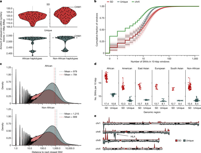
Fig. 1 Increased single-nucleotide variation in SDs. a, The portion of the human genome analysed for SD (red) and unique (blue) regions among African and non-African genomes. Shown are the number of megabase pairs aligned in 1:1 syntenic blocks to T2T-CHM13 v1.1 for each assembled haplotype. Data are shown as both a single point per haplotype originating from a single individual and a smoothed violin plot to represent the population distribution. b, Empirical cumulative distribution showing the number of SNVs in 10-kbp windows in the syntenic regions stratified by unique (grey), SD (red) and the X chromosome (chrX; green). Dashed lines represent individual haplotypes and thick lines represent the average trend of all the data. c, Distribution of the average distance to the next closest SNV in SD (red) and unique (grey) space separating African (top) and non-African (bottom) samples. Dashed vertical lines are drawn at the mean of each distribution. d, Average number of SNVs per 10-kbp window in SD (red) versus unique (grey) space by superpopulation and with mean value shown underneath each violin. The non-African column represents an aggregation of the data from all non-African populations in this study. e, Density of SNVs in 10 bp of each other for SD (top, red) and unique (bottom, grey) regions for chromosomes 1, 6, 8 and X comparing the relative density of known (for example, HLA) and new hotspots of single-nucleotide variation.
Fig. 1 Increased single-nucleotide variation in SDs. a, The portion of the human genome analysed for SD (red) and unique (blue) regions among African and non-African genomes. Shown are the number of megabase pairs aligned in 1:1 syntenic blocks to T2T-CHM13 v1.1 for each assembled haplotype. Data are shown as both a single point per haplotype originating from a single individual and a smoothed violin plot to represent the population distribution. b, Empirical cumulative distribution showing the number of SNVs in 10-kbp windows in the syntenic regions stratified by unique (grey), SD (red) and the X chromosome (chrX; green). Dashed lines represent individual haplotypes and thick lines represent the average trend of all the data. c, Distribution of the average distance to the next closest SNV in SD (red) and unique (grey) space separating African (top) and non-African (bottom) samples. Dashed vertical lines are drawn at the mean of each distribution. d, Average number of SNVs per 10-kbp window in SD (red) versus unique (grey) space by superpopulation and with mean value shown underneath each violin. The non-African column represents an aggregation of the data from all non-African populations in this study. e, Density of SNVs in 10 bp of each other for SD (top, red) and unique (bottom, grey) regions for chromosomes 1, 6, 8 and X comparing the relative density of known (for example, HLA) and new hotspots of single-nucleotide variation.CC BY · PubMed Central
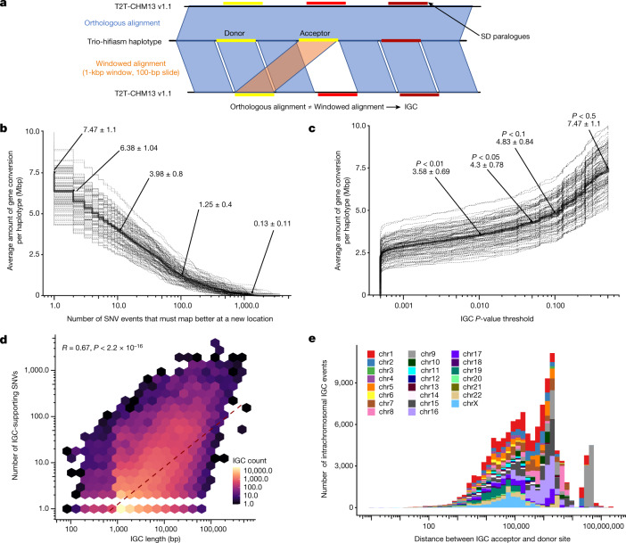
Fig. 2 Candidate IGC events. a, Method to detect IGC. The assembled human haplotype query sequence from 1:1 syntenic alignments was fragmented into 1-kbp windows in 100-bp increments and realigned back to T2T-CHM13 v1.1 independent of the flanking sequence information using minimap2 v2.24 to identify each window’s single best alignment position. These alignments were compared to their original syntenic alignment positions, and if they were not overlapping, we considered them to be candidate IGC windows. Candidate IGC windows were then merged into larger intervals and realigned when windows were overlapping in both the donor and the acceptor sequence. We then used the CIGAR string to identify the number of matching and mismatching bases at the ‘donor’ site and compared that to the number of matching and mismatching bases at the acceptor site determined by the syntenic alignment to calculate the number of supporting SNVs. b, The amount of SDs (in megabase pairs) predicted to be affected by IGC per haplotype, as a function of the minimum number of SNVs that support the IGC call. Dashed lines represent individual haplotypes and the solid line represents the average. c, Empirical cumulative distribution of the megabase pairs of candidate IGC observed in HPRC haplotypes, as a function of the minimum underlying P-value threshold used to define the IGC callset (see Methods for IGC P-value calculation). Dashed lines represent individual haplotypes and the solid line represents the average. d, Correlation between IGC length and the number of supporting SNVs. e, Distribution of the distance between predicted IGC acceptor and donor sites for intrachromosomal events by chromosome.
Fig. 2 Candidate IGC events. a, Method to detect IGC. The assembled human haplotype query sequence from 1:1 syntenic alignments was fragmented into 1-kbp windows in 100-bp increments and realigned back to T2T-CHM13 v1.1 independent of the flanking sequence information using minimap2 v2.24 to identify each window’s single best alignment position. These alignments were compared to their original syntenic alignment positions, and if they were not overlapping, we considered them to be candidate IGC windows. Candidate IGC windows were then merged into larger intervals and realigned when windows were overlapping in both the donor and the acceptor sequence. We then used the CIGAR string to identify the number of matching and mismatching bases at the ‘donor’ site and compared that to the number of matching and mismatching bases at the acceptor site determined by the syntenic alignment to calculate the number of supporting SNVs. b, The amount of SDs (in megabase pairs) predicted to be affected by IGC per haplotype, as a function of the minimum number of SNVs that support the IGC call. Dashed lines represent individual haplotypes and the solid line represents the average. c, Empirical cumulative distribution of the megabase pairs of candidate IGC observed in HPRC haplotypes, as a function of the minimum underlying P-value threshold used to define the IGC callset (see Methods for IGC P-value calculation). Dashed lines represent individual haplotypes and the solid line represents the average. d, Correlation between IGC length and the number of supporting SNVs. e, Distribution of the distance between predicted IGC acceptor and donor sites for intrachromosomal events by chromosome.CC BY · PubMed Central
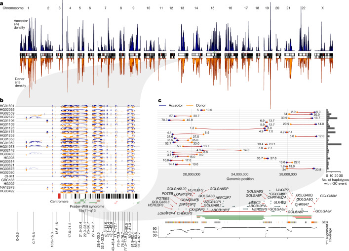
Fig. 3 IGC hotspots. a, Density of IGC acceptor (top, blue) and donor (bottom, orange) sites across the ‘SD genome’. The SD genome consists of all main SD regions (>50 kbp) minus the intervening unique sequences. b, All intrachromosomal IGC events on 24 human haplotypes analysed for chromosome 15. Arcs drawn in blue (top) have the acceptor site on the left-hand side and the donor site on the right. Arcs drawn in orange (bottom) are arranged oppositely. Protein-coding genes are drawn as vertical black lines above the ideogram, and large duplication (blue) and deletion (red) events associated with human diseases are drawn as horizontal lines just above the ideogram. c, Zoom of the 30 highest confidence (lowest P value) IGC events on chromosome 15 between 17 and 31 Mbp. The number to the left of each event shows its length (kbp) and that to the right shows its number of SNVs. Genes with IGC events are highlighted in red and associate with the breakpoint regions of Prader–Willi syndrome. An expanded graphic with all haplotypes is included in Extended Data Fig. 7.
Fig. 3 IGC hotspots. a, Density of IGC acceptor (top, blue) and donor (bottom, orange) sites across the ‘SD genome’. The SD genome consists of all main SD regions (>50 kbp) minus the intervening unique sequences. b, All intrachromosomal IGC events on 24 human haplotypes analysed for chromosome 15. Arcs drawn in blue (top) have the acceptor site on the left-hand side and the donor site on the right. Arcs drawn in orange (bottom) are arranged oppositely. Protein-coding genes are drawn as vertical black lines above the ideogram, and large duplication (blue) and deletion (red) events associated with human diseases are drawn as horizontal lines just above the ideogram. c, Zoom of the 30 highest confidence (lowest P value) IGC events on chromosome 15 between 17 and 31 Mbp. The number to the left of each event shows its length (kbp) and that to the right shows its number of SNVs. Genes with IGC events are highlighted in red and associate with the breakpoint regions of Prader–Willi syndrome. An expanded graphic with all haplotypes is included in Extended Data Fig. 7.CC BY · PubMed Central
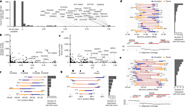
Fig. 4 Protein-coding genes affected by IGC. a, Number of putative IGC events intersecting exons of protein-coding genes as a function of a gene’s pLI. Of the 799 genes, 314 (39.3%) did not have a pLI score and are shown in the column labelled No pLI data available. b,c, Number of times a gene exon acts as an acceptor (b) or a donor (c) of an IGC event. d,e, IGC events at the complement factor locus, C4A and C4B (d), and the opsin middle- and long-wavelength-sensitive genes associated with colour blindness (OPN1MW and OPN1LW locus; e). Predicted donor (orange) and acceptor (blue) segments by length (number to left of event) and average number of supporting SNVs (number to right of event) are shown. The number of human haplotypes supporting each configuration is depicted by the histograms to the right. f,g, IGC events that reposition entire gene models for the FCGR (f) and TRIM (g) loci.
Fig. 4 Protein-coding genes affected by IGC. a, Number of putative IGC events intersecting exons of protein-coding genes as a function of a gene’s pLI. Of the 799 genes, 314 (39.3%) did not have a pLI score and are shown in the column labelled No pLI data available. b,c, Number of times a gene exon acts as an acceptor (b) or a donor (c) of an IGC event. d,e, IGC events at the complement factor locus, C4A and C4B (d), and the opsin middle- and long-wavelength-sensitive genes associated with colour blindness (OPN1MW and OPN1LW locus; e). Predicted donor (orange) and acceptor (blue) segments by length (number to left of event) and average number of supporting SNVs (number to right of event) are shown. The number of human haplotypes supporting each configuration is depicted by the histograms to the right. f,g, IGC events that reposition entire gene models for the FCGR (f) and TRIM (g) loci.CC BY · PubMed Central
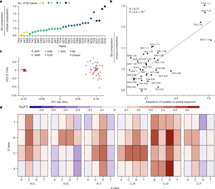
Fig. 5 Sequence composition and mutational spectra of SD SNVs. a, Compositional increase in GC-containing triplets in SD versus unique regions of the genome (coloured by GC content). b, Correlation between the enrichment of certain triplets in SDs compared to the mutability of that triplet in unique regions of the genome. Mutability is defined as the sum of all SNVs that change a triplet divided by the total count of that triplet in the genome. The enrichment ratio of SD over unique regions is indicated in text next to each triplet sequence. The text (upper left) indicates the value of the Pearson’s correlation coefficient and the P value from a two-sided t-test without adjustment for multiple comparisons. c, PCA of the mutational spectra of triplets in SD (circles) versus unique (triangles) regions polarized against a chimpanzee genome assembly and coloured by the continental superpopulation of the sample. AFR, African; AMR, American; EAS, East Asian; EUR, European; SAS, South Asian. d, The log[fold change] in triplet mutation frequency between SD and unique sequences. The y axis represents the 5′ base of the triplet context; the first level of the x axis shows which central base has changed and the second level of the x axis shows the 3′ base: heatmap depicts the log[fold change]. As an example, the top left corner shows the log[fold change] in frequency of TAA>TCA mutations in SD versus unique sequences.
Fig. 5 Sequence composition and mutational spectra of SD SNVs. a, Compositional increase in GC-containing triplets in SD versus unique regions of the genome (coloured by GC content). b, Correlation between the enrichment of certain triplets in SDs compared to the mutability of that triplet in unique regions of the genome. Mutability is defined as the sum of all SNVs that change a triplet divided by the total count of that triplet in the genome. The enrichment ratio of SD over unique regions is indicated in text next to each triplet sequence. The text (upper left) indicates the value of the Pearson’s correlation coefficient and the P value from a two-sided t-test without adjustment for multiple comparisons. c, PCA of the mutational spectra of triplets in SD (circles) versus unique (triangles) regions polarized against a chimpanzee genome assembly and coloured by the continental superpopulation of the sample. AFR, African; AMR, American; EAS, East Asian; EUR, European; SAS, South Asian. d, The log[fold change] in triplet mutation frequency between SD and unique sequences. The y axis represents the 5′ base of the triplet context; the first level of the x axis shows which central base has changed and the second level of the x axis shows the 3′ base: heatmap depicts the log[fold change]. As an example, the top left corner shows the log[fold change] in frequency of TAA>TCA mutations in SD versus unique sequences.CC BY · PubMed Central
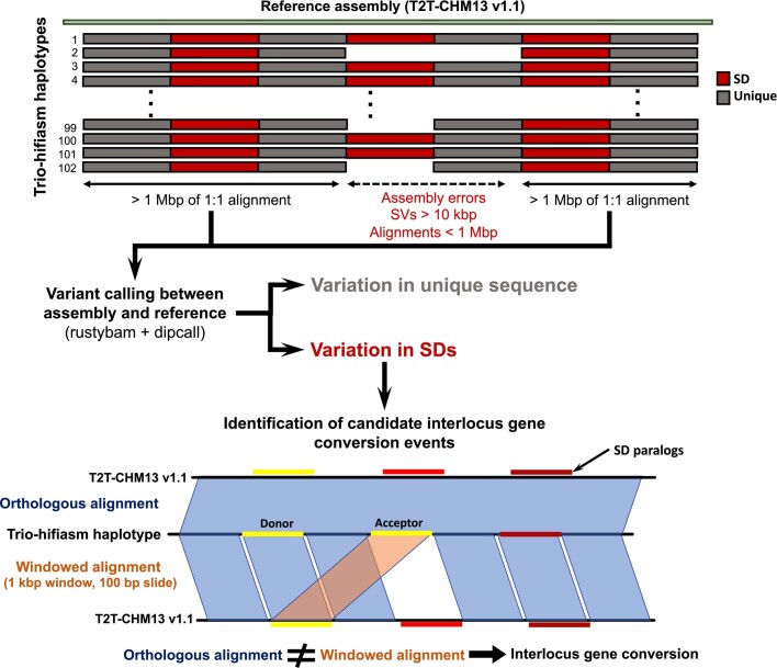
Extended Data Fig. 1 Analysis schema for variant and IGC calling. Whole-genome alignments were calculated for the HPRC assemblies against T2T-CHM13 v1.1 with a copy of GRCh38 chrY using minimap2 v2.24. The alignments were further processed to remove alignments that were redundant in query sequence or that had structural variants over 10 kbp in length. After these steps, the remaining alignments over 1 Mbp were defined to be syntenic and used in downstream analyses. We then counted all pairwise single-nucleotide differences between the haplotypes and the reference and stratified these results into unique regions versus SD regions based on the SD annotations from T2T-CHM13 v1.1. All variants intersecting tandem repeats were filtered to avoid spurious SNV calls. To detect candidate regions of IGC, the query sequence with syntenic alignments was fragmented into 1 kbp windows with a 100 bp slide and realigned back to T2T-CHM13 v1.1 independent of the flanking sequence using minimap2 v2.24 to identify each window’s single best alignment position. These alignments were compared to their original syntenic alignment positions, and if they were not overlapping, we considered them to be candidate IGC windows. Candidate IGC windows were then merged into larger intervals and realigned when windows were overlapping in both the donor and the acceptor sequence. We then used the CIGAR string to identify the number of matching and mismatching bases at the “donor” site and compared that to the number of matching and mismatching bases at the acceptor site determined by the syntenic alignment to calculate the number of supporting SNVs.
Extended Data Fig. 1 Analysis schema for variant and IGC calling. Whole-genome alignments were calculated for the HPRC assemblies against T2T-CHM13 v1.1 with a copy of GRCh38 chrY using minimap2 v2.24. The alignments were further processed to remove alignments that were redundant in query sequence or that had structural variants over 10 kbp in length. After these steps, the remaining alignments over 1 Mbp were defined to be syntenic and used in downstream analyses. We then counted all pairwise single-nucleotide differences between the haplotypes and the reference and stratified these results into unique regions versus SD regions based on the SD annotations from T2T-CHM13 v1.1. All variants intersecting tandem repeats were filtered to avoid spurious SNV calls. To detect candidate regions of IGC, the query sequence with syntenic alignments was fragmented into 1 kbp windows with a 100 bp slide and realigned back to T2T-CHM13 v1.1 independent of the flanking sequence using minimap2 v2.24 to identify each window’s single best alignment position. These alignments were compared to their original syntenic alignment positions, and if they were not overlapping, we considered them to be candidate IGC windows. Candidate IGC windows were then merged into larger intervals and realigned when windows were overlapping in both the donor and the acceptor sequence. We then used the CIGAR string to identify the number of matching and mismatching bases at the “donor” site and compared that to the number of matching and mismatching bases at the acceptor site determined by the syntenic alignment to calculate the number of supporting SNVs.CC BY · PubMed Central
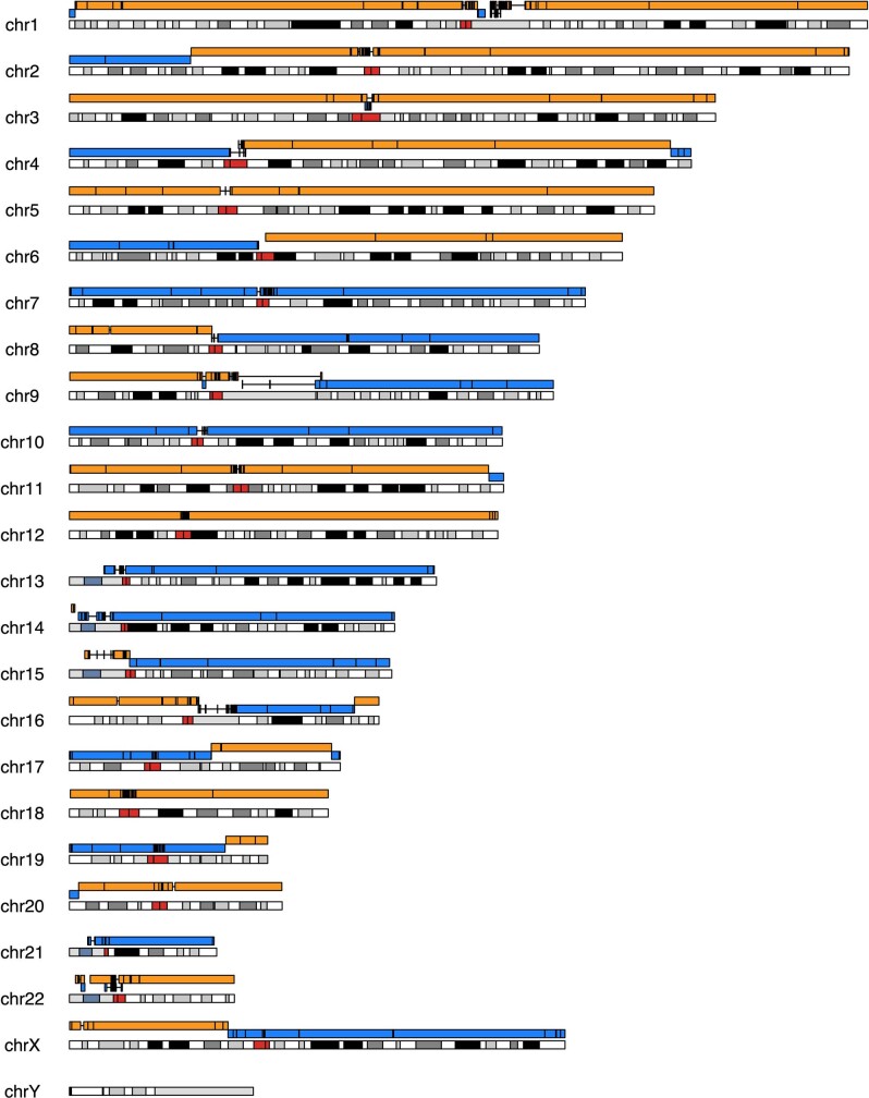
Extended Data Fig. 2 Ideogram of an assembly of CHM1 aligned to T2T-CHM13. The ideogram depicts the contiguity (alternating blue and orange contigs) of a CHM1 assembly generated by Verkko as compared to T2T-CHM13. The overall contig N50 is 105.2 Mbp providing near chromosome arm contiguity with the exception of breaks at the centromere (red) and other large satellite arrays. Because the sequence is derived from a monoploid complete hydatidiform mole, there is no opportunity for assembly errors due to inadvertent haplotype switching.
Extended Data Fig. 2 Ideogram of an assembly of CHM1 aligned to T2T-CHM13. The ideogram depicts the contiguity (alternating blue and orange contigs) of a CHM1 assembly generated by Verkko as compared to T2T-CHM13. The overall contig N50 is 105.2 Mbp providing near chromosome arm contiguity with the exception of breaks at the centromere (red) and other large satellite arrays. Because the sequence is derived from a monoploid complete hydatidiform mole, there is no opportunity for assembly errors due to inadvertent haplotype switching.CC BY · PubMed Central
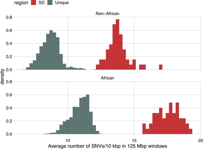
Extended Data Fig. 3 Increased variation in SD sequences and African haplotypes. Histograms of the average number of SNVs per 10 kbp over all 125 Mbp bins of unique (blue) and SD (red) sequence for all haplotypes. African haplotypes (bottom) are compared separately to non-African (top) haplotypes. All SD bins (125 Mbp each) have more SNVs than any unique bin irrespective of human superpopulation.
Extended Data Fig. 3 Increased variation in SD sequences and African haplotypes. Histograms of the average number of SNVs per 10 kbp over all 125 Mbp bins of unique (blue) and SD (red) sequence for all haplotypes. African haplotypes (bottom) are compared separately to non-African (top) haplotypes. All SD bins (125 Mbp each) have more SNVs than any unique bin irrespective of human superpopulation.CC BY · PubMed Central
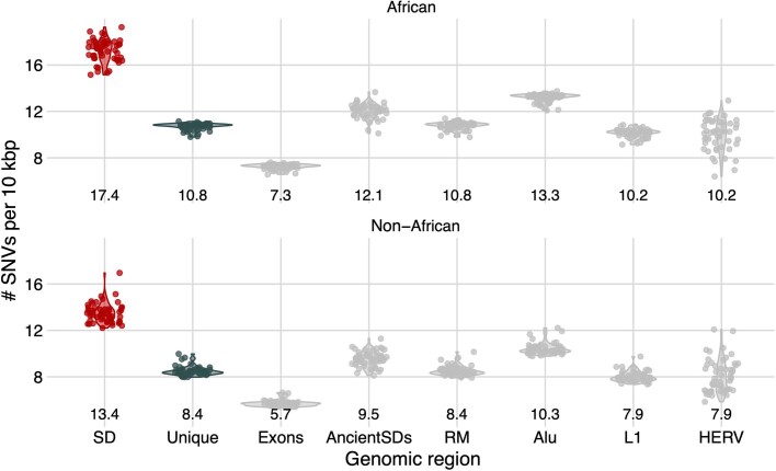
Extended Data Fig. 4 Average number of SNVs across different repeat classes. Shown are the average number of SNVs per 10 kbp within SDs (red), unique (blue), and additional sequence classes (gray) across the HPRC haplotypes. These classes include exonic regions, ancient SDs (SD with <90% sequence identity) and all elements identified by RepeatMasker (RM) with Alu, L1 LINE, and HERV elements broken out separately. Below each sequence class we show the average number of SNVs per 10 kbp for the median haplotype. Standard deviations and measurements for additional repeat classes are provided in Table S3.
Extended Data Fig. 4 Average number of SNVs across different repeat classes. Shown are the average number of SNVs per 10 kbp within SDs (red), unique (blue), and additional sequence classes (gray) across the HPRC haplotypes. These classes include exonic regions, ancient SDs (SD with <90% sequence identity) and all elements identified by RepeatMasker (RM) with Alu, L1 LINE, and HERV elements broken out separately. Below each sequence class we show the average number of SNVs per 10 kbp for the median haplotype. Standard deviations and measurements for additional repeat classes are provided in Table S3.CC BY · PubMed Central
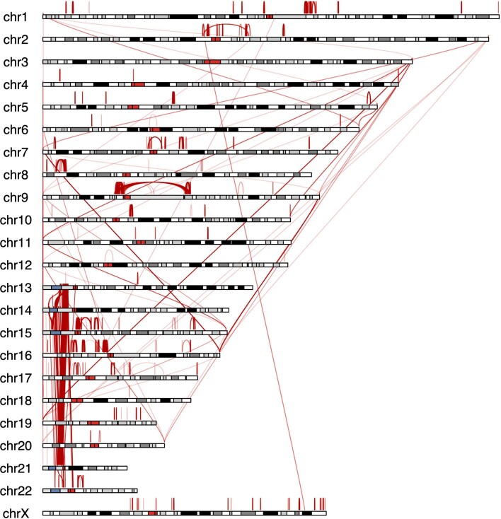
Extended Data Fig. 5 Largest IGC events in the human genome. The ideogram depicts as red arcs the positions of the largest IGC events between and within human chromosomes (top 10% of the length distribution).
Extended Data Fig. 5 Largest IGC events in the human genome. The ideogram depicts as red arcs the positions of the largest IGC events between and within human chromosomes (top 10% of the length distribution).CC BY · PubMed Central
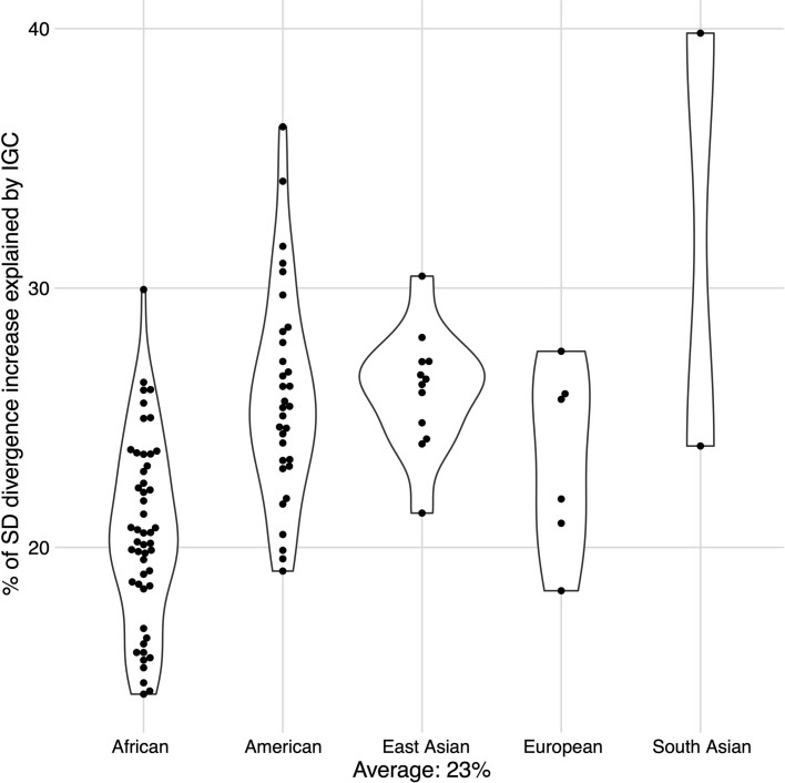
Extended Data Fig. 6 Percent of increased single-nucleotide variation explained by IGC. Shown is the fraction of the increased SNV diversity in SDs that can be attributed to IGC for each of the HPRC haplotypes stratified by global superpopulation. In text is the average across all haplotypes (23%).
Extended Data Fig. 6 Percent of increased single-nucleotide variation explained by IGC. Shown is the fraction of the increased SNV diversity in SDs that can be attributed to IGC for each of the HPRC haplotypes stratified by global superpopulation. In text is the average across all haplotypes (23%).CC BY · PubMed Central
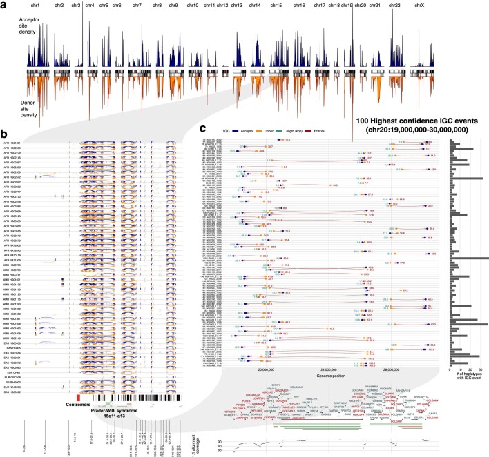
Extended Data Fig. 7 IGC hotspots. a) Density of IGC acceptor (top, blue) and donor (bottom, orange) sites across the “SD genome”. The SD genome consists of all main SD regions (>50 kbp) minus the intervening unique sequences. b) All intrachromosomal IGC events from 102 human haplotypes analyzed for chromosome 15. Arcs drawn in blue (top) have the acceptor site on the left-hand side and the donor site on the right. Arcs drawn in orange (bottom) are arranged oppositely. Protein-coding genes are drawn as vertical black lines above the ideogram, and large duplication (blue) and deletion (red) events associated with human diseases are drawn as horizontal lines just above the ideogram. c) Zoom of the 100 highest confidence (lowest p-value) IGC events identified on chromosome 15 between 17 and 31 Mbp. Genes that are intersected by IGC events are highlighted in red.
Extended Data Fig. 7 IGC hotspots. a) Density of IGC acceptor (top, blue) and donor (bottom, orange) sites across the “SD genome”. The SD genome consists of all main SD regions (>50 kbp) minus the intervening unique sequences. b) All intrachromosomal IGC events from 102 human haplotypes analyzed for chromosome 15. Arcs drawn in blue (top) have the acceptor site on the left-hand side and the donor site on the right. Arcs drawn in orange (bottom) are arranged oppositely. Protein-coding genes are drawn as vertical black lines above the ideogram, and large duplication (blue) and deletion (red) events associated with human diseases are drawn as horizontal lines just above the ideogram. c) Zoom of the 100 highest confidence (lowest p-value) IGC events identified on chromosome 15 between 17 and 31 Mbp. Genes that are intersected by IGC events are highlighted in red.CC BY · PubMed Central

Verification

Claims the paper makes

The claims and statistics of this paper haven’t been checked yet.

Run the check — free, no account needed. It reads the open-access text and checks the sources it cites.

A few free checks a day · papers already verified open instantly · results are public

Claims other papers make about this one

Each is checked against this paper’s own text: does it say what the citing paper says it does?

Self-check

Percentages, sums and reported differences re-computed from the paper’s own numbers.

The self-check needs the paper’s full text.

Citations

Cites

Show all 76 references

Research works printed but not linked: 1

  1. Sudmant, P. H. et al. Diversity of human copy number. Science 11184, 2–7 (2010).

Cited by

Sorted by citations

Items 101–103 of 103

Metadata

Authors
Vollger, M. R., Dishuck, P. C., Harvey, W. T., DeWitt, W. S., Guitart, X., Goldberg, M. E., Rozanski, A. N., Lucas, J., Asri, M., Human Pangenome Reference Consortium, Abel, H. J., Antonacci-Fulton, L. L., Baid, G., Baker, C. A., Belyaeva, A., Billis, K., Bourque, G., Buonaiuto, S., Carroll, A., Chaisson, M. J. P., Chang, P.-C., Chang, X. H., Cheng, H., Chu, J., Cody, S., Colonna, V., Cook, D. E., Cook-Deegan, R. M., Cornejo, O. E., Diekhans, M., Doerr, D., Ebert, P., Ebler, J., Eizenga, J. M., Fairley, S., Fedrigo, O., Felsenfeld, A. L., Feng, X., Fischer, C., Flicek, P., Formenti, G., Frankish, A., Fulton, R. S., Gao, Y., Garg, S., Garrison, E., Garrison, N. A., Giron, C. G., Green, R. E., Groza, C., Guarracino, A., Haggerty, L., Hall, I. M., Haukness, M., Haussler, D., Heumos, S., Hickey, G., Hourlier, T., Howe, K., Jain, M., Jarvis, E. D., Ji, H. P., Kenny, E. E., Koenig, B. A., Kolesnikov, A., Korbel, J. O., Kordosky, J., Koren, S., Lee, H., Li, H., Liao, W.-W., Lu, S., Lu, T.-Y., Lucas, J. K., Magalhães, H., Marco-Sola, S., Marijon, P., Markello, C., Marschall, T., Martin, F. J., McCartney, A., McDaniel, J., Miga, K. H., Mitchell, M. W., Monlong, J., Mountcastle, J., Mwaniki, M. N., Nattestad, M., Novak, A. M., Nurk, S., Olsen, H. E., Olson, N. D., Paten, B., Pesout, T., Phillippy, A. M., Popejoy, A. B., Prins, P., Puiu, D., Rautiainen, M., Regier, A. A., Rhie, A., Sacco, S., Sanders, A. D., Schneider, V. A., Schultz, B. I., Shafin, K., Sibbesen, J. A., Sirén, J., Smith, M. W., Sofia, H. J., Abou Tayoun, A. N., Thibaud-Nissen, F., Tomlinson, C., Tricomi, F. F., Villani, F., Vollger, M. R., Wagner, J., Walenz, B., Wang, T., Wood, J. M. D., Zimin, A. V., Zook, J. M., Munson, K. M., Lewis, A. P., Hoekzema, K., Logsdon, G. A., Porubsky, D., Paten, B., Harris, K., Hsieh, P., & Eichler, E. E.
Affiliations
  • University of Washington (US)
  • University of California, Santa Cruz (US)
  • Washington University in St. Louis (US)
  • Google (United States) (US)
  • European Bioinformatics Institute (GB)
  • Kyoto University (JP)
  • Institute of Genetics and Biophysics (IT)
  • University of Southern California (US)
  • Harvard University (US)
  • Dana-Farber Cancer Institute (US)
  • University of Tennessee Health Science Center (US)
  • Washington Center (US)
  • Heinrich Heine University Düsseldorf (DE)
  • Rockefeller University (US)
  • National Institutes of Health (US)
  • Children's Hospital of Philadelphia (US)
  • Novo Nordisk Foundation (DK)
  • University of California, Los Angeles (US)
  • McGill University (CA)
  • Yale University (US)
  • Howard Hughes Medical Institute (US)
  • Quantitative Biology Center (DE)
  • Wellcome Sanger Institute (GB)
  • Northeastern University (US)
  • Stanford Medicine (US)
  • Icahn School of Medicine at Mount Sinai (US)
  • University of California, San Francisco (US)
  • National Institute of Standards and Technology (US)
  • Coriell Institute For Medical Research (US)
  • University of Pisa (IT)
  • University of California, Davis (US)
  • Johns Hopkins University (US)
  • Max Delbrück Center (DE)
  • University of Copenhagen (DK)
Corresponding author
Eichler, Evan E.
ORCID
Venue
Nature
Published
2023-05-10
Volume
617
Issue
7960
Pages
325-334
ISSN
  • 0028-0836 (ISSN-L)
  • 1476-4687
OpenAlex
W4376133061
Cited by
104
References
76
Citations by year
2026: 29 · 2025: 26 · 2024: 35 · 2023: 14
FWCI
17.07
Type
journal article
Language
en
Open access
Locations
  • Nature (publishedVersion, cc-by)
  • PubMed Central (publishedVersion, cc-by)
  • PubMed (publishedVersion)
  • MDC Repository (Max-Delbrueck-Center for Molecular Medicine) (acceptedVersion)
  • RePEc: Research Papers in Economics (submittedVersion)
  • eScholarship (California Digital Library) (submittedVersion, cc-by)
  • Technical University of Denmark, DTU Orbit (Technical University of Denmark, DTU) (submittedVersion, cc-by)
  • UPCommons institutional repository (Universitat Politècnica de Catalunya) (submittedVersion, cc-by-nc-nd)
Funders
  • Howard Hughes Medical Institute
  • James S. McDonnell Foundation
  • National Institutes of Health (5r00hg011041-04)
  • National Human Genome Research Institute (R01HG010485, U01HG010973, U01HG010971, R00 HG011041, K99HG011041, R01HG002385, U24HG007234, U24HG010262)
  • National Institute of General Medical Sciences (R35GM133428, T32GM007454)
  • National Institute of Allergy and Infectious Diseases (F31AI150163)
Topics
  • Life Sciences › Biochemistry, Genetics and Molecular Biology › Genetics › Genomic variations and chromosomal abnormalities
  • Life Sciences › Biochemistry, Genetics and Molecular Biology › Genetics › Chromosomal and Genetic Variations
  • Life Sciences › Biochemistry, Genetics and Molecular Biology › Genetics › Genome Rearrangement Algorithms
Keywords
segmental duplication, single nucleotide variants, mutation spectrum, transversions, GC content, paralogs, coalescent analysis, genome rearrangement
MeSH terms
Cytosine, Exons, Gene Conversion, Guanine, Haplotypes, Humans, Mutation, Genome, Human, CpG Islands, Polymorphism, Single Nucleotide, Segmental Duplications, Genomic

About these checks

Registry, retraction, Cites and statistics results are computed, not judged, with one exception: a printed reference that no index lists is read once by a model before it counts as a citation error. Claims are extracted by a model and kept only when their quote appears verbatim in the text; each cited source is then checked, with a model’s help, for whether it says what the claim relies on. Those verdicts can be wrong — read the claim’s quote and the cited source before relying on one.

Statistics and arithmetic are recomputed, not judged.

Confidence bands are shown only for fields calibrated on our annotated test set. None is calibrated yet, so no band is shown.

What counts as a claim ›How we measure confidence ›How we check this ›

Are you an author, or do you see a problem with this page? Request a review