

The DOI matches its Crossref record; 51 of the 51 references it cites were found in a registry or an index; 6 of the 22 sources its claims cite were checked for support.
Phase I trial of hES cell-derived dopaminergic neurons for Parkinson’s disease
Open accessAbstract
Fluoro-DOPA positron emission tomography uptake increased, indicating graft survival. Secondary and exploratory clinical outcomes showed improvement or stability, including improvement in the Movement Disorder Society Unified Parkinson's Disease Rating Scale (MDS-UPDRS) Part III OFF scores by an average of 23 points in the high-dose cohort. There were no graft-induced dyskinesias. These data demonstrate safety and support future definitive clinical studies.
Overview
AI snapshot
Written from the paper’s full text.
TL;DR
In a first-in-human, open-label phase I trial, bilateral putaminal transplantation of the hES cell-derived dopaminergic neuron progenitor product bemdaneprocel met its 1-year safety and tolerability objectives in 12 patients with Parkinson’s disease. At 18 months, 18F-DOPA PET uptake increased in the putamen and MDS-UPDRS Part III OFF scores improved, especially in the high-dose cohort, with no graft-induced dyskinesias reported.
Key findings
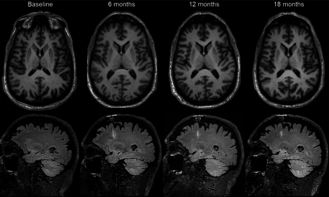
- Twelve patients were enrolled sequentially into low-dose (0.9 million cells per putamen, n = 5) and high-dose (2.7 million cells per putamen, n = 7) cohorts; all received 1 year of immunosuppression. The primary safety/tolerability criteria were satisfied at 12 months, with no adverse events related to the cell product and no deaths, tumours, abnormal tissue overgrowth or intracerebral haemorrhages detected by MRI.
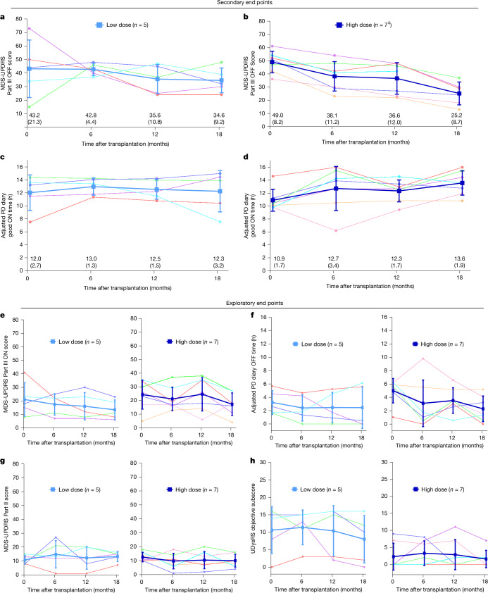
- At 18 months after transplantation, MDS-UPDRS Part III OFF scores decreased by a mean (s.d.) of 8.6 points (29.2) in the low-dose cohort and 23.0 points (7.9) in the high-dose cohort. Mean good ON times increased by 0.2 h (3.8 h) in the low-dose cohort and 2.7 h (1.6 h) in the high-dose cohort.
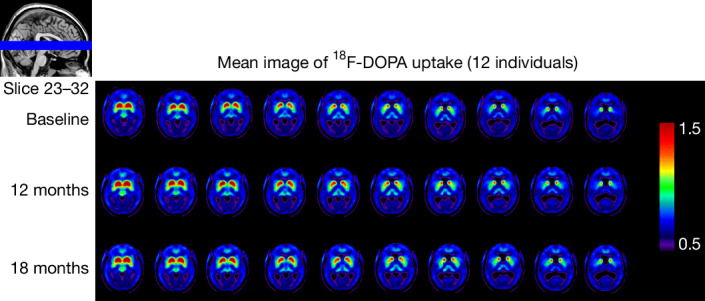
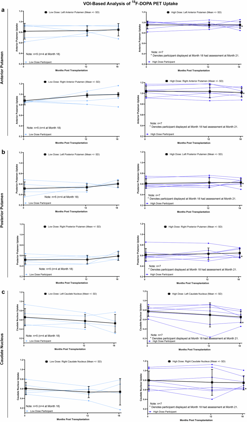
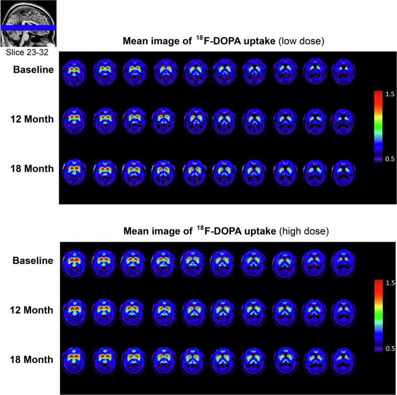
- Putaminal 18F-DOPA PET uptake increased bilaterally across cohorts, supporting graft survival through 18 months, 6 months after immunosuppression discontinuation; caudate uptake was stable to decreased. There were no reports of graft-induced dyskinesias through 18 months.
- Through 18 months, there were 78 cumulative treatment-emergent adverse events; most were mild or moderate, and none were related to bemdaneprocel. Two serious adverse events occurred at 12 months: one COVID-19 hospitalization and one seizure attributed to surgery; a later gastrointestinal haemorrhage at approximately 15 months was assessed as unrelated.
Methods
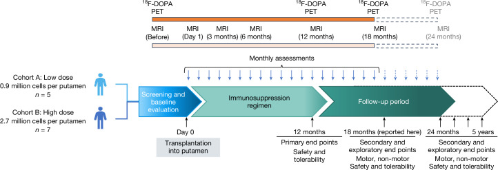
- First-in-human, multisite, open-label phase I trial (NCT04802733) of cryopreserved, off-the-shelf bemdaneprocel derived from hES cells, grafted bilaterally into the post-commissural putamen via nine deposits per putamen in a single surgical session.
- Participants had confirmed Parkinson’s disease; 12 were enrolled sequentially into low-dose then high-dose cohorts after a 30-day data safety monitoring committee pause. Median age was 67.0 years, 75% were male, and median time since diagnosis was 9 years.
- Outcomes included safety/tolerability at 1 year, 18F-DOPA PET imaging at baseline, 12 and 18 months, MDS-UPDRS Part III OFF and adjusted PD diary good ON times, plus exploratory motor, non-motor and quality-of-life measures. Analyses were descriptive by cohort; no inferential statistical testing was performed because of the small number of participants.
Limitations
- The paper states that the study was open-label, small, relatively short in observation, and not designed or powered to determine efficacy; expected PD symptom fluctuations and substantial placebo effects may influence results.
- The paper states that PET data have limitations in resolution, signal-to-noise ratio and variability across and within patients and between dose cohorts, and that correlation between 18F-DOPA PET uptake and clinical performance can be variable. It also notes unknown kinetics of graft maturation and disease vulnerability, complicating dose-finding.
- Evident from the design: there was no placebo or control arm and no blinding, and cohorts were enrolled sequentially rather than randomized, so efficacy signals cannot be attributed to the cell product alone.
Model-written summary. Check it against the paper before relying on it.
Integrity signals
Registry
Cites
Statistics
Figures
Verification
Claims the paper makes
39 Own result · 20 Own method · 6 No full text of the sources · 5 Own interpretation · 4 No basis given · 3 Partly supported · 1 Supported so far · 1 Sources not found · 1 Not checked yet · accuracy not yet measured
The cited sources support only part of this claim — read each source’s verdict below.
In the paper
“The high dose targets the estimated number of healthy DA neurons in the intact substantia nigra35 (300,000 DA neurons), and the lower dose targets the estimated minimal number of surviving grafted DA neurons required for clinical benefit36 (100,000 DA neurons).”
§ Phase I trial of hES cell-derived dopaminergic neurons for Parkinson’s disease > Main
What the sources say
- Partly supportedcites [35] Rudow, G. et al. Morphometry of the human substantia nigra in ageing and Parkinson’s disease. Acta Neuropathol. 115, 461–470 (2008). sourceExperimental — accuracy not yet measured
The Discussion reports a mean of 305,000 pigmented neurons on one side of the substantia nigra in healthy older controls, close to the cited 300,000. It reports 424,000 in young controls, however, and the 305,000 figure counts pigmented neurons rather than specifically DA neurons. The unqualified estimate of 300,000 healthy DA neurons is therefore more general than this measurement supports. (LLM, conf=0.87)
hedged- Asserted: passes — The authors put it forward as true.
- Checkable: passes — It can be checked against evidence.
- Load-bearing: passes — The paper’s conclusions depend on it.
- Based: passes — The paper gives a reason why it holds.
- Partly supported
The cited sources support only part of this claim — read each source’s verdict below.
In the paper
“The absence of GID in our study through 18 months is therefore particularly notable. This may relate to the lack of serotonergic neuron contaminants in bemdaneprocel, as preclinical studies confirmed the absence of serotonergic neurons in vitro and in vivo at 9 months after grafting34.”
§ Phase I trial of hES cell-derived dopaminergic neurons for Parkinson’s disease > Discussion
What the sources say
- Partly supportedcites [34] Piao, J. et al. Preclinical efficacy and safety of a human embryonic stem cell-derived midbrain dopamine progenitor product, MSK-DA01. Cell Stem Cell 28, 217–229 (2021). sourceExperimental — accuracy not yet measured
In Results and discussion, the Parkinsonian-rat graft analysis reports negative 5-HT staining, suggesting an absence of human serotonergic neurons in vivo. The cited paper does not establish their absence in vitro; its separate graft follow-up extends to day 266. The proposed link to the citing study’s absence of GID is the citing authors’ inference, not a finding attributed to this preclinical paper. (LLM, conf=0.85)
hedgedconditional- Asserted: passes — The authors put it forward as true.
- Checkable: passes — It can be checked against evidence.
- Load-bearing: passes — The paper’s conclusions depend on it.
- Based: passes — The paper gives a reason why it holds.
- Partly supported
A result the paper reports from its own data — it cites no other source for it, so there is none to check. The quote shows it is in the paper.
In the paper
“There were no graft-induced dyskinesias.”
- Also stated in: § Safety and tolerability · plain
“Notably, there were no reports of adverse events or clinical indications of graft-induced dyskinesias (GIDs) to date.”
§ Phase I trial of hES cell-derived dopaminergic neurons for Parkinson’s disease > Abstract
plain- Asserted: passes — The authors put it forward as true.
- Checkable: passes — It can be checked against evidence.
- Load-bearing: passes — The paper’s conclusions depend on it.
- Based: passes — The paper gives a reason why it holds.
- Also stated in: § Safety and tolerability · plain
A result the paper reports from its own data — it cites no other source for it, so there is none to check. The quote shows it is in the paper.
In the paper
“There were no deaths, no SAEs related to transplanted cells or immunosuppression, and there were no tumours, abnormal tissue overgrowth or intracerebral haemorrhages detected with MRI, which showed the expected postoperative changes (Extended Data Fig. 2).”
§ Phase I trial of hES cell-derived dopaminergic neurons for Parkinson’s disease > Safety and tolerability
plain- Asserted: passes — The authors put it forward as true.
- Checkable: passes — It can be checked against evidence.
- Load-bearing: passes — The paper’s conclusions depend on it.
- Based: passes — The paper gives a reason why it holds.
A result the paper reports from its own data — it cites no other source for it, so there is none to check. The quote shows it is in the paper.
In the paper
“The mean good ON times, based on adjusted PD diaries, increased by 0.2 h (3.8 h) from the baseline to 12.3 h (3.2 h) in the low-dose cohort and by 2.7 h (1.6 h) from the baseline to 13.6 h (1.9 h) in the high-dose cohort (Fig. 2c,d).”
§ Phase I trial of hES cell-derived dopaminergic neurons for Parkinson’s disease > Secondary and exploratory outcomes
plaindirect- Asserted: passes — The authors put it forward as true.
- Checkable: passes — It can be checked against evidence.
- Load-bearing: unclear — It is unclear whether the paper’s conclusions depend on it.
- Based: passes — The paper gives a reason why it holds.
A result the paper reports from its own data — it cites no other source for it, so there is none to check. The quote shows it is in the paper.
In the paper
“Across the total study population, ON times with troublesome dyskinesias (Extended Data Table 7) and use of antiparkinsonian medications were similar to the baseline (Extended Data Table 8).”
§ Phase I trial of hES cell-derived dopaminergic neurons for Parkinson’s disease > Secondary and exploratory outcomes
plain- Asserted: passes — The authors put it forward as true.
- Checkable: passes — It can be checked against evidence.
- Load-bearing: unclear — It is unclear whether the paper’s conclusions depend on it.
- Based: passes — The paper gives a reason why it holds.
A result the paper reports from its own data — it cites no other source for it, so there is none to check. The quote shows it is in the paper.
In the paper
“The mean (s.d.) PDQ-39 summary index scores increased (worsened) by 0.4 points (7.4) from the baseline to 25.4 (6.8) in the low-dose cohort and decreased (improved) by 4.2 points (8.2) from the baseline to 10.9 (8.7) in the high-dose cohort (Extended Data Table 6).”
§ Phase I trial of hES cell-derived dopaminergic neurons for Parkinson’s disease > Secondary and exploratory outcomes
plaindirect- Asserted: passes — The authors put it forward as true.
- Checkable: passes — It can be checked against evidence.
- Load-bearing: unclear — It is unclear whether the paper’s conclusions depend on it.
- Based: passes — The paper gives a reason why it holds.
A result the paper reports from its own data — it cites no other source for it, so there is none to check. The quote shows it is in the paper.
In the paper
“Safety data through 18 months continued to indicate a favourable safety profile.”
§ Phase I trial of hES cell-derived dopaminergic neurons for Parkinson’s disease > Discussion
hedged- Asserted: passes — The authors put it forward as true.
- Checkable: passes — It can be checked against evidence.
- Load-bearing: passes — The paper’s conclusions depend on it.
- Based: passes — The paper gives a reason why it holds.
A result the paper reports from its own data — it cites no other source for it, so there is none to check. The quote shows it is in the paper.
In the paper
“Among the exploratory end points (Fig. 2e–h), MDS-UPDRS Part III ON scores also improved with a decrease in the mean (s.d.) of 7.6 points (15.3) from the baseline to 13.4 (7.3) in the low-dose cohort and of 8.4 points (5.6) from the baseline to 15.9 (8.9) in the high-dose cohort (Fig. 2e).”
§ Phase I trial of hES cell-derived dopaminergic neurons for Parkinson’s disease > Secondary and exploratory outcomes
plaindirect- Asserted: passes — The authors put it forward as true.
- Checkable: passes — It can be checked against evidence.
- Load-bearing: unclear — It is unclear whether the paper’s conclusions depend on it.
- Based: passes — The paper gives a reason why it holds.
A result the paper reports from its own data — it cites no other source for it, so there is none to check. The quote shows it is in the paper.
In the paper
“with no adverse events related to the cell product.”
- Also stated in: § Safety and tolerability · plain
“No TEAEs were related to bemdaneprocel.”
§ Phase I trial of hES cell-derived dopaminergic neurons for Parkinson’s disease > Abstract
plainn = 12- Asserted: passes — The authors put it forward as true.
- Checkable: passes — It can be checked against evidence.
- Load-bearing: passes — The paper’s conclusions depend on it.
- Based: passes — The paper gives a reason why it holds.
- Also stated in: § Safety and tolerability · plain
- Probably not a substantial claim. 22
- It may state two separate claims as one. 26
The cited sources support only part of this claim — read each source’s verdict below.
In the paper
“the feasibility of allografting without long-term immunosuppression, coupled with cryopreservation, offers an off-the-shelf approach33,47 that could facilitate the broader application of this therapy in the future.”
§ Phase I trial of hES cell-derived dopaminergic neurons for Parkinson’s disease > Discussion
What the sources say
- Partly supportedcites [33] Kim, T. W. et al. Biphasic activation of WNT signaling facilitates the derivation of midbrain dopamine neurons from hESCs for translational use. Cell Stem Cell 28, 343–355 (2021). sourceExperimental — accuracy not yet measured
The Results section on cryopreserved, off-the-shelf mDA cells reports high post-thaw viability, and the Discussion says the protocol should be suitable for a scalable product for human translation. But the graft experiments used immunocompromised rats; the paper does not establish that allografting is feasible without long-term immunosuppression. Page numbers were not available in our copy. (LLM, conf=0.88)
- Partly supportedcites [47] Kirkeby, A. et al. Preclinical quality, safety, and efficacy of a human embryonic stem cell-derived product for the treatment of Parkinson’s disease, STEM-PD. Cell Stem Cell 30, 1299–1314 (2023). sourceExperimental — accuracy not yet measured
The Discussion says STEM-PD is an allogeneic therapy using a banked, cryopreserved product that makes clinical planning easier. But the trial design specifies immunotherapy for 12 months after grafting; the paper does not establish that grafts remain viable without immunosuppression thereafter or that broader application is feasible. (LLM, conf=0.85)
hedgedconditional- Asserted: passes — The authors put it forward as true.
- Checkable: passes — It can be checked against evidence.
- Load-bearing: fails — The paper’s conclusions may not depend on it.
- Based: passes — The paper gives a reason why it holds.
Probably not a substantial claim. The paper’s conclusions may not depend on it. It may state two separate claims as one.
- Partly supported
The sources with a verdict support this claim; the rest weren’t checked yet, have no full text we could read, or their verdicts aren’t rated yet.
In the paper
“This method had been previously validated49,50 and used successfully in the PET imaging studies of fetal cell transplantation25,42,51,52.”
§ Phase I trial of hES cell-derived dopaminergic neurons for Parkinson’s disease > Methods > Neuroimaging
What the sources say
- Not checked — no full text of the sourcecites [49] Dhawan, V. et al. Comparative analysis of striatal FDOPA uptake in Parkinson’s disease: ratio method versus graphical approach. J. Nucl. Med. 43, 1324–1330 (2002).
- Supportedcites [50] Jokinen, P. et al. Simple ratio analysis of 18F-fluorodopa uptake in striatal subregions separates patients with early Parkinson disease from healthy controls. J. Nucl. Med. 50, 893–899 (2009). source
The citation is attached to the method-validation part of the sentence, not the separate fetal-transplantation citations. In the Results, the paper reports excellent diagnostic accuracy for the 18F-FDOPA striatal-to-occipital ratio in separating patients with early Parkinson disease from healthy controls; the Conclusion says a single 15-minute scan is sufficient in a clinical research setting. The Discussion mentions fetal-transplant imaging but does not present it as this study's application. (LLM, conf=0.90)
- Not checked — no full text of the sourcecites [25] Olanow, C. W. et al. A double-blind controlled trial of bilateral fetal nigral transplantation in Parkinson’s disease. Ann. Neurol. 54, 403–414 (2003). source
- Supportedcites [42] Ma, Y. et al. Dopamine cell implantation in Parkinson’s disease: long-term clinical and 18F-FDOPA PET outcomes. J. Nucl. Med. 51, 7–15 (2010). source
The Materials and Methods, Volume-of-Interest Analysis section (page unavailable) describes the striatal-to-occipital activity ratio used for 18F-FDOPA PET and says the authors had previously shown it was comparable to an established uptake measure. The paper applies that ratio in its fetal dopamine cell transplantation PET study; the Results, 18F-FDOPA PET: VOI Analysis section reports significant posttransplantation uptake changes. (LLM, conf=0.88)
- Not checked — no full text of the sourcecites [51] Nakamura, T. et al. Blinded positron emission tomography study of dopamine cell implantation for Parkinson’s disease. Ann. Neurol. 50, 181–187 (2001). source
- Not checked — no full text of the sourcecites [52] Ma, Y. et al. Dyskinesia after fetal cell transplantation for parkinsonism: a PET study. Ann. Neurol. 52, 628–634 (2002). source
plain- Asserted: passes — The authors put it forward as true.
- Checkable: passes — It can be checked against evidence.
- Load-bearing: unclear — It is unclear whether the paper’s conclusions depend on it.
- Based: passes — The paper gives a reason why it holds.
It may state two separate claims as one.
A result the paper reports from its own data — it cites no other source for it, so there is none to check. The quote shows it is in the paper.
In the paper
“Most were mild or moderate in severity except for one fall that was deemed to be unrelated to treatment.”
§ Phase I trial of hES cell-derived dopaminergic neurons for Parkinson’s disease > Safety and tolerability
plain- Asserted: passes — The authors put it forward as true.
- Checkable: passes — It can be checked against evidence.
- Load-bearing: passes — The paper’s conclusions depend on it.
- Based: passes — The paper gives a reason why it holds.
It may state two separate claims as one.
A result the paper reports from its own data — it cites no other source for it, so there is none to check. The quote shows it is in the paper.
In the paper
“The NMSS scores varied across domains, with total scores trending towards stability in the high-dose cohort (Extended Data Table 9).”
§ Phase I trial of hES cell-derived dopaminergic neurons for Parkinson’s disease > Secondary and exploratory outcomes
plain- Asserted: passes — The authors put it forward as true.
- Checkable: passes — It can be checked against evidence.
- Load-bearing: fails — The paper’s conclusions may not depend on it.
- Based: passes — The paper gives a reason why it holds.
Probably not a substantial claim. The paper’s conclusions may not depend on it.
A result the paper reports from its own data — it cites no other source for it, so there is none to check. The quote shows it is in the paper.
In the paper
“The trial achieved its primary objectives of safety and tolerability one year after transplantation, with no adverse events related to the cell product.”
- Also stated in: § Safety and tolerability · plain
“Predefined safety criteria for the primary outcome were satisfied at 12 months of follow-up after transplantation (Table 1).”
- Also stated in: § Discussion · plain
“bilateral putaminal transplantation of bemdaneprocel, a hES cell-derived DA neuron progenitor cell product, was generally well tolerated and achieved predefined safety criteria at 12 months as a primary end point.”
§ Phase I trial of hES cell-derived dopaminergic neurons for Parkinson’s disease > Abstract
plainn = 12- Asserted: passes — The authors put it forward as true.
- Checkable: passes — It can be checked against evidence.
- Load-bearing: passes — The paper’s conclusions depend on it.
- Based: passes — The paper gives a reason why it holds.
It may state two separate claims as one.
- Also stated in: § Safety and tolerability · plain
A result the paper reports from its own data — it cites no other source for it, so there is none to check. The quote shows it is in the paper.
In the paper
“MRI imaging by 18 months did not show evidence of tumours or changes in the putaminal volume.”
§ Phase I trial of hES cell-derived dopaminergic neurons for Parkinson’s disease > Safety and tolerability
plain- Asserted: passes — The authors put it forward as true.
- Checkable: passes — It can be checked against evidence.
- Load-bearing: passes — The paper’s conclusions depend on it.
- Based: passes — The paper gives a reason why it holds.
It may state two separate claims as one.
A result the paper reports from its own data — it cites no other source for it, so there is none to check. The quote shows it is in the paper.
In the paper
“There were no deaths, no SAEs related to transplanted cells or immunosuppression, and there were no tumours, abnormal tissue overgrowth or intracerebral haemorrhages detected with MRI, which showed the expected postoperative changes (Extended Data Fig. 2).”
§ Phase I trial of hES cell-derived dopaminergic neurons for Parkinson’s disease > Safety and tolerability
plain- Asserted: passes — The authors put it forward as true.
- Checkable: passes — It can be checked against evidence.
- Load-bearing: passes — The paper’s conclusions depend on it.
- Based: passes — The paper gives a reason why it holds.
It may state two separate claims as one.
A result the paper reports from its own data — it cites no other source for it, so there is none to check. The quote shows it is in the paper.
In the paper
“The clinical data also show a greater amplitude of changes in UPDRS motor scores and reduction in OFF times, and an earlier onset of improvement, in the high-dose group.”
§ Phase I trial of hES cell-derived dopaminergic neurons for Parkinson’s disease > Discussion
plaincorrelational- Asserted: passes — The authors put it forward as true.
- Checkable: passes — It can be checked against evidence.
- Load-bearing: passes — The paper’s conclusions depend on it.
- Based: passes — The paper gives a reason why it holds.
It may state two separate claims as one.
The paper states this without a source or data behind it here, so there is nothing to check it against. The quote shows it is in the paper.
In the paper
“Transplant feasibility was achieved, with intraoperative delivery of the total number of all intended cell deposits in all of the participants.”
§ Phase I trial of hES cell-derived dopaminergic neurons for Parkinson’s disease > Safety and tolerability
plain- Asserted: passes — The authors put it forward as true.
- Checkable: passes — It can be checked against evidence.
- Load-bearing: passes — The paper’s conclusions depend on it.
- Based: passes — The paper gives a reason why it holds.
It may state two separate claims as one.
A result the paper reports from its own data — it cites no other source for it, so there is none to check. The quote shows it is in the paper.
In the paper
“There were no deaths, no SAEs related to transplanted cells or immunosuppression, and there were no tumours, abnormal tissue overgrowth or intracerebral haemorrhages detected with MRI, which showed the expected postoperative changes (Extended Data Fig. 2).”
§ Phase I trial of hES cell-derived dopaminergic neurons for Parkinson’s disease > Safety and tolerability
plain- Asserted: passes — The authors put it forward as true.
- Checkable: passes — It can be checked against evidence.
- Load-bearing: passes — The paper’s conclusions depend on it.
- Based: passes — The paper gives a reason why it holds.
It may state two separate claims as one.
A result the paper reports from its own data — it cites no other source for it, so there is none to check. The quote shows it is in the paper.
In the paper
“Aside from two severe TEAEs (one fall deemed unrelated to treatment and the aforementioned SAE of gastrointestinal haemorrhage), all others were mild or moderate in severity”
§ Phase I trial of hES cell-derived dopaminergic neurons for Parkinson’s disease > Methods > TEAEs
plain- Asserted: passes — The authors put it forward as true.
- Checkable: passes — It can be checked against evidence.
- Load-bearing: unclear — It is unclear whether the paper’s conclusions depend on it.
- Based: passes — The paper gives a reason why it holds.
It may state two separate claims as one.
A result the paper reports from its own data — it cites no other source for it, so there is none to check. The quote shows it is in the paper.
In the paper
“The participants had a median age of 67.0 years (quartile 1 (Q1), Q3: 64.5, 70.0); 67% were white and 75% were male”
§ Phase I trial of hES cell-derived dopaminergic neurons for Parkinson’s disease > Methods > Demographics
plainn = 12- Asserted: passes — The authors put it forward as true.
- Checkable: passes — It can be checked against evidence.
- Load-bearing: fails — The paper’s conclusions may not depend on it.
- Based: passes — The paper gives a reason why it holds.
Probably not a substantial claim. The paper’s conclusions may not depend on it. It may state two separate claims as one.
A result the paper reports from its own data — it cites no other source for it, so there is none to check. The quote shows it is in the paper.
In the paper
“The median (Q1, Q3) time since diagnosis was 9.0 (5.9, 11.5) years.”
§ Phase I trial of hES cell-derived dopaminergic neurons for Parkinson’s disease > Methods > Demographics
plainn = 12- Asserted: passes — The authors put it forward as true.
- Checkable: passes — It can be checked against evidence.
- Load-bearing: fails — The paper’s conclusions may not depend on it.
- Based: passes — The paper gives a reason why it holds.
Probably not a substantial claim. The paper’s conclusions may not depend on it.
A result the paper reports from its own data — it cites no other source for it, so there is none to check. The quote shows it is in the paper.
In the paper
“TEAEs most frequently experienced by participants (Extended Data Table 6) were in the nervous system disorders (11 TEAEs in eight participants; Extended Data Table 7) and infections and infestations (10 TEAEs in seven participants; Extended Data Table 8) system/organ classes.”
§ Phase I trial of hES cell-derived dopaminergic neurons for Parkinson’s disease > Methods > TEAEs
plainn = 11- Asserted: passes — The authors put it forward as true.
- Checkable: passes — It can be checked against evidence.
- Load-bearing: unclear — It is unclear whether the paper’s conclusions depend on it.
- Based: passes — The paper gives a reason why it holds.
It may state two separate claims as one.
A result the paper reports from its own data — it cites no other source for it, so there is none to check. The quote shows it is in the paper.
In the paper
“At the baseline, all of the participants had a Hoehn–Yahr ON score of 2; 11 participants had a Hoehn–Yahr OFF score of 3 and one participant (low-dose cohort; Canada) had a score of 2.”
§ Phase I trial of hES cell-derived dopaminergic neurons for Parkinson’s disease > Methods > Demographics
plainn = 12- Asserted: passes — The authors put it forward as true.
- Checkable: passes — It can be checked against evidence.
- Load-bearing: fails — The paper’s conclusions may not depend on it.
- Based: passes — The paper gives a reason why it holds.
Probably not a substantial claim. The paper’s conclusions may not depend on it. It may state two separate claims as one.
A claim is checked only against the cited paper’s full text, never its abstract, and none is available for its sources.
In the paper
“Fetal grafting studies showed evidence of Lewy body formation within the graft; however, such disease transmission occurred only in later stages (more than a decade) after transplantation and affected only a minority of grafted DA neurons43,44.”
§ Phase I trial of hES cell-derived dopaminergic neurons for Parkinson’s disease > Discussion
What the sources say
- Not checked — no full text of the sourcecites [43] Li, J. Y. et al. Lewy bodies in grafted neurons in subjects with Parkinson’s disease suggest host-to-graft disease propagation. Nat. Med. 14, 501–503 (2008). source
- Not checked — no full text of the sourcecites [44] Kordower, J. H., Chu, Y., Hauser, R. A., Freeman, T. B. & Olanow, C. W. Lewy body-like pathology in long-term embryonic nigral transplants in Parkinson’s disease. Nat. Med. 14, 504–506 (2008). source
plaindirect- Asserted: passes — The authors put it forward as true.
- Checkable: passes — It can be checked against evidence.
- Load-bearing: passes — The paper’s conclusions depend on it.
- Based: passes — The paper gives a reason why it holds.
It may state two separate claims as one.
In the paper
“This scale was selected as an outcome measure as it is the most widely used PD scale; it is well validated with high inter-rater reliability, and is sensitive to clinical changes40.”
§ Phase I trial of hES cell-derived dopaminergic neurons for Parkinson’s disease > Discussion
What the sources say
- Not matchedcites [40] Martinez–Martin, P., Rodriguez-Blazquez, C. & Forjaz, M. J. in Neuroscience and Biobehavioral Psychology 8–16 (Elsevier, 2017).
Couldn’t be matched to a paper in Crossref, so it can’t be checked.
boosted- Asserted: passes — The authors put it forward as true.
- Checkable: passes — It can be checked against evidence.
- Load-bearing: passes — The paper’s conclusions depend on it.
- Based: passes — The paper gives a reason why it holds.
It may state two separate claims as one.
- Not matched
What the paper did — it cites no source for this, so there is none to check. The quote shows it is in the paper.
In the paper
“Enrolment occurred sequentially, first into the low-dose cohort, then into the high-dose cohort after a 30-day pause for evaluation by the data safety monitoring committee.”
- Also stated in: § Study design and participants · plain
“Enrolment occurred sequentially, first into the low-dose cohort, then into the high-dose cohort after a 30-day pause for evaluation by the data safety monitoring committee.”
§ Phase I trial of hES cell-derived dopaminergic neurons for Parkinson’s disease > Main
plain- Asserted: passes — The authors put it forward as true.
- Checkable: passes — It can be checked against evidence.
- Load-bearing: fails — The paper’s conclusions may not depend on it.
- Based: passes — The paper gives a reason why it holds.
Probably not a substantial claim. The paper’s conclusions may not depend on it. It may state two separate claims as one.
- Also stated in: § Study design and participants · plain
What the paper did — it cites no source for this, so there is none to check. The quote shows it is in the paper.
In the paper
“18F-DOPA PET studies and analysis were performed at a single imaging site by investigators blinded to clinical and dose information.”
§ Phase I trial of hES cell-derived dopaminergic neurons for Parkinson’s disease > Methods > Neuroimaging
plain- Asserted: passes — The authors put it forward as true.
- Checkable: passes — It can be checked against evidence.
- Load-bearing: passes — The paper’s conclusions depend on it.
- Based: passes — The paper gives a reason why it holds.
Probably not a substantial claim. It may state two separate claims as one.
What the paper did — it cites no source for this, so there is none to check. The quote shows it is in the paper.
In the paper
“However, these data need to be interpreted cautiously in view of the open-label study design, the small patient cohort, the relatively short observation period, the expected fluctuations in PD symptoms and the possibility of placebo effects, which can be substantial in studies of PD.”
§ Phase I trial of hES cell-derived dopaminergic neurons for Parkinson’s disease > Discussion
plain- Asserted: passes — The authors put it forward as true.
- Checkable: passes — It can be checked against evidence.
- Load-bearing: passes — The paper’s conclusions depend on it.
- Based: passes — The paper gives a reason why it holds.
Probably not a substantial claim.
- Rests on an assumption: Placebo effects can be substantial in studies of PD.
- Rests on an assumption: PD symptoms fluctuate expectedly.
A conclusion the paper draws from its own results — it cites no other source for it, so there is none to check. The quote shows it is in the paper.
In the paper
“Taken together, our data support moving towards larger definitive clinical studies.”
§ Phase I trial of hES cell-derived dopaminergic neurons for Parkinson’s disease > Discussion
plain- Asserted: passes — The authors put it forward as true.
- Checkable: passes — It can be checked against evidence.
- Load-bearing: passes — The paper’s conclusions depend on it.
- Based: passes — The paper gives a reason why it holds.
Probably not a substantial claim.
A conclusion the paper draws from its own results — it cites no other source for it, so there is none to check. The quote shows it is in the paper.
In the paper
“Our results suggest that the dosing and duration of immunosuppression were sufficient, well-tolerated and protected cell survival and function; longer-term follow-up data will be critical to confirm these findings.”
§ Phase I trial of hES cell-derived dopaminergic neurons for Parkinson’s disease > Discussion
hedgedconditional- Asserted: passes — The authors put it forward as true.
- Checkable: passes — It can be checked against evidence.
- Load-bearing: passes — The paper’s conclusions depend on it.
- Based: passes — The paper gives a reason why it holds.
It may state two separate claims as one.
The paper states this without a source or data behind it here, so there is nothing to check it against. The quote shows it is in the paper.
In the paper
“consistently manufacturing and releasing an autologous product poses new challenges”
§ Phase I trial of hES cell-derived dopaminergic neurons for Parkinson’s disease > Discussion
plain- Asserted: passes — The authors put it forward as true.
- Checkable: passes — It can be checked against evidence.
- Load-bearing: fails — The paper’s conclusions may not depend on it.
- Based: passes — The paper gives a reason why it holds.
Probably not a substantial claim. The paper’s conclusions may not depend on it.
What the paper did — it cites no source for this, so there is none to check. The quote shows it is in the paper.
In the paper
“All safety data from all of the participants were used to conduct the formal analysis of the primary end point at 1 year after transplantation.”
§ Phase I trial of hES cell-derived dopaminergic neurons for Parkinson’s disease > Main
plain- Asserted: passes — The authors put it forward as true.
- Checkable: passes — It can be checked against evidence.
- Load-bearing: fails — The paper’s conclusions may not depend on it.
- Based: passes — The paper gives a reason why it holds.
Probably not a substantial claim. The paper’s conclusions may not depend on it.
What the paper did — it cites no source for this, so there is none to check. The quote shows it is in the paper.
In the paper
“Although this phase I study is not designed nor powered for determining efficacy, there is nonetheless a possibility of improvement in the key secondary outcomes, including the MDS-UPDRS OFF motor scores, reflecting improved motor function without the benefit of anti-Parkinsonian medication.”
§ Phase I trial of hES cell-derived dopaminergic neurons for Parkinson’s disease > Discussion
plain- Asserted: passes — The authors put it forward as true.
- Checkable: passes — It can be checked against evidence.
- Load-bearing: passes — The paper’s conclusions depend on it.
- Based: passes — The paper gives a reason why it holds.
It may state two separate claims as one.
What the paper did — it cites no source for this, so there is none to check. The quote shows it is in the paper.
In the paper
“our findings should be interpreted carefully within the context of a small and unblinded safety and tolerability study”
§ Phase I trial of hES cell-derived dopaminergic neurons for Parkinson’s disease > Discussion
plain- Asserted: passes — The authors put it forward as true.
- Checkable: passes — It can be checked against evidence.
- Load-bearing: passes — The paper’s conclusions depend on it.
- Based: passes — The paper gives a reason why it holds.
It may state two separate claims as one.
What the paper did — it cites no source for this, so there is none to check. The quote shows it is in the paper.
In the paper
“The cells were manufactured under cGMP conditions to establish working cell banks that were further certified by Waisman Biomanufacturing.”
§ Phase I trial of hES cell-derived dopaminergic neurons for Parkinson’s disease > Methods > Cell preparation
plain- Asserted: passes — The authors put it forward as true.
- Checkable: passes — It can be checked against evidence.
- Load-bearing: fails — The paper’s conclusions may not depend on it.
- Based: passes — The paper gives a reason why it holds.
Probably not a substantial claim. The paper’s conclusions may not depend on it. It may state two separate claims as one.
What the paper did — it cites no source for this, so there is none to check. The quote shows it is in the paper.
In the paper
“However, PET data should be interpreted cautiously owing to limitations in the resolution, signal-to-noise ratio and variability across and within patients as well as between the low- and high-dose cohorts.”
§ Phase I trial of hES cell-derived dopaminergic neurons for Parkinson’s disease > Discussion
plain- Asserted: passes — The authors put it forward as true.
- Checkable: fails — It may be too vague to check against evidence.
- Load-bearing: passes — The paper’s conclusions depend on it.
- Based: passes — The paper gives a reason why it holds.
Probably not a substantial claim. It may be too vague to check against evidence.
- Rests on an assumption: The correlation between 18F-DOPA PET uptake and clinical performance can be variable.
- Rests on an assumption: Graft survival alone is not sufficient to predict long-term patient benefit.
What the paper did — it cites no source for this, so there is none to check. The quote shows it is in the paper.
In the paper
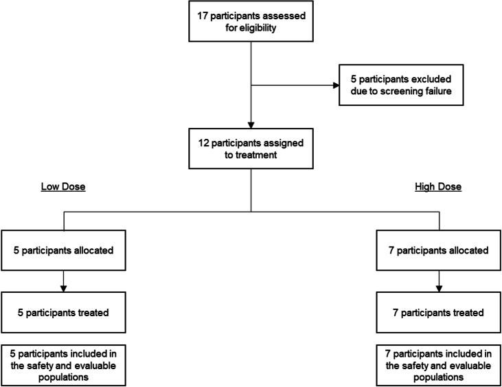
“Between 3 May 2021 and 30 March 2022, 17 participants were assessed for eligibility; five were excluded due to screening failure.”
§ Phase I trial of hES cell-derived dopaminergic neurons for Parkinson’s disease > Methods > Demographics
plainn = 17- Asserted: passes — The authors put it forward as true.
- Checkable: passes — It can be checked against evidence.
- Load-bearing: fails — The paper’s conclusions may not depend on it.
- Based: passes — The paper gives a reason why it holds.
Probably not a substantial claim. The paper’s conclusions may not depend on it. It may state two separate claims as one.
What the paper did — it cites no source for this, so there is none to check. The quote shows it is in the paper.
In the paper
“All of the participants signed the informed consent.”
§ Phase I trial of hES cell-derived dopaminergic neurons for Parkinson’s disease > Methods > Trial oversight
plain- Asserted: passes — The authors put it forward as true.
- Checkable: passes — It can be checked against evidence.
- Load-bearing: fails — The paper’s conclusions may not depend on it.
- Based: passes — The paper gives a reason why it holds.
Probably not a substantial claim. The paper’s conclusions may not depend on it.
What the paper did — it cites no source for this, so there is none to check. The quote shows it is in the paper.
In the paper
“The trial was conducted in accordance with the protocol and all applicable local government laws, regulations and guidance, including policies with foundations in the World Medical Association Declaration of Helsinki, the International Council for Harmonisation of Technical Requirements for Pharmaceuticals for Human Use.”
§ Phase I trial of hES cell-derived dopaminergic neurons for Parkinson’s disease > Methods > Trial oversight
plain- Asserted: passes — The authors put it forward as true.
- Checkable: passes — It can be checked against evidence.
- Load-bearing: fails — The paper’s conclusions may not depend on it.
- Based: passes — The paper gives a reason why it holds.
Probably not a substantial claim. The paper’s conclusions may not depend on it. It may state two separate claims as one.
What the paper did — it cites no source for this, so there is none to check. The quote shows it is in the paper.
In the paper
“Inferential statistical testing was not performed due to the small number of participants.”
§ Phase I trial of hES cell-derived dopaminergic neurons for Parkinson’s disease > Methods > Statistics
plaindirect- Asserted: passes — The authors put it forward as true.
- Checkable: passes — It can be checked against evidence.
- Load-bearing: fails — The paper’s conclusions may not depend on it.
- Based: passes — The paper gives a reason why it holds.
Probably not a substantial claim. The paper’s conclusions may not depend on it.
What the paper did — it cites no source for this, so there is none to check. The quote shows it is in the paper.
In the paper
“The remaining 12 participants were sequentially enrolled into the low-dose (n = 5) or high-dose (n = 7) cohort”
§ Phase I trial of hES cell-derived dopaminergic neurons for Parkinson’s disease > Methods > Demographics
plainn = 12- Asserted: passes — The authors put it forward as true.
- Checkable: passes — It can be checked against evidence.
- Load-bearing: fails — The paper’s conclusions may not depend on it.
- Based: passes — The paper gives a reason why it holds.
Probably not a substantial claim. The paper’s conclusions may not depend on it.
What the paper did — it cites no source for this, so there is none to check. The quote shows it is in the paper.
In the paper
“A data safety monitoring committee and the institutional review boards monitored trial progression and data handling.”
§ Phase I trial of hES cell-derived dopaminergic neurons for Parkinson’s disease > Methods > Trial oversight
plain- Asserted: passes — The authors put it forward as true.
- Checkable: passes — It can be checked against evidence.
- Load-bearing: fails — The paper’s conclusions may not depend on it.
- Based: passes — The paper gives a reason why it holds.
Probably not a substantial claim. The paper’s conclusions may not depend on it.
What the paper did — it cites no source for this, so there is none to check. The quote shows it is in the paper.
In the paper
“The Tri-institutional Human Embryonic Stem Cell Research Oversight committee (ESCRO) at MSKCC and WCM approved the study and the consent on 11 October 2019.”
§ Phase I trial of hES cell-derived dopaminergic neurons for Parkinson’s disease > Methods > Trial oversight
plain- Asserted: passes — The authors put it forward as true.
- Checkable: passes — It can be checked against evidence.
- Load-bearing: fails — The paper’s conclusions may not depend on it.
- Based: passes — The paper gives a reason why it holds.
Probably not a substantial claim. The paper’s conclusions may not depend on it. It may state two separate claims as one.
What the paper did — it cites no source for this, so there is none to check. The quote shows it is in the paper.
In the paper
“An institutional review board at Memorial Sloan Kettering Cancer Center and Weill Cornell Medicine reviewed and approved the clinical protocol (submitted with the Article) and the patient consent form.”
§ Phase I trial of hES cell-derived dopaminergic neurons for Parkinson’s disease > Methods > Trial oversight
plain- Asserted: passes — The authors put it forward as true.
- Checkable: passes — It can be checked against evidence.
- Load-bearing: fails — The paper’s conclusions may not depend on it.
- Based: passes — The paper gives a reason why it holds.
Probably not a substantial claim. The paper’s conclusions may not depend on it. It may state two separate claims as one.
What the paper did — it cites no source for this, so there is none to check. The quote shows it is in the paper.
In the paper
“At the time of the last participant completing the 18-month visit, the median (min, max) follow-up time was 21.2 months (18.5, 24.7) and all of the participants were still engaged in clinical follow-up.”
§ Phase I trial of hES cell-derived dopaminergic neurons for Parkinson’s disease > Methods > Demographics
plainn = 12- Asserted: passes — The authors put it forward as true.
- Checkable: passes — It can be checked against evidence.
- Load-bearing: fails — The paper’s conclusions may not depend on it.
- Based: passes — The paper gives a reason why it holds.
Probably not a substantial claim. The paper’s conclusions may not depend on it. It may state two separate claims as one.
The paper relies on this without support.
The correlation between 18F-DOPA PET uptake and clinical performance can be variable.
“Furthermore, the correlation between 18F-DOPA PET uptake and clinical performance can be variable, and graft survival alone is not sufficient to predict long-term patient benefit.”
The paper relies on this without support.
Graft survival alone is not sufficient to predict long-term patient benefit.
“Furthermore, the correlation between 18F-DOPA PET uptake and clinical performance can be variable, and graft survival alone is not sufficient to predict long-term patient benefit.”
The paper relies on this without support.
Placebo effects can be substantial in studies of PD.
“the possibility of placebo effects, which can be substantial in studies of PD.”
The paper relies on this without support.
PD symptoms fluctuate expectedly.
“the expected fluctuations in PD symptoms”
Self-check
Percentages, sums and reported differences re-computed from the paper’s own numbers.
Each contradiction between two passages listed here was confirmed by an independent check.
Rated minor: it would not touch a result — a value no result rests on, a p-value that stays on the same side of the significance level, or a conflict that is clerical, resolved elsewhere in the paper, or outside its own findings.
In the paper
§ Phase I trial of hES cell-derived dopaminergic neurons for Parkinson’s disease > Safety and tolerability
“Most were mild or moderate in severity except for one fall that was deemed to be unrelated to treatment.”
§ Phase I trial of hES cell-derived dopaminergic neurons for Parkinson’s disease > Methods > TEAEs
“Aside from two severe TEAEs (one fall deemed unrelated to treatment and the aforementioned SAE of gastrointestinal haemorrhage), all others were mild or moderate in severity (Extended Data Table 4).”
Citations
Cites
Poewe, W., Seppi, K., Tanner, C. M., Halliday, G. M., Brundin, P., Volkmann, J., Schrag, A.-E., & Lang, A. E. (2017) · Nature Reviews Disease Primers · doi:10.1038/nrdp.2017.13 · Cited by 5,415
Diagnosis and Treatment of Parkinson Disease: A Review
Armstrong, M. J., & Okun, M. S. (2020) · JAMA · doi:10.1001/jama.2019.22360 · Cited by 2,924
Transplantation of Embryonic Dopamine Neurons for Severe Parkinson's Disease
Freed, C. R., Greene, P. E., Breeze, R. E., Tsai, W.-Y., DuMouchel, W., Kao, R., Dillon, S., Winfield, H., Culver, S., Trojanowski, J. Q., et al. (2001) · New England Journal of Medicine · doi:10.1056/nejm200103083441002 · Cited by 2,366
Self-citationA Bayesian model of shape and appearance for subcortical brain segmentation
Patenaude, B., Smith, S. M., Kennedy, D. N., & Jenkinson, M. (2011) · NeuroImage · doi:10.1016/j.neuroimage.2011.02.046 · Cited by 2,287
Kriks, S., Shim, J., Piao, J., Ganat, Y. M., Wakeman, D. R., Xie, Z., Carrillo‐Reid, L., Auyeung, G., Antonacci, C. L., Buch, A., et al. (2011) · Nature · doi:10.1038/nature10648 · Cited by 1,951
Self-citationLi, J.-Y., Englund, E., Holton, J. L., Soulet, D., Hagell, P., Lees, A. J., Lashley, T., Quinn, N. P., Rehncrona, S., Björklund, A., et al. (2008) · Nature Medicine · doi:10.1038/nm1746 · Cited by 1,845
Lewy body–like pathology in long-term embryonic nigral transplants in Parkinson's disease
Kordower, J. H., Chu, Y., Hauser, R. A., Freeman, T. B., & Olanow, C. W. (2008) · Nature Medicine · doi:10.1038/nm1747 · Cited by 1,722
Goetz, C. G., Fahn, S., Martínez‐Martín, P., Poewe, W. H., Sampaio, C., Stebbins, G. T., Stern, M. B., Tilley, B. C., Dodel, R. C., Dubois, B., et al. (2006) · Movement Disorders · doi:10.1002/mds.21198 · Cited by 1,443
A double‐blind controlled trial of bilateral fetal nigral transplantation in Parkinson's disease
Olanow, C. W., Goetz, C. G., Kordower, J. H., Stoessl, A. J., Sossi, V., Brin, M. F., Shannon, K. M., Nauert, G. M., Perl, D. P., Godbold, J., et al. (2003) · Annals of Neurology · doi:10.1002/ana.10720 · Cited by 1,307
The pathogenesis of Parkinson's disease
Morris, H. R., Spillantini, M. G., Sue, C. M., & Williams‐Gray, C. H. (2024) · The Lancet · doi:10.1016/s0140-6736(23)01478-2 · Cited by 1,164
Research works printed but not linked: 1
- Martinez–Martin, P., Rodriguez-Blazquez, C. & Forjaz, M. J. in Neuroscience and Biobehavioral Psychology 8–16 (Elsevier, 2017).
Cited by
Sorted by citations
Items 101–181 of 181
Advances and unmet needs in fluid and tissue biomarkers in Parkinson’s disease
Rajmohan, R., & Henchcliffe, C. (2026) · Neural Regeneration Research · doi:10.4103/nrr.nrr-d-25-01239
Self-citationNurturing eggs with hiPSC-derived cells to improve outcomes in in vitro fertilization
Fang, F., & Pera, R. A. R. (2026) · Cell stem cell · doi:10.1016/j.stem.2026.01.005
Lee, S. W., Kim, Y.-J., Kang, S., Cha, H., & Kim, H. J. (2026) · Chemical Engineering Journal · doi:10.1016/j.cej.2026.174074
Parsaeian, A., Naserisalehabad, P., & Najmoddin, N. (2025) · National and International Iranian Conference on Biomedical Engineering (ICBME) · doi:10.1109/icbme68496.2025.11392575
Hunt, C. P., Thek, K. K., Durnall, J. C., McDougall, S. J., Parish, C. L., & Gantner, C. W. (2026) · Scientific Reports · doi:10.1038/s41598-026-42112-y
Tang, Q. B., Sun, J., Xue, J., Qiu, C., Wang, T., Matveeva, T., Wu, Y., Li, X. Y., Cheng, T., Seo, H., et al. (2026) · Stem Cell Reports · doi:10.1016/j.stemcr.2026.102830
Ajibare, A. J., Asuku, A. O., & Akintoye, O. O. (2026) · Neuroprotection/Neuroprotection (Chichester, England. Print) · doi:10.1002/nep3.70030
Bridging Regenerative and Restorative Stem Cell Therapies in Parkinson’s Disease
Onuigbo, C. C., Martínez-Lemus, J. D., Tharp, E., & Schiess, M. C. (2026) · Cells · doi:10.3390/cells15070578
Vgll2 and Tead1 Govern Generation of Mouse and Human Hypothalamic Hypocretin (Orexin) NeuronsPreprint
Wei, R., Sheikhshahrokh, A., Saeidi, E., Gómez‐Inclán, C., Gopalakrishnan, A., Balderson, B., Mikael Bodén, A., Piper, M., & Thor, S. (2026) · bioRxiv (Cold Spring Harbor Laboratory) · doi:10.64898/2026.03.30.715421
Engineering craniofacial skeletal organoids from human pluripotent stem cells via neural crest cells
Motoike, S., & Ikeya, M. (2026) · Cell Biomaterials · doi:10.1016/j.celbio.2026.100442
2026 Watch List: Regenerative Medicine
CDA-AMC (2026) · Canadian Journal of Health Technologies · doi:10.51731/cjht.2026.1420
Zhu, Y., Zhang, H., Zhang, Z., Song, G., Fu, J., & Wang, H. (2026) · Neuroscience · doi:10.1016/j.neuroscience.2026.04.024
Limitations of Stem Cell Therapy for Parkinson’s Disease
Li, Z., Peng, S., Jia, O. Y., & Liu, R. (2026) · Cells · doi:10.3390/cells15090839
Self-citationHomogenous Cell Transplantation Trials in Parkinson's Disease: A Systematic Review and Meta‐Analysis
Reinisch, N., Mancini, A., Elsherbini, A., Yuen, J., & Lozano, A. (2026) · Movement Disorders Clinical Practice · doi:10.1002/mdc3.70647
Self-citationLi, Y., Guo, L., Yu, X., & Peng, C. (2026) · Ageing and Neurodegenerative Diseases · doi:10.20517/and.2025.31
Overgaard, A., Molnar, K., Jurtz, V. I., Lie, M. E. K., & Nyengaard, J. R. (2026) · Computers in Biology and Medicine · doi:10.1016/j.compbiomed.2026.111738
Liu, Q., Tasaki, S., Bennett, D. A. A., Seyfried, N. T., Jager, P. L. D., Menon, V., Buchman, A. S., & Yang, J. (2026) · medRxiv · doi:10.64898/2026.05.23.26353951
The Therapeutic Potential of Mesenchymal Stem Cells in Post-Stroke Depression
Fan, M., Deng, Q., Li, Z., Wang, G., & Lu, M. (2026) · International Journal of Molecular Sciences · doi:10.3390/ijms27114796
Chang, C.-Y., Chiang, C.-Y., Ting, H.-C., Tsai, Y.-J., Chan, P.-C., Chen, M.-F., Guo, Y., Liu, Y., Chen, Y.-S., Chuang, C., et al. (2026) · npj Parkinson s Disease · doi:10.1038/s41531-026-01399-4
Translational stem cell therapy for neurodegeneration and CNS trauma: a focused review
Khosrofar, R., Nafei, P., Zare, F., Sadrmohammadbagheri, A., Zargari, F., Rastgar, N., Radvar, S., Eskandani, N. A., Hoseinzade, F., & Akhavan‐Sigari, R. (2026) · Molecular Biology Reports · doi:10.1007/s11033-026-12011-6
Liu, Q., Tasaki, S., Bennett, D. A. A., Seyfried, N. T., Jager, P. L. D., Menon, V., Buchman, A. S., & Yang, J. (2026) · Research Square · doi:10.21203/rs.3.rs-9622597/v1
Neurosurgical treatments for Parkinson's disease: A narrative review
Winkelmann, L. S., Astradsson, A., Pereira, E. A. C., Ágústsdóttir, D. H., Meyer, M., & Einarsson, H. B. (2026) · Interdisciplinary Neurosurgery · doi:10.1016/j.inat.2026.102291
Pavan, C., de Lichtenberg, K. H., Gantner, C. W., Frausin, S., Chen, J., Hunt, C. P., Xuereb, B., Hart, E., Chatterton, Z., Halliday, G. M., et al. (2026) · bioRxiv (Cold Spring Harbor Laboratory) · doi:10.64898/2026.06.01.729439
Stem Cell Therapy for Parkinson’s Disease: A Mechanistically Distinct Role for Muse Cells
Mesches, M. H., Granholm, A., Paredes, D., Coleman-Mesches, K., Oguma, Y., & Dezawa, M. (2026) · Journal of Clinical Medicine · doi:10.3390/jcm15114370
Lineage-selective suicide gene system enables post-engraftment editing of cell therapy compositionPreprint
Jin, J., Pavan, C., Moriarty, N., Ovchinnikov, D. A., Farrell, G., Quattrocchi, A. T., Hunt, C. P., & Parish, C. L. (2026) · bioRxiv (Cold Spring Harbor Laboratory) · doi:10.64898/2026.06.03.729995
Yurek, D. M., Quintero, J. E., Welleford, A. S., Seblani, N. E., Powell, D. K., Pomerleau, F., Lundgren, K. H., Seroogy, K. B., van Horne, C. G., & Gerhardt, G. A. (2026) · Journal of Neuroscience Methods · doi:10.1016/j.jneumeth.2026.110825
Liang, Z., Liu, Y., & Shang, P. (2026) · Advanced technology in neuroscience . · doi:10.4103/atn.atn-d-25-00027
Microglial advances in Parkinson’s disease
Debasa‐Mouce, M., Ouro, A., Leon, J. D., Gulkarov, S., Reiss, A. B., & Bougea, A. M. (2026) · Advances in immunology · doi:10.1016/bs.ai.2026.06.003
The regulation of stem cell fate and its application in neural regeneration
He, Y., Chen, X., He, M., & Li, Z. Y. (2026) · Interdisciplinary medicine · doi:10.1002/inmd.70137
A Defined Xeno-Free Matrix Supports Midbrain Dopaminergic Cell Differentiation
Wang, T., Zeng, Y., Xue, J., Liu, R., Sun, J., Bian, X., Qiu, C., Lan, Q., Wu, Y., Tang, Q. B., et al. (2026) · ACS Nano · doi:10.1021/acsnano.6c04239
Barbero, J., Vo, A., Strohl, J. J., Vacca, S., Tang, C. C., Nguyen, N., Unadkat, P., Ma, Y., Peng, S., Niethammer, M., et al. (2026) · Research Square · doi:10.21203/rs.3.rs-10119103/v1
Self-citationOzzy Osbourne and Parkinson's disease: from darkness to awareness
Paleare, L. F. F., Pedroso, J. L., Rolim, F. D. P. S., de Araújo, N. R. A., Camargo, C. H. F., Teive, H. A. G., & Franklin, G. L. (2026) · Arquivos de Neuro-Psiquiatria · doi:10.1055/s-0046-1823655
Novel treatments for Parkinson’s disease
Yin, Y., Gao, J., Zhang, K., Meng, F. D., & Feng, T. (2026) · Chinese Medical Journal · doi:10.1097/cm9.0000000000004163
Nrf2 Activators in Parkinson’s Disease: Modulating Mitophagy and Regulating Cuproptosis
Xu, J., Xiang, J., Zhang, Y., & Liu, X. (2026) · Molecular Neurobiology · doi:10.1007/s12035-026-06036-y
Evangelisti, A., Phillips, S. M., Jungverdorben, J., Walsh, R. M., Wu, Y., Bocchi, V. D., Zhou, T., & Studer, L. (2026) · bioRxiv (Cold Spring Harbor Laboratory) · doi:10.64898/2026.06.30.729930
Self-citationMussin, N. M., Myssayev, A., Zhilisbayeva, K. R., Kurmanalina, M. A., & Tamadon, A. (2026) · Медицинский журнал Западного Казахстана · doi:10.4103/wkmj.wkmj_66_26
Epigenomic analyses identify novel regulators of dopaminergic neuron identity
Valceschini, E. (2026) · Open Repository and Bibliography (University of Luxembourg)
Wang, G., Huang, C., Luo, X., Pan, J., Pan, S., Xu, A., Liu, Q., Chen, J., Sun, L., Duan, J., et al. (2026) · International Journal of Biological Macromolecules · doi:10.1016/j.ijbiomac.2026.153252
Halabian, N., Park, C., Omoto, L., Bocca, L. F., Ramos-Palacios, G., Wu, K., Hamani, C., Rabin, J. S., Abrahão, A., Davidson, B., et al. (2026) · Biological Psychiatry · doi:10.1016/j.biopsych.2026.07.008
Hemeda, S., Janszky, J., Pintér, D., & Kovács, N. (2026) · Journal of Central Nervous System Disease · doi:10.1177/11795735261463495
The transformative impact of stem cell core facilities in biomedical research
Corneo, B., Fagà, G., Figueira, I., Zasso, J., Yang, W., Neuman, K., Johansson, P. A., Healy, L. E., Daheron, L. M., Generali, M., et al. (2026) · Stem Cell Reports · doi:10.1016/j.stemcr.2026.103047
Therapeutic landscape of Parkinson's disease: Recent advances in saponin-mediated interventions
Tripathy, D., Sarma, S. S., Kumar, D., & Chakraborty, J. (2026) · Neurochemistry International · doi:10.1016/j.neuint.2026.106239
Ali, S., Quintero, J. E., Richert, M., Koehl, L., Anderson-Mooney, A. J., Gurwell, J. A., Manning, K. S., Hulou, S., Hines, T. K., Slevin, J. T., et al. (2026) · Journal of neurosurgery · doi:10.3171/2026.3.jns251474
Regenerative medicine for neurodegenerative diseases: history, strategies, and clinical advances
Li, J., Duan, H., Hao, P., Zhao, W., Gao, Y., Yang, Z., & Li, X. (2026) · Translational research · doi:10.1016/j.trsl.2026.08.005
Gutierrez-Arratia, J. D., Awad, P. S., Schuh, A. F. S., & Cornejo‐Olivas, M. (2026) · Journal of Parkinson s Disease · doi:10.1177/1877718x261478730
Azulay, J. (2026) · Bulletin de l Académie Nationale de Médecine · doi:10.1016/j.banm.2026.03.018
Aktuelle Entwicklungen in der Parkinsontherapie
Höglinger, G. U. (2026) · DNP – Die Neurologie & Psychiatrie · doi:10.1007/s15202-026-6741-9
Xi, Y.-T., Jia, C.-X., Wang, H. Q., Liu, P., Li, P., Niu, Q., & Yin, Y. (2026) · Ageing Research Reviews · doi:10.1016/j.arr.2026.103358
Parkinson’s Disease: Current Knowledge and Future Focus
Pantazis, C. B., Sherer, T., & Riley, E. A. U. (2026) · Springer eBooks · doi:10.1007/978-3-032-31497-0_7
王, 兴. (2026) · Traditional Chinese Medicine · doi:10.12677/tcm.2026.159490
Emerging therapies expected to make a difference in Parkinson's disease management
An, Y. A., & Kalia, L. V. (2026) · Parkinsonism & Related Disorders · doi:10.1016/j.parkreldis.2026.108962
Mitochondrial Modulation as a Therapeutic Entry Point in Neurodegeneration
Mamala, M. B., Skowron, I., Marczuk, K. M., Bujas, A., Harasiuk, J., & Kulbacka, J. (2026) · Cells · doi:10.3390/cells15181683
Bratt‐Leal, A. M., Gorba, T., Williams, R. M., Mossman, J. A., Hills, R., Tran, H., Barken, D. M., Beauvais, G., Fremgen, D. M., Zhang, A., et al. (2026) · Cell stem cell · doi:10.1016/j.stem.2026.08.014
Ichinose, H., Sato, Y., & MURAMATSU, S.-I. (2026) · Expert Opinion on Biological Therapy · doi:10.1080/14712598.2026.2738706
Yang, D., Choi, S., Piao, J., Evangelisti, A., Yang, Z., Bocchi, V. D., Donohue, S. A., Claros, N., Jungverdorben, J., Sidharta, M., et al. (2026) · bioRxiv (Cold Spring Harbor Laboratory) · doi:10.64898/2026.09.23.753784
Self-citationSuspension culture drives metabolic rewiring in human pluripotent stem cellsPreprint
Agger, A. S. P., Kafkia, E., Larsen, M. B., Möritz, T., Madsen, C. T., de Lichtenberg, K. H., Halloin, C., Serup, P., Żylicz, J. J., & Honoré, C. (2026) · bioRxiv (Cold Spring Harbor Laboratory) · doi:10.64898/2026.09.24.753129
SpiraMed – A Stereotactic Helix-based Therapy Delivery system for the Human BrainPreprint
Bulstrode, H. J. C. J., Young, A., Huysman, I., Seevaratnam, D., Man, K. K., Pak, H. L., Wilson, G., Stamper, A., Wang, X., Wang, Y., et al. (2026) · bioRxiv (Cold Spring Harbor Laboratory) · doi:10.64898/2026.09.27.754823
Li, H., Jiang, S., Liu, X., Zhang, J., Hu, Y., Liu, B., Wu, M., Li, H., & Li, B. (2026) · International Journal of Nanomedicine · doi:10.2147/ijn.s636187
Early spinogenesis in postnatally-born hippocampal granule cells
Frey, L., Kirscht, S., Ebert, J., Hanauske, T., Deller, T., Schwarzacher, S. W., & Jungenitz, T. (2026) · Frontiers in Neuroanatomy · doi:10.3389/fnana.2026.1899044
Verlaufsmodifizierende Therapien der Parkinsonkrankheit
Schäfer, N., & Weise, D. (2026) · DNP – Die Neurologie & Psychiatrie · doi:10.1007/s15202-026-6784-y
van der Walle, C. F. (2026) · Elsevier eBooks · doi:10.1016/b978-0-443-45791-3.00006-8
Cha, Y., & Kim, K. (2026) · Cell Reports Medicine · doi:10.1016/j.xcrm.2026.103104
Syaifudin, Wildan, Mu’azzaroh, Prasetyo, & Ningrum (2025) · Journal of Regenerative Medicine and Molecular Innovation
Ren, S.-X., Cai, H., Xing, R.-Y., Liu, J.-J., Jia, F., Guo, H.-D., & Cui, G.-H. (2026) · Pharmacological Research · doi:10.1016/j.phrs.2026.109670
Berger, C., Wiora, L., Holmes, T. L., Walz, F., Sauter, C. M., Baraban, L., Schwebler, J., Beer, G., Lauke, S., Keller, J., et al. (2026) · bioRxiv (Cold Spring Harbor Laboratory) · doi:10.64898/2026.10.07.757018
Marlonsson, A., Quintero, J. E., Olofsson, H. E., Granholm, A.-C., van Horne, C. G., & Gerhardt, G. A. (2026) · Cell Transplantation · doi:10.1177/09636897261495817
Peng, K., Guan, Q., Hong, R., He, Y., Jiao, F., Zhang, T., Wu, P., Zhao, X., Zeng, Q., Zhang, J., et al. (2026) · Cell Transplantation · doi:10.1177/09636897261494929
Emerging stem cell strategies for neurodegenerative diseases
Hardjo, M., & Sophian, A. (2026) · Transplant Immunology · doi:10.1016/j.trim.2026.102465
Timed binary WNT activity is sufficient to model the early human neural axisPreprint
Hackland, J. O. S., Anand, G., Vijayakumar, R., Wang, X., Bocchi, V. D., Rowland, E. S., Yang, D., Wu, Y., Mehta, S., Zhong, A., et al. (2026) · bioRxiv (Cold Spring Harbor Laboratory) · doi:10.64898/2026.10.17.759682
Self-citationOh, S.-Y., Park, Y. S., Choi, B. H., Kim, H. S., Jung, S.-C., & Jo, I. (2026) · Pharmacology & Therapeutics · doi:10.1016/j.pharmthera.2026.109133
Stem cell therapies in modern medicine: Progress and challenges
Ahmed, N., Khan, M. S. I., Kodeeswaran, M., Rajabov, T., & Chaurasia, B. K. (2026) · Regenesis repair rehabilitation. · doi:10.1016/j.rerere.2026.10.002
He, Z., Zeng, J., Wang, G., Li, Y., & Zhang, F. (2026) · MedComm · doi:10.1002/mco2.71090
Kipanyula, M. J. (2026) · Ageing Research Reviews · doi:10.1016/j.arr.2026.103429
Γενετική βάση της νόσου του Parkinson: σύγχρονες προσεγγίσεις και θεραπευτικές προοπτικές
Μπλαζιάκη, Α.-Ι. Ν. (2026) · University of Thessaly Institutional Repository (University of Thessaly)
Zhao, Z., Jia, A., Ma, W., Xu, X., Zhou, M.-G., & Yuan, J.-L. (2026) · Military Medical Research · doi:10.1016/j.mmr.2026.100077
Recent Advances in Clinical Neuromodulation for Brain Disorders
刘秀云, Du, Y., Guo, Q., Li, S., Wang, W., Peng, R., Zhao, Z., Song, X., Zhi, H., Li, H., et al. (2026) · Engineering · doi:10.1016/j.eng.2026.10.011
The 2025 Yearbook of Neurorestoratology
Huang, H., Wang, J., He, J., Hu, Y., Sanberg, P. R., Sharma, H. S., Siniscalco, D., Chen, X., Bach, J. R., Andrews, R. J., et al. (2026) · Journal of Neurorestoratology · doi:10.1016/j.jnrt.2026.100348
Gong, D., Li, G., Li, M., Zhang, B., Wang, W., Chen, J., & Zhao, Y. (2026) · PLoS ONE · doi:10.1371/journal.pone.0362081
Replacing the neurons that Parkinson’s destroys
Dattaro, L. (2026) · Nature · doi:10.1038/d41586-026-03166-0
Metadata
- Authors
- Tabar, V., Sarva, H., Lozano, A. M., Fasano, A., Kalia, S. K., Yu, K. K. H., Brennan, C., Ma, Y., Peng, S., Eidelberg, D., Tomishima, M., Irion, S., Stemple, W., Abid, N., Lampron, A., Studer, L., & Henchcliffe, C.
- Affiliations
- Memorial Sloan Kettering Cancer Center (US)
- Weill Cornell Medicine (US)
- University of Toronto (CA)
- University Health Network (CA)
- Feinstein Institute for Medical Research (US)
- University of California, Irvine (US)
- Corresponding author
- Tabar, V.
- ORCID
- Venue
- Nature
- Published
- 2025-04-16
- Volume
- 641
- Issue
- 8064
- Pages
- 978-983
- ISSN
- 0028-0836 (ISSN-L)
- 1476-4687
- PMC
- PMC12095069
- OpenAlex
- W4409502153
- Cited by
- 181
- References
- 51
- Citations by year
- 2026: 124 · 2025: 57
- FWCI
- 97.18
- Type
- journal article
- Language
- en
- Open access
- hybrid
- doi.org
- Licence
- Locations
- Nature (publishedVersion, cc-by-nc-nd)
- PubMed Central (publishedVersion, cc-by-nc-nd)
- PubMed (publishedVersion)
- RePEc: Research Papers in Economics (submittedVersion)
- Funders
- National Cancer Institute (P30CA008748)
- National Institute of Neurological Disorders and Stroke (R01NS118067)
- Topics
- Health Sciences › Medicine › Clinical Neurology › Parkinson's Disease Mechanisms and Treatments
- Health Sciences › Medicine › Clinical Neurology › Deep Brain Stimulation and Movement Disorders
- Life Sciences › Biochemistry, Genetics and Molecular Biology › Molecular Biology › Pluripotent Stem Cells Research
- Keywords
- Parkinson's disease, human embryonic stem cells, dopaminergic neurons, cell therapy, graft survival, dyskinesia, safety
- MeSH terms
- Human Embryonic Stem Cells, Adult, Aged, Female, Graft Survival, Humans, Male, Middle Aged, Parkinson Disease, Putamen, Cohort Studies, Cryopreservation, Treatment Outcome, Stem Cell Transplantation, Positron-Emission Tomography, Dopaminergic Neurons
About these checks
Registry, retraction, Cites and statistics results are computed, not judged, with one exception: a printed reference that no index lists is read once by a model before it counts as a citation error. Claims are extracted by a model and kept only when their quote appears verbatim in the text; each cited source is then checked, with a model’s help, for whether it says what the claim relies on. Those verdicts can be wrong — read the claim’s quote and the cited source before relying on one.
A language model drew the claims from the paper on 2026-11-09, under version 3 of the claim definition (model deepseek-v4.1-flash, prompt extract-v3).
A second model labelled each claim on 2026-11-09 (model jev-1.13-20260917, definition revision gold-guideline-v0.2).
Cited sources are checked with a language model’s help (gpt-6-sol).
Statistics and arithmetic are recomputed, not judged.
Confidence bands are shown only for fields calibrated on our annotated test set. None is calibrated yet, so no band is shown.
What counts as a claim ›How we measure confidence ›How we check this ›
Are you an author, or do you see a problem with this page? Request a review
