

The DOI matches its Crossref record; 105 of the 106 references it cites were found in a registry or an index.
Functional, metabolic and transcriptional maturation of human pancreatic islets derived from stem cells
Open accessPublished version of a preprint
Abstract
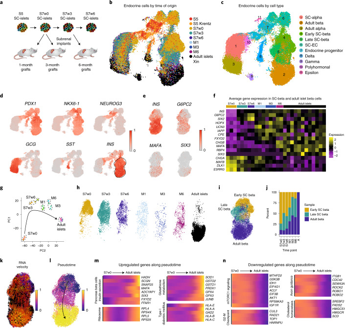
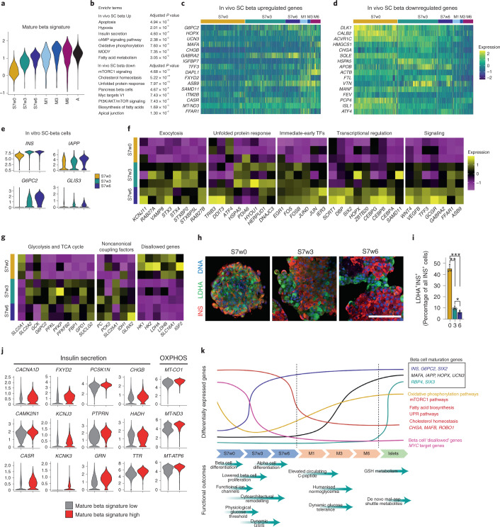
Transplantation of pancreatic islet cells derived from human pluripotent stem cells is a promising treatment for diabetes. Despite progress in the generation of stem-cell-derived islets (SC-islets), no detailed characterization of their functional properties has been conducted. Here, we generated functionally mature SC-islets using an optimized protocol and benchmarked them comprehensively against primary adult islets. Biphasic glucose-stimulated insulin secretion developed during in vitro maturation, associated with cytoarchitectural reorganization and the increasing presence of alpha cells. Electrophysiology, signaling and exocytosis of SC-islets were similar to those of adult islets. Glucose-responsive insulin secretion was achieved despite differences in glycolytic and mitochondrial glucose metabolism. Single-cell transcriptomics of SC-islets in vitro and throughout 6 months of engraftment in mice revealed a continuous maturation trajectory culminating in a transcriptional landscape closely resembling that of primary islets. Our thorough evaluation of SC-islet maturation highlights their advanced degree of functionality and supports their use in further efforts to understand and combat diabetes.
Overview
The claims and statistics of this paper haven’t been checked yet.
Run the check — free, no account needed. It reads the open-access text and checks the sources it cites.
Integrity signals
Registry
Cites
Statistics
Figures
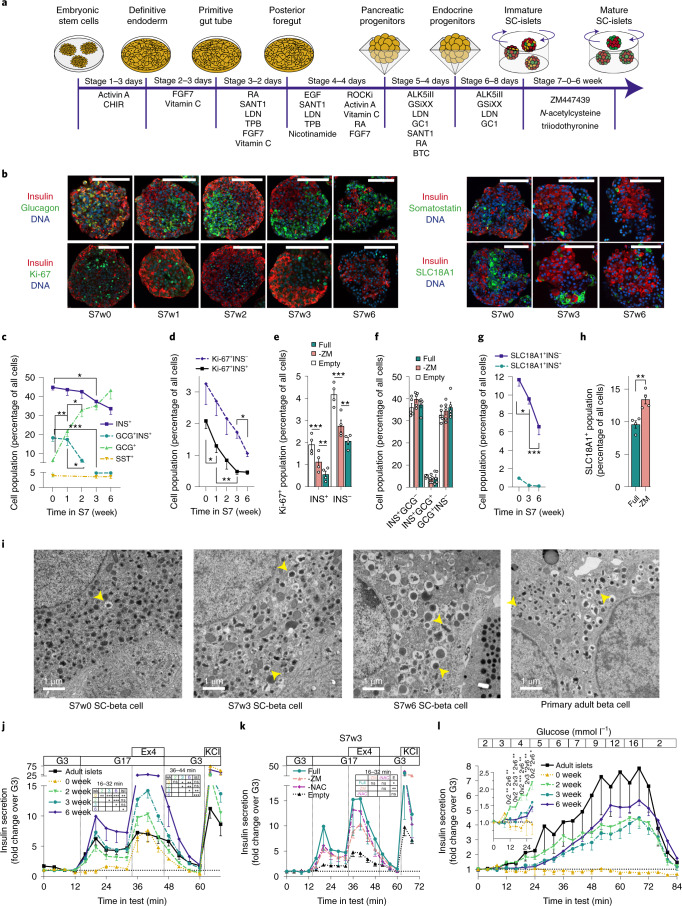
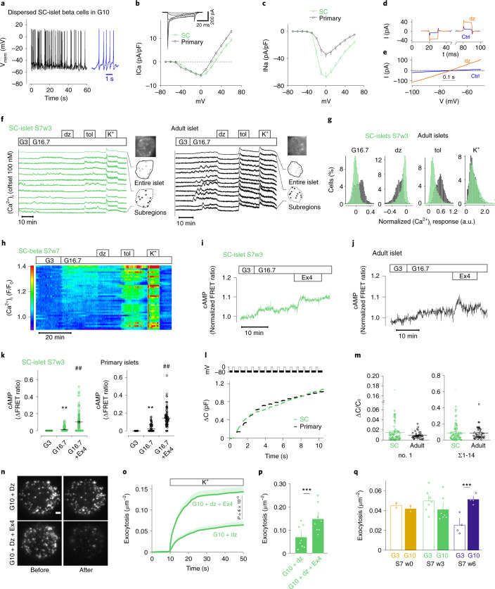
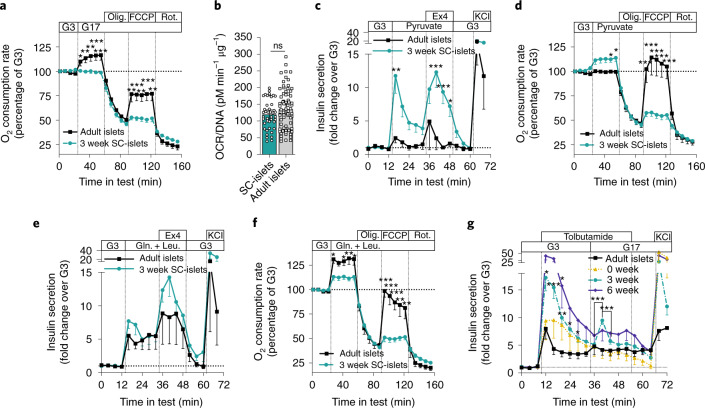
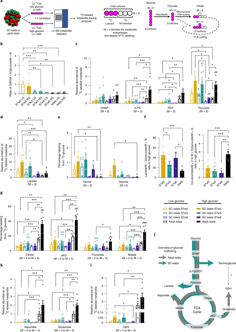
Verification
Claims the paper makes
The claims and statistics of this paper haven’t been checked yet.
Run the check — free, no account needed. It reads the open-access text and checks the sources it cites.
Claims other papers make about this one
Each is checked against this paper’s own text: does it say what the citing paper says it does?
The source backs part of the claim, not all of it — or backs it for a different population, condition or comparison.
In the citing paper
“These cells have been subsequently identified in SC-islets generated from various differentiation protocols and stem cell lines by multiple groups.”
From Developments in stem cell-derived islet replacement therapy for treating type 1 diabetes (2023) · cites this paper as [43]
Experimental — accuracy not yet measuredThe source backs part of the claim, not all of it — or backs it for a different population, condition or comparison.
In the citing paper
“Specifically, known markers of β cell maturation, such as INS, MAFA, IAPP, MNX1, and G6PC2, became more highly expressed in a greater proportion of SC-islet cells after transplantation.”
From Developments in stem cell-derived islet replacement therapy for treating type 1 diabetes (2023) · cites this paper as [43]
Experimental — accuracy not yet measuredThe source backs part of the claim, not all of it — or backs it for a different population, condition or comparison.
In the citing paper
“Transplantation of SC-islets into mice improves their insulin secretion and glucose responsiveness, suggesting that SC-islet immaturity can be resolved.”
From Developments in stem cell-derived islet replacement therapy for treating type 1 diabetes (2023) · cites this paper as [43]
Experimental — accuracy not yet measuredThe source backs part of the claim, not all of it — or backs it for a different population, condition or comparison.
In the citing paper
“These transcriptional changes appeared to occur temporally, as some genes (e.g., G6PC2) increased in expression shortly after transplantation, while others (e.g., MAFA) took several months to upregulate.”
From Developments in stem cell-derived islet replacement therapy for treating type 1 diabetes (2023) · cites this paper as [43]
Experimental — accuracy not yet measuredThe source backs part of the claim, not all of it — or backs it for a different population, condition or comparison.
In the citing paper
“Overall, comparison of scRNA-seq and snATAC-seq data from transplanted SC-islets and primary islets has indicated that SC-islets become more similar to human islets overtime in vivo in terms of both their transcriptional and chromatin accessibility profiles, though some important differences still remain.”
From Developments in stem cell-derived islet replacement therapy for treating type 1 diabetes (2023) · cites this paper as [43]
Experimental — accuracy not yet measured
The cited source says what the claim attributes to it.
In the citing paper
“These cells have been shown to improve glycemic control in diabetic mice, highlighting their potential utility in vivo.36,37,39–44”
From Developments in stem cell-derived islet replacement therapy for treating type 1 diabetes (2023) · cites this paper as [43]
The cited source says what the claim attributes to it.
In the citing paper
“and recent advances in single-cell sequencing technologies have allowed for unprecedented characterization of these cells to further elucidate ways to refine SC-β cell differentiation strategies.43,47,50,51”
From Developments in stem cell-derived islet replacement therapy for treating type 1 diabetes (2023) · cites this paper as [43]
The cited source says what the claim attributes to it.
In the citing paper
“scRNA-seq analysis of transplanted SC-islets revealed that improved function corresponded with transcriptional maturation.”
From Developments in stem cell-derived islet replacement therapy for treating type 1 diabetes (2023) · cites this paper as [43]
The cited source says what the claim attributes to it.
In the citing paper
“This differentiation methodology takes approximately a month or more to complete, and functional maturation can continue for months once the cells are transplanted in vivo.43,51”
From Developments in stem cell-derived islet replacement therapy for treating type 1 diabetes (2023) · cites this paper as [43]
The cited source says what the claim attributes to it.
In the citing paper
“Despite transcriptional improvements, SC-islets nonetheless had significantly reduced activity of metabolic pathways compared to primary islets.”
From Developments in stem cell-derived islet replacement therapy for treating type 1 diabetes (2023) · cites this paper as [43]
Self-check
Percentages, sums and reported differences re-computed from the paper’s own numbers.
The self-check needs the paper’s full text.
Citations
Cites
Metascape provides a biologist-oriented resource for the analysis of systems-level datasets
Zhou, Y., Zhou, B., Pache, L., Chang, M., Khodabakhshi, A. H., Tanaseichuk, O., Benner, C., & Chanda, S. K. (2019) · Nature Communications · doi:10.1038/s41467-019-09234-6 · Cited by 14,611
Integrating single-cell transcriptomic data across different conditions, technologies, and species
Butler, A., Hoffman, P., Smibert, P., Papalexi, E., & Satija, R. (2018) · Nature Biotechnology · doi:10.1038/nbt.4096 · Cited by 13,174
Enrichr: a comprehensive gene set enrichment analysis web server 2016 update
Kuleshov, M. V., Jones, M. R., Rouillard, A. D., Fernandez, N. F., Duan, Q., Wang, Z., Koplev, S., Jenkins, S. L., Jagodnik, K. M., Lachmann, A., et al. (2016) · Nucleic Acids Research · doi:10.1093/nar/gkw377 · Cited by 10,799
Fast, sensitive and accurate integration of single-cell data with Harmony
Korsunsky, I., Millard, N., Fan, J., Slowikowski, K., Zhang, F., Wei, K., Baglaenko, Y., Brenner, M., Loh, P.-R., & Raychaudhuri, S. (2019) · Nature Methods · doi:10.1038/s41592-019-0619-0 · Cited by 10,488
Trapnell, C., Cacchiarelli, D., Grimsby, J., Pokharel, P., Li, S., Morse, M., Lennon, N. J., Livak, K. J., Mikkelsen, T. S., & Rinn, J. L. (2014) · Nature Biotechnology · doi:10.1038/nbt.2859 · Cited by 7,314
La Manno, G., Soldatov, R., Zeisel, A., Braun, E., Hochgerner, H., Petukhov, V., Lidschreiber, K., Kastriti, M. E., Lönnerberg, P., Furlan, A., et al. (2018) · Nature · doi:10.1038/s41586-018-0414-6 · Cited by 4,118
Efremova, M., Vento-Tormo, M., Teichmann, S. A., & Vento-Tormo, R. (2020) · Nature Protocols · doi:10.1038/s41596-020-0292-x · Cited by 3,449
Finak, G., McDavid, A., Yajima, M., Deng, J., Gersuk, V., Shalek, A. K., Slichter, C. K., Miller, H. W., McElrath, M. J., Prlic, M., et al. (2015) · Genome Biology · doi:10.1186/s13059-015-0844-5 · Cited by 3,159
Generalizing RNA velocity to transient cell states through dynamical modeling
Bergen, V., Lange, M., Peidli, S., Wolf, F. A., & Theis, F. J. (2020) · Nature Biotechnology · doi:10.1038/s41587-020-0591-3 · Cited by 2,951
Biodiversity on the Rocks: Macrofauna Inhabiting Authigenic Carbonate at Costa Rica Methane Seeps
Levin, L. A., Mendoza, G., Grupe, B., Gonzalez, J. P., Jellison, B. M., Rouse, G. W., Thurber, A. R., & Warén, A. (2015) · PLoS ONE · doi:10.1371/journal.pone.0131080 · Cited by 2,166
Cited by
Sorted by citations
Items 401–441 of 441
Martín-Díaz, L., Patrikainen, M. S., Chongtham, C., Gupta, A., Dotsenko, V., Ojanen, M., Martins, F. T. A., George, J. J., Davidsson, R., Hakanen, S., et al. (2026) · bioRxiv (Cold Spring Harbor Laboratory) · doi:10.64898/2026.05.28.728433
Self-citationWang, X., Carcamo, S., Lambertini, L., Wang, P., Liu, H.-T., Paulsen, J. D., Brody, R., Fernandez-Ranvier, G., Hasson, D., Stewart, A., et al. (2026) · bioRxiv · doi:10.64898/2026.05.29.728759
Deng, L., & Krishnamurthy, M. (2026) · Frontiers in Endocrinology · doi:10.3389/fendo.2026.1819394
Wang, X., Lee, H., Le, A., Turhan, B., Hu, N., Garcia, P. S., Cao, X., Liu, D. K., Ali, T. A., Zhang, N., et al. (2026) · bioRxiv (Cold Spring Harbor Laboratory) · doi:10.64898/2026.06.12.731989
Parikh, S., Strobl, D. C., Jiménez, S., Beckmann, J., Arnoldt, L., Roellin, E., Vandenbempt, V., Sterr, M., Aije, M., Vu, H. T. H., et al. (2026) · bioRxiv (Cold Spring Harbor Laboratory) · doi:10.64898/2026.06.22.733853
Self-citationYaseen, Z., & Mothilal, M. (2026) · Stem Cell Reviews and Reports · doi:10.1007/s12015-026-11184-8
Press, C., Damour, K., Mamrak, N. E., Pepperl, D., Kutner, R. H., Colleluori, D. M., Graham, M. L., Brehm, M. A., Hussain, N., Latres, E., et al. (2026) · Stem Cell Reports · doi:10.1016/j.stemcr.2026.102998
Vitamin A and its relationship with insulin secretion and diabetes mellitus: a review
Vial-Dahmer, D. D. S., Spada, E. C. D. L., Neto, E. W. A., da Silva, W. B., Latorraca, M. Q., & de França, S. A. (2026) · Anais da Academia Brasileira de Ciências · doi:10.1590/0001-3765202620250426
Huang, X., chen, Q., & Lü, H. (2026) · Frontiers in Immunology · doi:10.3389/fimmu.2026.1856373
Islet ultrastructure: past achievements and future directions
Müller, A., Duran, S. A., Santos, P. I., Weigel, A. V., Espinosa-Medina, I., & Solimena, M. (2026) · Diabetologia · doi:10.1007/s00125-026-06782-7
Mitochondrial Function Changes in hiPSCs-Derived Vascular Smooth Muscle Cells
Fang, L., Sun, X.-H., Shu, L., Chen, L., Liu, J., Zhou, J., Chen, W., Liang, Y. N., & Lin, Z. (2026) · ACS Omega · doi:10.1021/acsomega.6c02360
Applications of Stem Cell–Derived Islets and Emerging Experimental Platforms to Understand Diabetes
Mukherjee, N., & Millman, J. R. (2026) · Diabetes · doi:10.2337/db26-0013
Varghese, S. S., Peña, A. G. H.-D. L., Wang, L., Ruiz, V., Tixi, W., Pandey, A., Anchondo, L. M., Chiu, A., Wu, X., Georgia, S. K., et al. (2026) · Diabetes · doi:10.2337/db25-1027
Moreno‐Castro, C., Arroyo, M. N., Benabdallah, K., Olazagoitia‐Garmendia, A., Santacreu, B. J., Sawatani, T., Green, J., Pachera, N., Cosentino, C., Communi, D., et al. (2026) · SSRN Electronic Journal · doi:10.2139/ssrn.7198220
Stem Cell–Derived Islet Therapy for Type 1 Diabetes
Barsby, T., Balboa, D., & Otonkoski, T. (2026) · Elsevier eBooks · doi:10.1016/b978-0-443-11985-9.00025-1
Self-citationPancreas and Islet Cell Transplantation for Diabetes
Czarnecka, Z., & Shapiro, A. M. J. (2026) · Elsevier eBooks · doi:10.1016/b978-0-443-11985-9.00024-x
Abuarqoub, D. A., Mohammad, M., Albarghouthi, R., AbuOun, M., Alnatour, M., Alhawarat, F., & Jaradat, A. (2026) · PLoS ONE · doi:10.1371/journal.pone.0354587
Lithovius, V., Montaser, H., Saarimäki‐Vire, J., Ibrahim, H., Barsby, T., Balboa, D., & Otonkoski, T. (2026) · Stem Cells Translational Medicine · doi:10.1093/stcltm/szag055
Self-citationMohamed, N. A., Salem, A. M. H., El-Erian, A. M., Mahmoud, M., Ahmed, A. E., Hozayen, W. G., Azmy, O. M., & Abu-Shahba, N. (2026) · The International Journal of Biochemistry & Cell Biology · doi:10.1016/j.biocel.2026.107003
Tsvetkova, V. V., & Todorova, K. N. (2026) · International Journal of Molecular Sciences · doi:10.3390/ijms27167083
Essaouiba, A., Vicogne, J., Stella, A., Danoy, M., de Peredo, A. G., Legallais, C., Sakai, Y., Jellali, R., & Leclerc, É. (2026) · Molecular Omics · doi:10.1093/molecular-omics/aaiag021
Beta Cell Proinsulin Response to Amino Acid Availability
Ruan, Y., Gleason, N. F., & Arvan, P. (2026) · Cells · doi:10.3390/cells15171584
Transcriptional coactivator MED15 is required to maintain β-cell maturityPreprint
Spencer, R. J., Dan, M., Cristiano, S., Verchere, C. B., Lynn, F. C., & Taubert, S. (2026) · bioRxiv (Cold Spring Harbor Laboratory) · doi:10.64898/2026.08.26.745805
Stem cell strategies for cell replacement therapy in endocrine diseases
Mi, C., Xie, C., Tang, J., Gao, Y., Zhang, J., Yang, Y., Tan, G., Zheng, C., & Liu, H. (2026) · Cell Transplantation · doi:10.1177/09636897261486242
The comparative cell physiology of stem cell-derived and primary human β cells
Maghera, J., Chu, C. M. J., Schaaf, C., Bruin, J. E., Johnson, J. D., Lynn, F. C., & MacDonald, P. E. (2026) · Nature Reviews Endocrinology · doi:10.1038/s41574-026-01295-0 · Cited by 1
Veronese-Paniagua, D., Banks, C. E., Bradley, K., Mukherjee, N., Gale, S. E., Hinshaw, K. E., Meacham, A. M., Iwaloye, O. F., Tse, H. M., Mathews, C. E., et al. (2026) · iScience · doi:10.1016/j.isci.2026.117560
A DPP6 ‐Targeted PET Tracer for Non‐Invasive Imaging of Transplanted Human Islets
Khalil, A., Roca-Rivada, A., Vasylovska, S., Balestri, L. J. I., Velikyan, I., Jawurek, S., Title, A. C., Yesildag, B., Odell, L. R., Lau, J., et al. (2026) · Diabetes Obesity and Metabolism · doi:10.1111/dom.71343
Self-citationWang, P., Kumar, K., Lambertini, L., Liu, H., Wood, O., CHEN, S., Pillard, A., Scott, D. K., Paulo, J. A., Gygi, S. P., et al. (2026) · Diabetologia · doi:10.1007/s00125-026-06857-5
Sodium tungstate promotes vascularization to support beta cell replacement in diabetes
Garcia-Alaman, A., Fontcuberta-PiSunyer, M., Saarimäki‐Vire, J., Asumaa, N., Perea-Atienzar, M., Rebeca, F.-R., Alves-Figueiredo, H., Broca, C., Servitja, J., Gomis, R., et al. (2026) · EMBO Molecular Medicine · doi:10.1038/s44321-026-00523-4
Self-citationHirani, K., Blaschke, C., Olloqui, C., & Mittal, R. (2026) · Frontiers in Public Health · doi:10.3389/fpubh.2026.1915502
CD98hc marks a precursor of human stem cell-derived alpha cells primed for functional maturation
Lewis-Brinkman, S., Salomon, T., Knofczynski, K. R., Duffy, C., & Peterson, Q. P. (2026) · Frontiers in Endocrinology · doi:10.3389/fendo.2026.1923474
Cochrane, V. A., Sterr, M., Aniag, M., Jiménez, S., Wang, M. S., Liu, Y., Boos, A., Merl‐Pham, J., Anand, A., Hoang, R. E., et al. (2026) · bioRxiv (Cold Spring Harbor Laboratory) · doi:10.64898/2026.09.29.755533
Odorico, J. S., Citro, A., de Koning, E. J. P., Kay, T., Latres, E., Millman, J. R., Nostro, M. C., Piemonti, L., Rickels, M. R., Russ, H. A., et al. (2026) · Transplantation · doi:10.1097/tp.0000000000005783
Self-citationToward adaptive cyborg tissues.
Li, Q., Lunshof, J., & Liu, J. (2026) · Trends in Biotechnology · doi:10.1016/j.tibtech.2026.09.009
Chen, L., Hu, X., Zheng, C., Huang, Q., Xu, J., You, X., & Wang, C. (2026) · Frontiers in Endocrinology · doi:10.3389/fendo.2026.1981945
Alvarez‐Dominguez, J. R., Liu, M., Sawan, C., Soleimanpour, S. A., Liu, C., Naji, A., & Rickels, M. R. (2026) · Transplant International · doi:10.3389/ti.2026.17176
Fibroblast growth factor in pancreatic development, homeostasis, and disease
Dou, Z., Jian, Y., Liu, X., Liu, Z., 刘会生, Lin, H., & Guo, Y. (2026) · Cytokine & Growth Factor Reviews · doi:10.1016/j.cytogfr.2026.101535
Wang, J., Pélissier, A., Lin, J. S. H., Quintard, C., TAMAKHINA, A., Wang, J., Jiao, J., Werschler, N., An, M., Kozieradzki, I., et al. (2026) · Cell stem cell · doi:10.1016/j.stem.2026.09.011 · Cited by 1
Saber, L., Kozawa, K., Karampelias, C., Jing, C., Jaki, J., Canan, M. A., Sterr, M., Pereira, O. R., Czarnecki, O., Korbutt, G. S., et al. (2026) · Diabetologia · doi:10.1007/s00125-026-06905-0
Sullivan, R., Gatward, L. F. D., Hong, T., Lyu, Z., Whines, W. T. B., Duff, C. E., Arora, R., Lonsane, A., Zhao, M., Salem, V., et al. (2026) · Diabetic Medicine · doi:10.1111/dme.70507
Zhao, J., Liang, S., Cen, H. H., Baker, R. K., & Kieffer, T. J. (2026) · Developmental Cell · doi:10.1016/j.devcel.2026.10.003
Metadata
- Authors
- Balboa, D., Barsby, T., Lithovius, V., Saarimäki-Vire, J., Omar-Hmeadi, M., Dyachok, O., Montaser, H., Lund, P.-E., Yang, M., Ibrahim, H., Näätänen, A., Chandra, V., Vihinen, H., Jokitalo, E., Kvist, J., Ustinov, J., Nieminen, A. I., Kuuluvainen, E., Hietakangas, V., Katajisto, P., Lau, J., Carlsson, P.-O., Barg, S., Tengholm, A., & Otonkoski, T.
- Affiliations
- University of Helsinki (FI)
- Uppsala University (SE)
- Institute for Molecular Medicine Finland (FI)
- Corresponding author
- Otonkoski, Timo
- ORCID
- Balboa, Diego
- Barsby, Tom
- Lithovius, Väinö
- Saarimäki-Vire, Jonna
- Omar-Hmeadi, Muhmmad
- Dyachok, Oleg
- Montaser, Hossam
- Lund, Per-Eric
- Yang, Mingyu
- Ibrahim, Hazem
- Chandra, Vikash
- Vihinen, Helena
- Jokitalo, Eija
- Kvist, Jouni
- Nieminen, Anni I.
- Kuuluvainen, Emilia
- Hietakangas, Ville
- Katajisto, Pekka
- Lau, Joey
- Carlsson, Per-Ola
- Barg, Sebastian
- Tengholm, Anders
- Otonkoski, Timo
- Venue
- Nature Biotechnology
- Published
- 2022-03-03
- Volume
- 40
- Issue
- 7
- Pages
- 1042-1055
- ISSN
- 0733-222X (ISSN-L)
- 1087-0156
- 1546-1696
- PMC
- PMC9287162
- OpenAlex
- W4214950345
- Cited by
- 433
- References
- 106
- Citations by year
- 2026: 95 · 2025: 96 · 2024: 113 · 2023: 88 · 2022: 35
- FWCI
- 81.41
- Type
- journal article
- Language
- en
- Open access
- hybrid
- doi.org
- Locations
- Nature Biotechnology (publishedVersion, cc-by)
- bioRxiv (Cold Spring Harbor Laboratory) (acceptedVersion)
- PubMed Central (publishedVersion, cc-by)
- PubMed (publishedVersion)
- KTH Publication Database DiVA (KTH Royal Institute of Technology) (submittedVersion, other-oa)
- Työväentutkimus Vuosikirja (submittedVersion)
- Repositori digital de la UPF (Universitat Pompeu Fabra) (submittedVersion, cc-by)
- Funders
- Leona M. and Harry B. Helmsley Charitable Trust (115797)
- European Federation of Pharmaceutical Industries and Associations (115797)
- University of Alberta
- Academy of Finland (297466, 312437)
- Novo Nordisk
- Vetenskapsrådet
- Barndiabetesfonden
- Sigrid Juséliuksen Säätiö
- Stiftelsen Familjen Ernfors Fond
- Novo Nordisk Fonden (NNF20OC0063654, NNF15OC0016260, NNF20OC0064000, NNF19OC0057286, NNF18OC0034450, NNF14OC0010001, NNF17OC0027344)
- Innovative Medicines Initiative (115797], 945268)
- Diabetes Wellness Suomi Säätiö
- Biocenter Finland
- Helsinki Institute of Life Science, Helsingin Yliopisto
- Topics
- Health Sciences › Medicine › Endocrinology, Diabetes and Metabolism › Pancreatic function and diabetes
- Life Sciences › Biochemistry, Genetics and Molecular Biology › Molecular Biology › Pluripotent Stem Cells Research
- Life Sciences › Biochemistry, Genetics and Molecular Biology › Genetics › Diabetes and associated disorders
- Keywords
- islet maturation, diabetes mellitus, glucose-stimulated insulin secretion, alpha cells, exocytosis, glycolytic metabolism, single-cell transcriptomics, islet engraftment, islet transplantation, pluripotent stem cells
- MeSH terms
- Animals, Glucose, Humans, Insulin, Islets of Langerhans, Islets of Langerhans Transplantation, Pluripotent Stem Cells, Mice
About these checks
Registry, retraction, Cites and statistics results are computed, not judged, with one exception: a printed reference that no index lists is read once by a model before it counts as a citation error. Claims are extracted by a model and kept only when their quote appears verbatim in the text; each cited source is then checked, with a model’s help, for whether it says what the claim relies on. Those verdicts can be wrong — read the claim’s quote and the cited source before relying on one.
Statistics and arithmetic are recomputed, not judged.
Confidence bands are shown only for fields calibrated on our annotated test set. None is calibrated yet, so no band is shown.
What counts as a claim ›How we measure confidence ›How we check this ›
Are you an author, or do you see a problem with this page? Request a review
