

The DOI matches its Crossref record; 56 of the 56 references it cites were found in a registry or an index.
High-definition spatial transcriptomic profiling of immune cell populations in colorectal cancer
Open accessAbstract
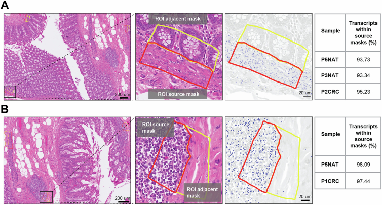
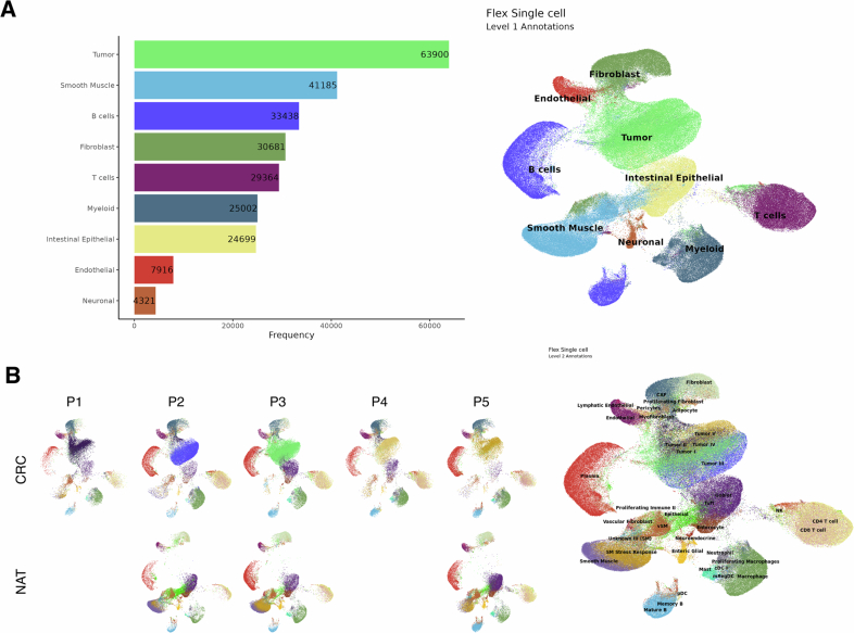
A comprehensive understanding of cellular behavior and response to the tumor microenvironment (TME) in colorectal cancer (CRC) remains elusive. Here, we introduce the high-definition Visium spatial transcriptomic technology (Visium HD) and investigate formalin-fixed paraffin-embedded human CRC samples (n = 5). We demonstrate the high sensitivity, single-cell-scale resolution and spatial accuracy of Visium HD, generating a highly refined whole-transcriptome spatial profile of CRC samples. We identify transcriptomically distinct macrophage subpopulations in different spatial niches with potential pro-tumor and anti-tumor functions via interactions with tumor and T cells. In situ gene expression analysis validates our findings and localizes a clonally expanded T cell population close to macrophages with anti-tumor features. Our study demonstrates the power of high-resolution spatial technologies to understand cellular interactions in the TME and paves the way for larger studies that will unravel mechanisms and biomarkers of CRC biology, improving diagnosis and disease management strategies.
Overview
The claims and statistics of this paper haven’t been checked yet.
Run the check — free, no account needed. It reads the open-access text and checks the sources it cites.
Integrity signals
Registry
Cites
Statistics
Figures
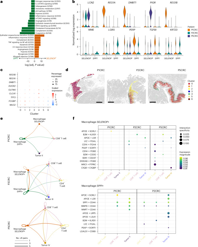
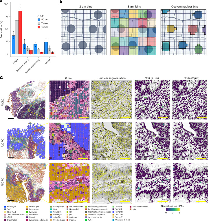
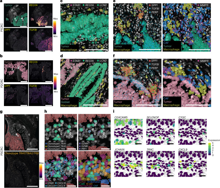
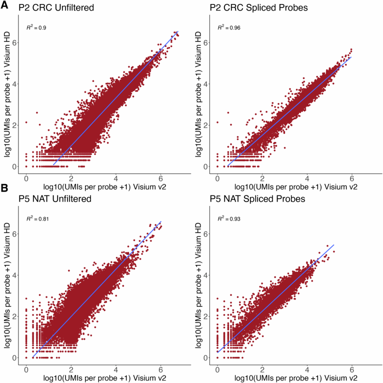
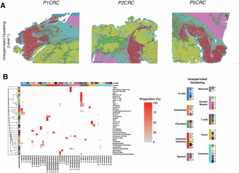
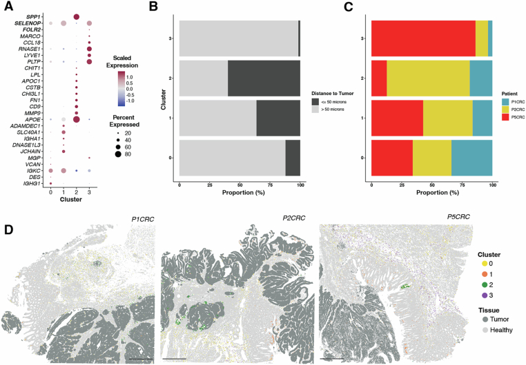
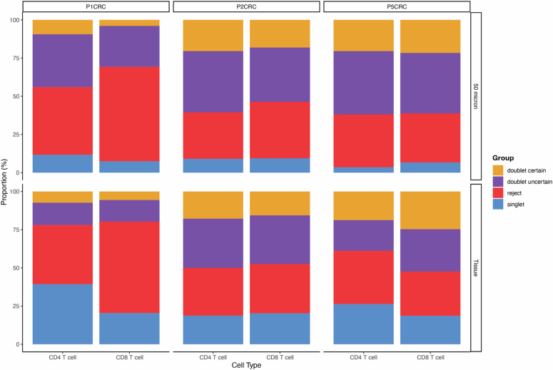
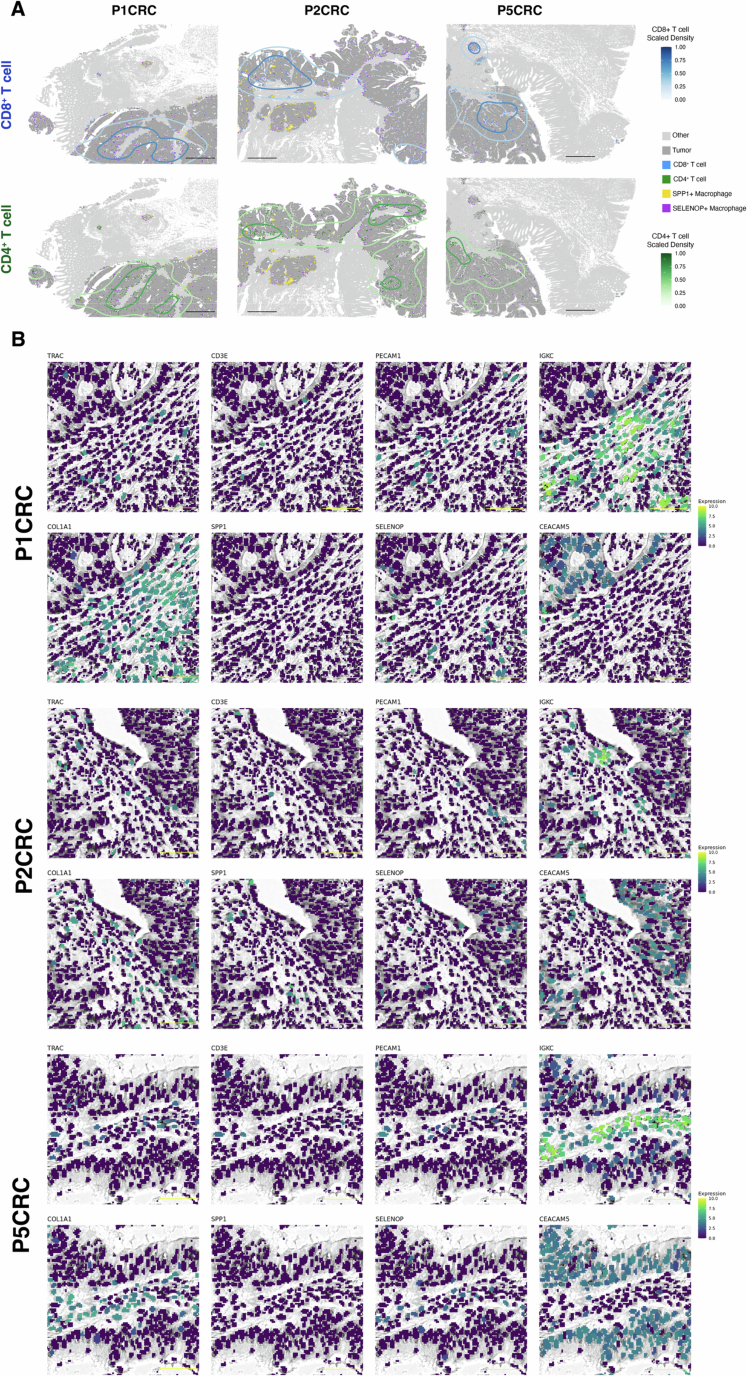
Verification
Claims the paper makes
The claims and statistics of this paper haven’t been checked yet.
Run the check — free, no account needed. It reads the open-access text and checks the sources it cites.
Claims other papers make about this one
Each is checked against this paper’s own text: does it say what the citing paper says it does?
The source backs part of the claim, not all of it — or backs it for a different population, condition or comparison.
In the citing paper
“Moreover, vascular-immune interactions were primarily inferred from histological proximity, flow cytometry and gene-expression patterns; spatial transcriptomics and single-cell multiomics approaches will be valuable for elucidating cell-type-specific crosstalk at single-cell resolution 54,55 .”
From Vascularized human skin assembloids model compartmental skin organization and immune responsiveness in vitro (2026) · cites this paper as [54]
Experimental — accuracy not yet measured
Self-check
Percentages, sums and reported differences re-computed from the paper’s own numbers.
The self-check needs the paper’s full text.
Citations
Cites
The Molecular Signatures Database Hallmark Gene Set Collection
Liberzon, A., Birger, C., Thorvaldsdóttir, H., Ghandi, M., Mesirov, J. P., & Tamayo, P. (2015) · Cell Systems · doi:10.1016/j.cels.2015.12.004 · Cited by 13,807
QuPath: Open source software for digital pathology image analysis
Bankhead, P., Loughrey, M. B., Fernández, J. A., Dombrowski, Y., McArt, D. G., Dunne, P. D., McQuaid, S., Gray, R. T., Murray, L. J., Coleman, H. G., et al. (2017) · Scientific Reports · doi:10.1038/s41598-017-17204-5 · Cited by 8,908
Dictionary learning for integrative, multimodal and scalable single-cell analysis
Hao, Y., Stuart, T., Kowalski, M. H., Choudhary, S., Hoffman, P., Hartman, A., Srivastava, A., Molla, G., Madad, S., Fernandez-Granda, C., et al. (2024) · Nature Biotechnology · doi:10.1038/s41587-023-01767-y · Cited by 5,631
The consensus molecular subtypes of colorectal cancer
Guinney, J., Dienstmann, R., Wang, X., de Reyniès, A., Schlicker, A., Soneson, C., Marisa, L., Roepman, P., Nyamundanda, G., Angelino, P., et al. (2015) · Nature Medicine · doi:10.1038/nm.3967 · Cited by 5,170
Global colorectal cancer burden in 2020 and projections to 2040
Xi, Y., & Xu, P. (2021) · Translational Oncology · doi:10.1016/j.tranon.2021.101174 · Cited by 2,633
Immunotherapy in colorectal cancer: Rationale, challenges and potential
Ganesh, K., Stadler, Z. K., Cercek, A., Mendelsohn, R. B., Shia, J., Segal, N. H., & Diaz, L. A. (2019) · Nature Reviews Gastroenterology & Hepatology · doi:10.1038/s41575-019-0126-x · Cited by 1,954 · cited 1×
“Figure 7D has been updated to show SELENOP +/ STAB1 + macrophages near REG1A + cells.”
CXCL9, CXCL10, CXCL11/CXCR3 axis for immune activation - a target for novel cancer therapy
Tokunaga, R., Zhang, W., Naseem, M., Puccini, A., Berger, M. D., Soni, S., McSkane, M., Baba, H., & Lenz, H.-J. (2018) · Cancer Treatment Reviews · doi:10.1016/j.ctrv.2017.11.007 · Cited by 1,627 · cited 2×
“Figure 7D has been updated to show SELENOP +/ STAB1 + macrophages near REG1A + cells.”
Single-Cell Analyses Inform Mechanisms of Myeloid-Targeted Therapies in Colon Cancer
Zhang, L., Li, Z., Skrzypczynska, K. M., Fang, Q., Zhang, W., O’Brien, S. A., He, Y., Wang, L., Zhang, Q., Kim, A., et al. (2020) · Cell · doi:10.1016/j.cell.2020.03.048 · Cited by 1,620 · cited 1×
“These cells were localized in microenvironments with a higher density of SELENOP +/ STAB1 + macrophages (Figure 4D) .”
Colon Cancer Survival Rates With the New American Joint Committee on Cancer Sixth Edition Staging
O'Connell, J. B., Maggard, M. A., & Ko, C. Y. (2004) · JNCI Journal of the National Cancer Institute · doi:10.1093/jnci/djh275 · Cited by 1,598 · cited 1×
“Additionally, comparing these metrics with other high-resolution methods like Stereo-seq or Seq-scope could enhance the manuscript's appeal and provide a clearer benchmark for readers.”
Robust decomposition of cell type mixtures in spatial transcriptomics
Cable, D. M., Murray, E., Zou, L. S., Goeva, A., Macosko, E. Z., Chen, F., & Irizarry, R. A. (2022) · Nature Biotechnology · doi:10.1038/s41587-021-00830-w · Cited by 1,487
High-definition spatial transcriptomics for in situ tissue profiling
Vickovic, S., Eraslan, G., Salmén, F., Klughammer, J., Stenbeck, L., Schapiro, D., Äijö, T., Bonneau, R., Bergenstråhle, L., Navarro, J. F., et al. (2019) · Nature Methods · doi:10.1038/s41592-019-0548-y · Cited by 1,150
Li, H., Courtois, E. T., Sengupta, D., Tan, Y., Chen, K. H., Goh, J. J. L., Kong, S. L., Chua, C., Hon, L. K., Tan, W. S., et al. (2017) · Nature Genetics · doi:10.1038/ng.3818 · Cited by 1,080
Qi, J., Sun, H., Zhang, Y., Wang, Z., Xun, Z., Li, Z., Ding, X., Bao, R., Hong, L., Jia, W., et al. (2022) · Nature Communications · doi:10.1038/s41467-022-29366-6 · Cited by 997
High-Spatial-Resolution Multi-Omics Sequencing via Deterministic Barcoding in Tissue
Liu, Y., Yang, M., Deng, Y., Su, G., Enninful, A., Guo, C. C., Tebaldi, T., Zhang, D., Kim, D., Bai, Z., et al. (2020) · Cell · doi:10.1016/j.cell.2020.10.026 · Cited by 966
Lineage-dependent gene expression programs influence the immune landscape of colorectal cancer
Lee, H., Hong, Y., Etlioglu, H. E., Cho, Y. B., Pomella, V., den Bosch, B. V., Vanhecke, J., Verbandt, S., Hong, H.-K., Min, J.-W., et al. (2020) · Nature Genetics · doi:10.1038/s41588-020-0636-z · Cited by 934 · cited 1×
“Cell type identification without single-cell data: While the authors claim that cell types can be identified using Visium HD data alone (line 215), the resolution, even at the bin level, still contains multiple cell types (Figure 3C).”
Hossain, M. S., Karuniawati, H., Jairoun, A. A., Urbi, Z., Ooi, D. J., John, A., Lim, Y. C., Kibria, K. M. K., Mohiuddin, A. K. M., Ming, L. C., et al. (2022) · Cancers · doi:10.3390/cancers14071732 · Cited by 920 · cited 1×
“Additionally, comparing these metrics with other high-resolution methods like Stereo-seq or Seq-scope could enhance the manuscript's appeal and provide a clearer benchmark for readers.”
Spatially organized multicellular immune hubs in human colorectal cancer
Pelka, K., Hofree, M., Chen, J. H., Sarkizova, S., Pirl, J. D., Jorgji, V., Bejnood, A., Dionne, D. A., Ge, W. H., Xu, K. H., et al. (2021) · Cell · doi:10.1016/j.cell.2021.08.003 · Cited by 917
Janesick, A., Shelansky, R. I., Gottscho, A. D., Wagner, F., Williams, S. R., Rouault, M., Beliakoff, G., Morrison, C. A., Oliveira, M. F., Sicherman, J., et al. (2023) · Nature Communications · doi:10.1038/s41467-023-43458-x · Cited by 909 · cited 1×
“The cell type annotation for Visium HD, similar to other spatial transcriptomics methods, is based on deconvolution.”
Self-citationAspects of the Tumor Microenvironment Involved in Immune Resistance and Drug Resistance
Khalaf, K., Hana, D., Chou, J. T.-T., Singh, C., Mackiewicz, A. A., & Kaczmarek, M. (2021) · Frontiers in Immunology · doi:10.3389/fimmu.2021.656364 · Cited by 551 · cited 1×
“The authors selected 50 µm as the tumor peripheral region but did not justify why this distance was chosen over others, such as 100 µm.”
The expanding vistas of spatial transcriptomics
Tian, L., Chen, F., & Macosko, E. Z. (2023) · Nature Biotechnology · doi:10.1038/s41587-022-01448-2 · Cited by 539 · cited 1×
“If, as the authors claim, it is possible to identify cell types solely with Visium HD data, then by jointly clustering different samples and removing batch effects, uniform labels can be assigned and different samples could be compared without the need for single-cell data, as well (line 217).”
Microscopic examination of spatial transcriptome using Seq-Scope
Cho, C.-S., Xi, J., Si, Y., Park, S.-R., Hsu, J.-E., Kim, M., Jun, G., Kang, H. M., & Lee, J. H. (2021) · Cell · doi:10.1016/j.cell.2021.05.010 · Cited by 520
Membrane Cholesterol Efflux Drives Tumor-Associated Macrophage Reprogramming and Tumor Progression
Goossens, P., Rodriguez-Vita, J., Etzerodt, A., Masse, M., Rastoin, O., Gouirand, V., Ulas, T., Papantonopoulou, O., Van Eck, M., Auphan-Anezin, N., et al. (2019) · Cell Metabolism · doi:10.1016/j.cmet.2019.02.016 · Cited by 502 · cited 1×
“Figure 7D has been updated to show SELENOP +/ STAB1 + macrophages near REG1A + cells.”
Joanito, I., Wirapati, P., Zhao, N., Nawaz, Z., Yeo, G., Lee, F., Eng, C. L. P., Macalinao, D. C., Kahraman, M., Srinivasan, H., et al. (2022) · Nature Genetics · doi:10.1038/s41588-022-01100-4 · Cited by 434
An osteopontin/CD44 immune checkpoint controls CD8+ T cell activation and tumor immune evasion
Klement, J. D., Paschall, A. V., Redd, P. S., Ibrahim, M. L., Lu, C., Yang, D., Celis, E., Abrams, S. I., Ozato, K., & Liu, K. (2018) · Journal of Clinical Investigation · doi:10.1172/jci123360 · Cited by 372 · cited 1×
“Figure 7D has been updated to show SELENOP +/ STAB1 + macrophages near REG1A + cells.”
Becker, W. R., Nevins, S. A., Chen, D. C., Chiu, R., Horning, A. M., Guha, T. K., Laquindanum, R., Mills, M. A., Chaı̈b, H., Ladabaum, U., et al. (2022) · Nature Genetics · doi:10.1038/s41588-022-01088-x · Cited by 333
LIANA+ Provides an All-in-One Framework for Cell–Cell Communication Inference
Dimitrov, D., Schäfer, P. S. L., Farr, E., Rodriguez-Mier, P., Lobentanzer, S., Badia-i-Mompel, P., Dugourd, A., Tanevski, J., Ramirez Flores, R. O., & Saez-Rodriguez, J. (2024) · Nature Cell Biology · doi:10.1038/s41556-024-01469-w · Cited by 263
Wang, F., Long, J., Li, L., Wu, Z., Da, T.-T., Wang, X., Huang, C., Jiang, Y., Yao, X., Ma, H., et al. (2023) · Science Advances · doi:10.1126/sciadv.adf5464 · Cited by 263 · cited 12×
“Is there any explanation for this, such as whether these cells exhibit cancerous features or other distinctive characteristics from other goblet cells not expressing REG1A?”
InfluentialOpen-ST: High-resolution spatial transcriptomics in 3D
Schott, M., León-Periñán, D., Splendiani, E., Strenger, L., Licha, J. R., Pentimalli, T. M., Schallenberg, S., Alles, J., Tagliaferro, S. S., Boltengagen, A., et al. (2024) · Cell · doi:10.1016/j.cell.2024.05.055 · Cited by 253
Refining colorectal cancer classification and clinical stratification through a single-cell atlas
Khaliq, A. M., Erdoğan, C., Kurt, Z., Turgut, S. S., Grunvald, M. W., Rand, T., Khare, S., Borgia, J. A., Hayden, D. M., Pappas, S. G., et al. (2022) · Genome biology · doi:10.1186/s13059-022-02677-z · Cited by 246
Ma, C., Rong, Y., Radiloff, D. R., Datto, M., Centeno, B. A., Bao, S., Cheng, A. W. M., Lin, F., Jiang, S., Yeatman, T. J., et al. (2008) · Genes & Development · doi:10.1101/gad.1632008 · Cited by 225
Nuclei Instance Segmentation and Classification in Histopathology Images with Stardist
WEIGERT, M. G., & Schmidt, U. (2022) · doi:10.1109/isbic56247.2022.9854534 · Cited by 198
Molecular subtyping of colorectal cancer: Recent progress, new challenges and emerging opportunities
Wang, W., Kandimalla, R., Huang, H., Zhu, L., Li, Y., Gao, F., Goel, A., & Wang, X. (2018) · Seminars in Cancer Biology · doi:10.1016/j.semcancer.2018.05.002 · Cited by 192 · cited 2×
“The claim that Visium HD controls analyte transfer through a controlled environment (line 160) is not substantiated by the provided reference.”
Geometric Sketching Compactly Summarizes the Single-Cell Transcriptomic Landscape
Hie, B., Cho, H., DeMeo, B., Bryson, B. D., & Berger, B. (2019) · Cell Systems · doi:10.1016/j.cels.2019.05.003 · Cited by 170
Reconstruction of the tumor spatial microenvironment along the malignant-boundary-nonmalignant axis
Xun, Z., Ding, X., Zhang, Y., Zhang, B., Lai, S., Zou, D., Zheng, J., Chen, G., Su, B., Han, L., et al. (2023) · Nature Communications · doi:10.1038/s41467-023-36560-7 · Cited by 169
Singh, M. P., Rai, S., Pandey, A., Singh, N. K., & Srivastava, S. (2019) · Genes & Diseases · doi:10.1016/j.gendis.2019.10.013 · Cited by 167
Polony gels enable amplifiable DNA stamping and spatial transcriptomics of chronic pain
Fu, X., Sun, L., Dong, R., Chen, J. Y., Silakit, R., Condon, L. F., Lin, Y., Lin, S., Palmiter, R. D., & Gu, L. (2022) · Cell · doi:10.1016/j.cell.2022.10.021 · Cited by 161
Biological Functions and Therapeutic Potential of Lipocalin 2 in Cancer
Santiago-Sánchez, G. S., Pita-Grisanti, V., Quiñones‐Díaz, B. I., Gumpper, K., Cruz‐Monserrate, Z., & Vivas‐Mejía, P. E. (2020) · International Journal of Molecular Sciences · doi:10.3390/ijms21124365 · Cited by 159
Ozato, Y., Kojima, Y., Kobayashi, Y., Hisamatsu, Y., Toshima, T., Yonemura, Y., Masuda, T., Kagawa, K., Goto, Y., Utou, M., et al. (2023) · Cell Reports · doi:10.1016/j.celrep.2022.111929 · Cited by 144 · cited 3×
“These cells were localized in microenvironments with a higher density of SELENOP +/ STAB1 + macrophages (Figure 4D) .”
Peng, Z., Ye, M., Ding, H., Feng, Z., & Hu, K. (2022) · Journal of Translational Medicine · doi:10.1186/s12967-022-03510-8 · Cited by 133 · cited 1×
“Is there any explanation for this, such as whether these cells exhibit cancerous features or other distinctive characteristics from other goblet cells not expressing REG1A?”
Lee, Y., Bogdanoff, D., Wang, Y., Hartoularos, G. C., Woo, J. M., Mowery, C. T., Nisonoff, H. M., Lee, D. S., Sun, Y., Lee, J., et al. (2021) · Science Advances · doi:10.1126/sciadv.abg4755 · Cited by 110
T-cell infiltration and its regulatory mechanisms in cancers: insights at single-cell resolution
Yang, W., Liu, S., Mao, M., Gong, Y., Li, X., Lei, T., Liu, C., Wu, S., & Hu, Q. (2024) · Journal of Experimental & Clinical Cancer Research · doi:10.1186/s13046-024-02960-w · Cited by 95 · cited 2×
“Figure 7D has been updated to show SELENOP +/ STAB1 + macrophages near REG1A + cells.”
Hamid, M. A., Wang, R., Yao, X., Fan, P., Li, X., Chang, X.-M., Feng, Y., Jones, S., Maldonado‐Pérez, D., Waugh, C., et al. (2019) · Cancer Immunology Research · doi:10.1158/2326-6066.cir-18-0885 · Cited by 92
Eugène, J., Jouand, N., Ducoin, K., Dansette, D., Oger, R., Deleine, C., Leveque, E., Meurette, G., Podevin, J., Matysiak‐Budnik, T., et al. (2019) · Modern Pathology · doi:10.1038/s41379-019-0322-9 · Cited by 81
Astrosini, C., Röefzaad, C., Dai, Y., Dieckgraefe, B. K., Jöns, T., & Kemmner, W. (2008) · International Journal of Cancer · doi:10.1002/ijc.23466 · Cited by 67
Investigation of colorectal cancer in accordance with consensus molecular subtype classification
Sawayama, H., Miyamoto, Y., Ogawa, K., Yoshida, N., & Baba, H. (2020) · Annals of Gastroenterological Surgery · doi:10.1002/ags3.12362 · Cited by 60 · cited 1×
“The claim that Visium HD controls analyte transfer through a controlled environment (line 160) is not substantiated by the provided reference.”
FICTURE: scalable segmentation-free analysis of submicron-resolution spatial transcriptomics
Si, Y., Lee, C., Hwang, Y., Yun, J. H., Cheng, W., Cho, C., Quirós, M., Nusrat, A., Zhang, W., Jun, G., et al. (2024) · Nature Methods · doi:10.1038/s41592-024-02415-2 · Cited by 59
Tian, X., Ye, C., Yang, Y., Guan, X., Dong, B., Zhao, M., & Hao, C. (2015) · Journal of Translational Medicine · doi:10.1186/s12967-015-0702-y · Cited by 45
Zhou, M., He, J., Li, Y., Jiang, L., Ran, J., Wang, C., Ju, C., Du, D., Xu, X., Wang, X., et al. (2023) · Cell Death and Disease · doi:10.1038/s41419-023-06067-6 · Cited by 38 · cited 1×
“Figure 7D has been updated to show SELENOP +/ STAB1 + macrophages near REG1A + cells.”
Nova-ST: Nano-patterned ultra-dense platform for spatial transcriptomics
Poovathingal, S. K., Davie, K., Borm, L. E., Vandepoel, R., Poulvellarie, N., Verfaillie, A., Corthout, N., & Aerts, S. (2024) · Cell Reports Methods · doi:10.1016/j.crmeth.2024.100831 · Cited by 37
Benchmarking spatial transcriptomics technologies with the multi-sample SpatialBenchVisium dataset
Du, M. R. M., Wang, C., Law, C. W., Amann‐Zalcenstein, D., Anttila, C. J. A., Ling, L., Hickey, P. F., Sargeant, C. J., Chen, Y., Ioannidis, L. J., et al. (2025) · Genome biology · doi:10.1186/s13059-025-03543-4 · Cited by 37 · cited 1×
“If, as the authors claim, it is possible to identify cell types solely with Visium HD data, then by jointly clustering different samples and removing batch effects, uniform labels can be assigned and different samples could be compared without the need for single-cell data, as well (line 217).”
Wen, R., Zhou, L., Peng, Z., Fan, H., Zhang, T., Jia, H., Gao, X., Hao, L., Lou, Z., Cao, F., et al. (2023) · Frontiers in Immunology · doi:10.3389/fimmu.2023.1175343 · Cited by 27 · cited 1×
“Cell type identification without single-cell data: While the authors claim that cell types can be identified using Visium HD data alone (line 215), the resolution, even at the bin level, still contains multiple cell types (Figure 3C).”
Spatially resolved transcriptomics revealed local invasion-related genes in colorectal cancer
Liu, H., Chen, S., Peng, L., Li, Z. A., Zhou, L., Liao, S.-Q., Chen, Z., Wang, Q., He, S., & Zhou, Z. (2023) · Frontiers in Oncology · doi:10.3389/fonc.2023.1089090 · Cited by 18
Dasgupta, A., Arneson‐Wissink, P. C., Schmitt, R. E., Cho, D. S., Ducharme, A. M., Hogenson, T. L., Krueger, E. W., Bamlet, W. R., Zhang, L., Razidlo, G. L., et al. (2021) · JCI Insight · doi:10.1172/jci.insight.153842 · Cited by 17 · cited 1×
“Figure 7D has been updated to show SELENOP +/ STAB1 + macrophages near REG1A + cells.”
Wu, L., Xia, W., Hua, Y., Fan, K., Lu, Y., Wang, M., Jin, Y., Zhang, W., & Pan, S. (2023) · Frontiers in Pharmacology · doi:10.3389/fphar.2023.1295442 · Cited by 10 · cited 1×
“These cells were localized in microenvironments with a higher density of SELENOP +/ STAB1 + macrophages (Figure 4D) .”
Dolan, B., & Hansson, G. C. (2021) · Elsevier eBooks · doi:10.1016/b978-0-12-821618-7.00005-5 · Cited by 3
States), 1. G. (. (2025) · Zenodo (CERN European Organization for Nuclear Research) · doi:10.5281/zenodo.15042462 · Cited by 1
Research works printed but not linked: 1
- Avraham-Davidi, I. et al. Spatially defined multicellular functional units in colorectal cancer revealed from single cell and spatial transcriptomics. eLife 14, RP104815 (2025).
Cited by
Sorted by citations
Schroeder, A. R., Loth, M., Luo, C., Yao, S., Yan, H., Zhang, D., Piya, S., Plowey, E. D., Hu, W., Clemenceau, J. R. V., et al. (2025) · Nature Methods · doi:10.1038/s41592-025-02770-8 · Cited by 37
Tian, R., Xue, Z., Chen, Y., Qi, Y., Zhang, J., Yuan, J., Ruan, D., Lin, J., Liu, J., Wang, D., et al. (2025) · Nature Communications · doi:10.1038/s41467-025-63915-z · Cited by 38
Non-invasive profiling of the tumour microenvironment with spatial ecotypes
Zhang, W., Brown, E. L., Usmani, A., Earland, N. J., Kang, M., Olelewe, C., Viswanathan, A., Chauhan, P. S., Steen, C. B., Jeon, H. S., et al. (2026) · Nature · doi:10.1038/s41586-026-10452-4 · Cited by 35
“For these samples, Visium HD bins generally yielded robust cell-type discrimination, in line with a previous report 78 .”
Thor: a platform for cell-level investigation of spatial transcriptomics and histology
Zhang, P., Chen, W., Tran, T. H., Zhou, M., Carter, K. N., Kandel, I., Li, S., Hoi, X. P., Sun, Y., Lai, L., et al. (2025) · Nature Communications · doi:10.1038/s41467-025-62593-1 · Cited by 25
Tang, J., Fan, W., Ruan, Y., Liu, X., Qiu, F., Feng, J., Huang, G., Yan, M., Wang, H., Mu, Q., et al. (2025) · Cancer Cell · doi:10.1016/j.ccell.2025.08.006 · Cited by 25
Metadata
- Authors
- Oliveira, M. F. D., Romero, J. P., Chung, M., Williams, S. R., Gottscho, A. D., Gupta, A., Pilipauskas, S. E., Mohabbat, S., Raman, N., Sukovich, D. J., Patterson, D. M., Visium HD Development Team, de Oliveira, M. F., Romero, J. P., Williams, S. R., Patterson, D. M., Taylor, S. E. B., & Taylor, S. E. B.
- Affiliations
- 10X Genomics (United States) (US)
- Venue
- Nature Genetics
- Published
- 2025-06
- Volume
- 57
- Issue
- 6
- Pages
- 1512-1523
- ISSN
- 1061-4036 (ISSN-L)
- 1546-1718
- PMC
- PMC12165841
- OpenAlex
- W4411069266
- Cited by
- 321
- References
- 56
- Citations by year
- 2026: 253 · 2025: 68
- FWCI
- 95.95
- Type
- journal article
- Language
- en
- Open access
- hybrid
- doi.org
- Licence
- Locations
- Nature Genetics (publishedVersion, cc-by-nc-nd)
- Europe PMC (PubMed Central) (publishedVersion, cc-by-nc-nd)
- PubMed Central (publishedVersion, cc-by-nc-nd)
- PubMed (publishedVersion)
- Topics
- Life Sciences › Biochemistry, Genetics and Molecular Biology › Molecular Biology › Single-cell and spatial transcriptomics
- Health Sciences › Medicine › Oncology › Cancer Immunotherapy and Biomarkers
- Life Sciences › Immunology and Microbiology › Immunology › Immune cells in cancer
- Keywords
- colorectal cancer, tumor microenvironment, spatial transcriptomics, Visium HD, macrophage subpopulations, T cell clonal expansion, immune cell interactions, formalin-fixed paraffin-embedded tissue, in situ gene expression, antitumor immunity
- MeSH terms
- Female, Humans, Macrophages, Male, T-Lymphocytes, Biomarkers, Tumor, Colorectal Neoplasms, Gene Expression Regulation, Neoplastic, Gene Expression Profiling, Single-Cell Analysis, Tumor Microenvironment, Transcriptome
About these checks
Registry, retraction, Cites and statistics results are computed, not judged, with one exception: a printed reference that no index lists is read once by a model before it counts as a citation error. Claims are extracted by a model and kept only when their quote appears verbatim in the text; each cited source is then checked, with a model’s help, for whether it says what the claim relies on. Those verdicts can be wrong — read the claim’s quote and the cited source before relying on one.
Statistics and arithmetic are recomputed, not judged.
Confidence bands are shown only for fields calibrated on our annotated test set. None is calibrated yet, so no band is shown.
What counts as a claim ›How we measure confidence ›How we check this ›
Are you an author, or do you see a problem with this page? Request a review
