

The DOI matches its Crossref record; 56 of the 56 references it cites were found in a registry or an index.
High-definition spatial transcriptomic profiling of immune cell populations in colorectal cancer
Open accessAbstract
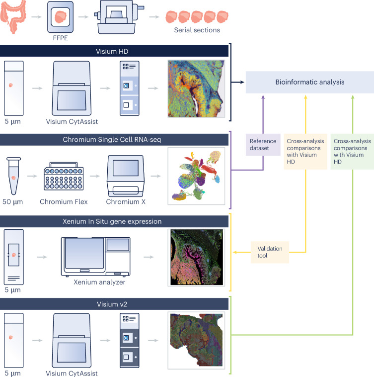
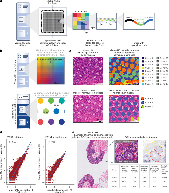
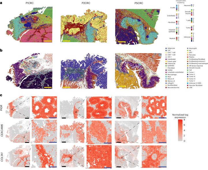
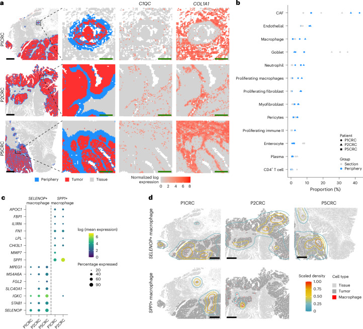
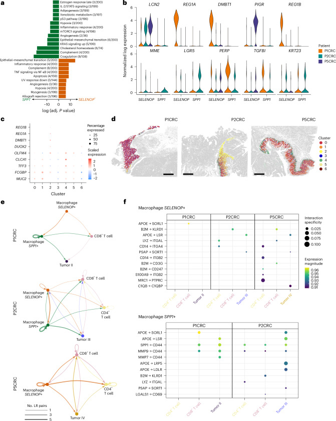
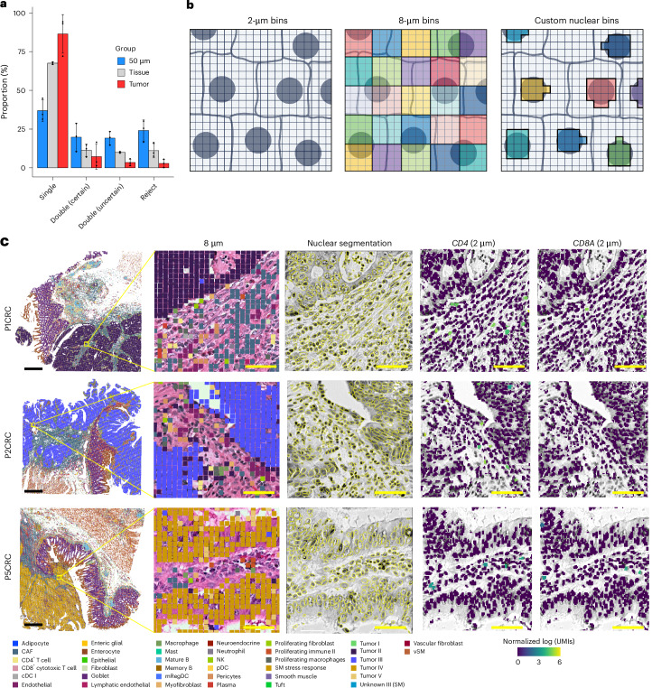
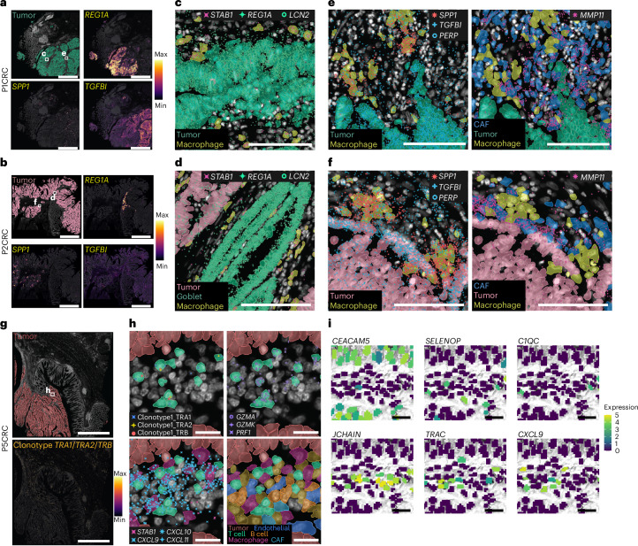
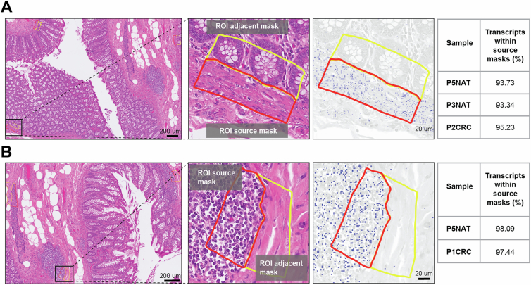
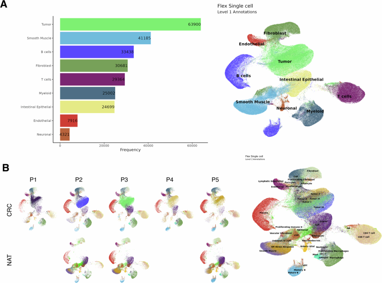
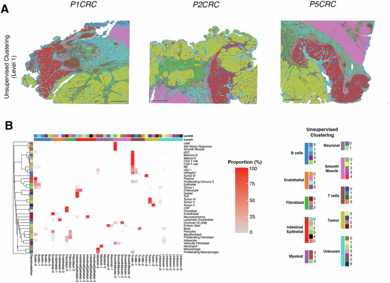
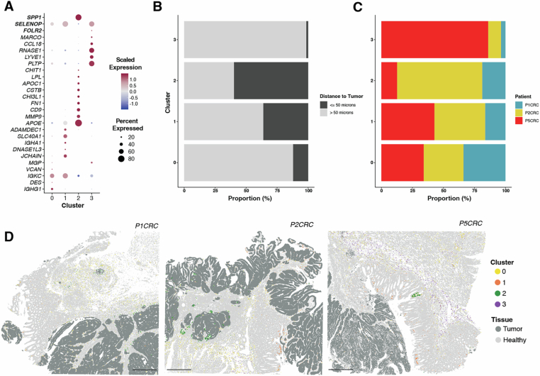
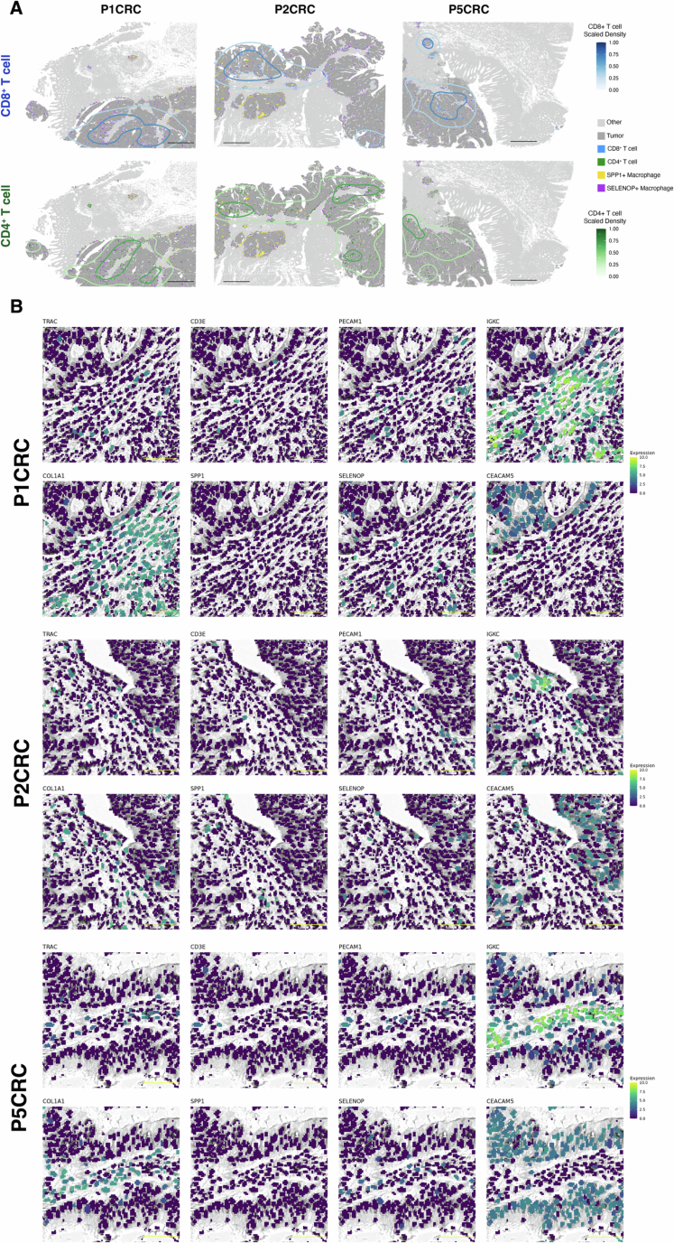
A comprehensive understanding of cellular behavior and response to the tumor microenvironment (TME) in colorectal cancer (CRC) remains elusive. Here, we introduce the high-definition Visium spatial transcriptomic technology (Visium HD) and investigate formalin-fixed paraffin-embedded human CRC samples (n = 5). We demonstrate the high sensitivity, single-cell-scale resolution and spatial accuracy of Visium HD, generating a highly refined whole-transcriptome spatial profile of CRC samples. We identify transcriptomically distinct macrophage subpopulations in different spatial niches with potential pro-tumor and anti-tumor functions via interactions with tumor and T cells. In situ gene expression analysis validates our findings and localizes a clonally expanded T cell population close to macrophages with anti-tumor features. Our study demonstrates the power of high-resolution spatial technologies to understand cellular interactions in the TME and paves the way for larger studies that will unravel mechanisms and biomarkers of CRC biology, improving diagnosis and disease management strategies.
Overview
The claims and statistics of this paper haven’t been checked yet.
Run the check — free, no account needed. It reads the open-access text and checks the sources it cites.
Integrity signals
Registry
Cites
Statistics
Figures
Verification
Claims the paper makes
The claims and statistics of this paper haven’t been checked yet.
Run the check — free, no account needed. It reads the open-access text and checks the sources it cites.
Claims other papers make about this one
Each is checked against this paper’s own text: does it say what the citing paper says it does?
The source backs part of the claim, not all of it — or backs it for a different population, condition or comparison.
In the citing paper
“Moreover, vascular-immune interactions were primarily inferred from histological proximity, flow cytometry and gene-expression patterns; spatial transcriptomics and single-cell multiomics approaches will be valuable for elucidating cell-type-specific crosstalk at single-cell resolution 54,55 .”
From Vascularized human skin assembloids model compartmental skin organization and immune responsiveness in vitro (2026) · cites this paper as [54]
Experimental — accuracy not yet measured
Self-check
Percentages, sums and reported differences re-computed from the paper’s own numbers.
The self-check needs the paper’s full text.
Citations
Cites
The Molecular Signatures Database Hallmark Gene Set Collection
Liberzon, A., Birger, C., Thorvaldsdóttir, H., Ghandi, M., Mesirov, J. P., & Tamayo, P. (2015) · Cell Systems · doi:10.1016/j.cels.2015.12.004 · Cited by 13,807
QuPath: Open source software for digital pathology image analysis
Bankhead, P., Loughrey, M. B., Fernández, J. A., Dombrowski, Y., McArt, D. G., Dunne, P. D., McQuaid, S., Gray, R. T., Murray, L. J., Coleman, H. G., et al. (2017) · Scientific Reports · doi:10.1038/s41598-017-17204-5 · Cited by 8,908
Dictionary learning for integrative, multimodal and scalable single-cell analysis
Hao, Y., Stuart, T., Kowalski, M. H., Choudhary, S., Hoffman, P., Hartman, A., Srivastava, A., Molla, G., Madad, S., Fernandez-Granda, C., et al. (2024) · Nature Biotechnology · doi:10.1038/s41587-023-01767-y · Cited by 5,631
The consensus molecular subtypes of colorectal cancer
Guinney, J., Dienstmann, R., Wang, X., de Reyniès, A., Schlicker, A., Soneson, C., Marisa, L., Roepman, P., Nyamundanda, G., Angelino, P., et al. (2015) · Nature Medicine · doi:10.1038/nm.3967 · Cited by 5,170
Global colorectal cancer burden in 2020 and projections to 2040
Xi, Y., & Xu, P. (2021) · Translational Oncology · doi:10.1016/j.tranon.2021.101174 · Cited by 2,633
Immunotherapy in colorectal cancer: Rationale, challenges and potential
Ganesh, K., Stadler, Z. K., Cercek, A., Mendelsohn, R. B., Shia, J., Segal, N. H., & Diaz, L. A. (2019) · Nature Reviews Gastroenterology & Hepatology · doi:10.1038/s41575-019-0126-x · Cited by 1,954 · cited 1×
“Figure 7D has been updated to show SELENOP +/ STAB1 + macrophages near REG1A + cells.”
CXCL9, CXCL10, CXCL11/CXCR3 axis for immune activation - a target for novel cancer therapy
Tokunaga, R., Zhang, W., Naseem, M., Puccini, A., Berger, M. D., Soni, S., McSkane, M., Baba, H., & Lenz, H.-J. (2018) · Cancer Treatment Reviews · doi:10.1016/j.ctrv.2017.11.007 · Cited by 1,627 · cited 2×
“Figure 7D has been updated to show SELENOP +/ STAB1 + macrophages near REG1A + cells.”
Single-Cell Analyses Inform Mechanisms of Myeloid-Targeted Therapies in Colon Cancer
Zhang, L., Li, Z., Skrzypczynska, K. M., Fang, Q., Zhang, W., O’Brien, S. A., He, Y., Wang, L., Zhang, Q., Kim, A., et al. (2020) · Cell · doi:10.1016/j.cell.2020.03.048 · Cited by 1,620 · cited 1×
“These cells were localized in microenvironments with a higher density of SELENOP +/ STAB1 + macrophages (Figure 4D) .”
Colon Cancer Survival Rates With the New American Joint Committee on Cancer Sixth Edition Staging
O'Connell, J. B., Maggard, M. A., & Ko, C. Y. (2004) · JNCI Journal of the National Cancer Institute · doi:10.1093/jnci/djh275 · Cited by 1,598 · cited 1×
“Additionally, comparing these metrics with other high-resolution methods like Stereo-seq or Seq-scope could enhance the manuscript's appeal and provide a clearer benchmark for readers.”
Robust decomposition of cell type mixtures in spatial transcriptomics
Cable, D. M., Murray, E., Zou, L. S., Goeva, A., Macosko, E. Z., Chen, F., & Irizarry, R. A. (2022) · Nature Biotechnology · doi:10.1038/s41587-021-00830-w · Cited by 1,487
Research works printed but not linked: 1
- Avraham-Davidi, I. et al. Spatially defined multicellular functional units in colorectal cancer revealed from single cell and spatial transcriptomics. eLife 14, RP104815 (2025).
Cited by
Sorted by citations
Schroeder, A. R., Loth, M., Luo, C., Yao, S., Yan, H., Zhang, D., Piya, S., Plowey, E. D., Hu, W., Clemenceau, J. R. V., et al. (2025) · Nature Methods · doi:10.1038/s41592-025-02770-8 · Cited by 37
Tian, R., Xue, Z., Chen, Y., Qi, Y., Zhang, J., Yuan, J., Ruan, D., Lin, J., Liu, J., Wang, D., et al. (2025) · Nature Communications · doi:10.1038/s41467-025-63915-z · Cited by 38
Non-invasive profiling of the tumour microenvironment with spatial ecotypes
Zhang, W., Brown, E. L., Usmani, A., Earland, N. J., Kang, M., Olelewe, C., Viswanathan, A., Chauhan, P. S., Steen, C. B., Jeon, H. S., et al. (2026) · Nature · doi:10.1038/s41586-026-10452-4 · Cited by 35
“For these samples, Visium HD bins generally yielded robust cell-type discrimination, in line with a previous report 78 .”
Thor: a platform for cell-level investigation of spatial transcriptomics and histology
Zhang, P., Chen, W., Tran, T. H., Zhou, M., Carter, K. N., Kandel, I., Li, S., Hoi, X. P., Sun, Y., Lai, L., et al. (2025) · Nature Communications · doi:10.1038/s41467-025-62593-1 · Cited by 25
Tang, J., Fan, W., Ruan, Y., Liu, X., Qiu, F., Feng, J., Huang, G., Yan, M., Wang, H., Mu, Q., et al. (2025) · Cancer Cell · doi:10.1016/j.ccell.2025.08.006 · Cited by 25
A spatial atlas of the healthy human liver from live donors
Yakubovsky, O., Halpern, K. B., Shir, S., Novoselsky, R., Afriat, A., Egozi, A., Barkai, T., Harnik, Y., Hoefflin, R., Kohanim, Y. K., et al. (2026) · Nature · doi:10.1038/s41586-026-10377-y · Cited by 25
McKenzie, M. E., Irac, S. E., Chen, Z., Moradi, A., Jenner, A. L., Nguyen, Q. H., & Rashidieh, B. (2026) · Seminars in Cancer Biology · doi:10.1016/j.semcancer.2026.01.002 · Cited by 24
Du, L., & Yang, H. (2025) · Journal of Translational Medicine · doi:10.1186/s12967-025-07390-6 · Cited by 23
“For example, using 10× Visium HD (sub‐spot resolution), Romero et al. profiled CRC surgical specimens and identified diverse macrophage subsets occupying distinct niches [44].”
Li, Q., Luo, W., Li, X., Xu, X., Peng, X., Cheng, L., Zhou, X., & Zheng, X. M. (2025) · Journal of Oral Microbiology · doi:10.1080/20002297.2025.2544169 · Cited by 18
He, G., He, S., Jing, X., Dai, Y., Guo, X. N., Gao, J., & Zhang, W. (2025) · Oncogene · doi:10.1038/s41388-025-03635-2 · Cited by 19
Opposing functions of distinct regulatory T cell subsets in colorectal cancer
Huang, X., Feng, D., Mitra, S., Andretta, E. S., Hooshdaran, N., Ghelani, A. P., Wang, E. Y., Frost, J. N., Lawless, V. R., Vancheswaran, A., et al. (2025) · Immunity · doi:10.1016/j.immuni.2025.11.014 · Cited by 19
“45 The patients were then segregated into high (H) and low (L) abundance groups based on the predicted abundance of each tumoral Treg population.”
Yuan, M., Jin, K., Yan, H., Schroeder, A. R., Luo, C., Yao, S., Dumoulin, B., Levinsohn, J. L., Luo, T., Clemenceau, J. R. V., et al. (2025) · Nature Cell Biology · doi:10.1038/s41556-025-01811-w · Cited by 16
“2b, this ROI captured very few enteroendocrine (EE) cells and cells from communities 1, 2, 3 and 5.”
Spatial Imbalance of Innate-like T-Cell Niches Underlies Clinical Trajectories in Psoriasis
Bonilha, C. S. (2026) · International Journal of Molecular Sciences · doi:10.3390/ijms27020715 · Cited by 12
“These limitations do not detract from the overall framework but indicate that additional work integrating higher resolution spatial methods [32], longitudinal sampling and functional interrogation will be needed to refine the mechanistic links between γδ T and MAIT niches, their layer specific…”
Deciphering cell-fate trajectories using spatiotemporal single-cell transcriptomic data
Zhang, Z., Wang, Z., Sun, Y., Shen, J., Peng, Q., Li, T., & Zhou, P. (2025) · npj Systems Biology and Applications · doi:10.1038/s41540-025-00624-9 · Cited by 16
Ji, F., Chen, H., Li, H., et al. (2026) · Journal of Hematology & Oncology · doi:10.1186/s13045-025-01773-4 · Cited by 11
Heterogeneity and plasticity of cancer-associated fibroblasts
Pramod, S., Lavon, H., Scherz‐Shouval, R., & Sherman, M. H. (2026) · Nature Cancer · doi:10.1038/s43018-026-01146-x · Cited by 14
Chu, X., Ma, J., Li, S., Wang, M., Tian, Y., & Lv, C. (2026) · Molecular Cancer · doi:10.1186/s12943-026-02570-4 · Cited by 12
“It identified spatially distinct macrophage subpopulations with opposing tumor-promoting and tumor-suppressive functions, further validated by spatial correlation with in situ gene expression signatures [311].”
Visuomotor decision-making through multifeature convergence in the larval zebrafish hindbrain
Slangewal, K., Aimon, S., Capelle, M. Q., Kämpf, F., Naumann, H., Slanchev, K., Baier, H., & Bahl, A. (2026) · Nature Communications · doi:10.1038/s41467-026-69633-4 · Cited by 12
Chen, R., Yao, Y., Qian, J., Peng, X. X., Shao, X., & Fan, X. (2025) · iMeta · doi:10.1002/imt2.70084 · Cited by 9
“…approaches, which encode spatial coordinates with barcodes (e.g., ST [12], 10× Visium [13, 14], Slide ‐ seq [15, 16], Stereo ‐ seq [17], Visium HD [18]); and (2) imaging ‐ based approaches, which directly capture transcript localization (e.g., seqFISH+ [19], MERFISH [20], STARmap [21], CosMx…”
Ge, Q., Sheng, Y., Shan, Y., Yang, Y., Jiang, H., & Wang, R. (2025) · International Journal of Molecular Sciences · doi:10.3390/ijms262211076 · Cited by 10
Engineering tumor spatial heterogeneity in vitro
Chen, C., Zhao, Z., Seah, D. H., Wu, K. Z., Mohanaselvi, S., & Fong, E. L. S. (2025) · Advanced Drug Delivery Reviews · doi:10.1016/j.addr.2025.115757 · Cited by 9
Spatial transcriptomics: integrating platforms and computational approaches for clinical insights
Song, W., Wang, D., Li, J., & Zhang, R. (2025) · Biotechnology Advances · doi:10.1016/j.biotechadv.2025.108791 · Cited by 8
Ligand–receptor hotspots in dendritic–T cell niches expose targets in autoimmunity
Bonilha, C. S. (2026) · Translational research · doi:10.1016/j.trsl.2026.01.006 · Cited by 8
Immune triad microenvironments define coordinated activation architecture in psoriatic disease
Bonilha, C. S. (2026) · International Immunopharmacology · doi:10.1016/j.intimp.2026.116337 · Cited by 8
Li, L., Wang, T., Liang, Z., Yu, H., Ma, S., Yu, L., & Huang, Y. (2026) · Nature Communications · doi:10.1038/s41467-026-70528-7 · Cited by 8
Microbe-driven immune suppression in colorectal cancer: the Fusobacterium nucleatum playbook
Wuyi, W., & Tao, P. (2026) · Frontiers in Immunology · doi:10.3389/fimmu.2026.1795703 · Cited by 12
Single-cell TCR mapping reveals spatially coordinated T cell states in head and neck cancer
McCord, K. A., Kan, E., Hyslop, S., Xia, A. Y., Hofferek, C. J., Lewis, J. S., Wieland, A., Hernandez, D. J., Sandulache, V. C., & Hudson, W. H. (2026) · Science Immunology · doi:10.1126/sciimmunol.aec3133 · Cited by 8
Uncovering spatially resolved functional genomics with CRISPR screen sequencing
Zhang, H., Zhang, Z., Wang, P., Xu, T., Chen, X., Zhao, Y., Lin, S., Cai, W., Ren, P., Luo, C., et al. (2026) · Cell · doi:10.1016/j.cell.2026.04.049 · Cited by 9
Seq-Scope-eXpanded: spatial omics beyond optical resolution
Anacleto, A., Cheng, W., Feng, Q., Park, A., Cho, C., Hwang, Y., Kim, Y., Si, Y., Hsu, J.-E., Zhao, Q., et al. (2026) · Nature Communications · doi:10.1038/s41467-026-69346-8 · Cited by 11
Deutsch-Williams, R. J., Xie, Y., Rabinowitz, Z., Goemans, M. A., Wirapati, P., Vinegoni, C., Carlson, J. C., Pittet, M. J., & Weissleder, R. (2025) · Science Advances · doi:10.1126/sciadv.aeb5810 · Cited by 7
Prostate cancer cells converge to an inflammatory-like state upon metastatic dissemination
Keshavarzian, T., Furlano, K., Grillo, G., Mout, L., Arlidge, C., Hasan, F., Nand, A., Mikutenaite, M., Karadoulama, E., Goyal, A., et al. (2025) · Nature Communications · doi:10.1038/s41467-025-67856-5 · Cited by 7
“Whether TME and malignant epithelial populations with similar phenotypes congregate in distinct spatial pockets 53,54 and if local cellular interactions result in genetically distinct cells to adopt shared phenotypes remain to be explored.”
Yousef, N., Wu, W., Alajaji, S. A., Mahadevan, A., Sultan, A. S., Molloy, E. K., & Nguyen, J. T. (2026) · Head and Neck Pathology · doi:10.1007/s12105-025-01876-x · Cited by 7
Harnessing the potential of spatial statistics for spatial omics data with pasta
Emons, M., Gunz, S., Crowell, H. L., Mallona, I., Kuehl, M. B., Furrer, R., & Robinson, M. D. (2025) · Nucleic Acids Research · doi:10.1093/nar/gkaf870 · Cited by 11
“Similarly, HTS-based data with subcellular resolution (e.g. Visium HD [ 12 ]) can likewise be interpreted as a regular lattice of (sub)-cellular locations or irregular lattice from segmentation [ 39 ] or the reconstructed cells can then be approximated by their centroids as points and further…”
Miller, A. F., Potluru, S. S., Thormann, S., Wang, Y., & Nguyen, J. (2025) · Advanced Drug Delivery Reviews · doi:10.1016/j.addr.2025.115722 · Cited by 7
Wang, Z., Zeng, Y., Tan, Z., Chen, Y., Huang, X., Zhao, H., Lin, Z., & Yang, C. (2025) · Proceedings of the National Academy of Sciences · doi:10.1073/pnas.2503952122 · Cited by 6
Zhang, D., Zou, X., He, M., Sun, S., Yu, A., Shi, J., Ouyang, T., Lin, X., Yang, H., Bai, L., et al. (2025) · Cellular and Molecular Gastroenterology and Hepatology · doi:10.1016/j.jcmgh.2025.101701 · Cited by 6
“15 This approach preserves native spatial context and minimizes dissociation-induced artifacts (eg, cellular stress, apoptosis, and selective cell loss).”
sCellST predicts single-cell gene expression from H& E images
Chadoutaud, L., Lerousseau, M., Herrero-Saboya, D., Ostermaier, J., Fontugne, J., Barillot, E., & Walter, T. (2026) · Nature Communications · doi:10.1038/s41467-025-67965-1 · Cited by 12
Bonilha, C. S. (2026) · Journal of Molecular Medicine · doi:10.1007/s00109-026-02680-y · Cited by 6
Spatially resolving cancer: from cell states to therapy
Pei, G., Liu, Y., & Wang, L. (2025) · Trends in cancer · doi:10.1016/j.trecan.2025.09.002 · Cited by 7
Mapping multipathology via spatial omic integration
Palaganas, R. G., Sidiropoulos, D. N., Morris, M., & Stein-O’Brien, G. (2025) · Current Opinion in Biotechnology · doi:10.1016/j.copbio.2025.103398 · Cited by 6
Qi, H., Lv, G., Zhang, S., Xue, R., & Liu, M. (2026) · Cancer Letters · doi:10.1016/j.canlet.2026.218391 · Cited by 8
Spatial multi-omics landscape of colorectal cancer macro- and micrometastases
Liu, Y., Jadhav, A. S., Pan, Y., Liao, J., Khanduri, I., Liu, Y., Katkhuda, R., Lü, W., Cho, K. S., Tong, Z., et al. (2026) · Cancer Cell · doi:10.1016/j.ccell.2026.06.009 · Cited by 5
On metrics for subpopulation detection in single-cell and spatial omics data
Luo, S., Germain, P., von Meyenn, F., & Robinson, M. D. (2025) · Nucleic Acids Research · doi:10.1093/nar/gkaf921 · Cited by 6
“We used the Visium HD colon cancer dataset from Oliveira et al. [ 14 ] for benchmarking (see Supplementary Fig.”
Spatial transcriptomics in bone research: navigating hype and hurdles
Sokolowskei, D., Shah, A., Trostle, A. J., Juan, C., Sammarco, M. C., & Tower, R. J. (2025) · Pathology · doi:10.1016/j.pathol.2025.10.003 · Cited by 6
SpatialFusion: A lightweight multimodal foundation model for pathway-informed spatial niche mappingPreprint
Yates, J., Shavakhi, M., Choueiri, T. K., Allen, E. M. V., & Uhler, C. (2026) · bioRxiv (Cold Spring Harbor Laboratory) · doi:10.64898/2026.03.16.712056 · Cited by 5
“…pathologist annotations, and include distinct conditions that may drive shifts in tissue organization, we next applied the method to a Visium HD colorectal cancer (CRC) cohort comprising three tumor samples (P1CRC, P2CRC, P5CRC) and two normal-adjacent tissue (NAT) samples (P3NAT, P5NAT) 28 .”
Cheng, H.-C., Chang, C., Wu, C., Su, P., Hwang, D., Wu, C.-L., Hsu, S. N., Lim, S. Y., Liu, T., McGrath, J. A., et al. (2026) · Dermatologica Sinica · doi:10.4103/ds.ds-d-25-00298 · Cited by 4
Cancer in 4D: toward Spatiotemporal Hallmark Ecosystems
Sibai, M., Zacharias, M., McGranahan, N., Jamal‐Hanjani, M., & Porta‐Pardo, E. (2026) · Trends in cancer · doi:10.1016/j.trecan.2026.01.004 · Cited by 5
Zhou, J., Shi, G., Dai, S., Wu, S., Qian, F., Wu, Z., Zhang, J., Yang, Y., Xiao, B., Lu, Z., et al. (2026) · Cancer Letters · doi:10.1016/j.canlet.2026.218613 · Cited by 4
Spatial omics of immunity: Mapping cellular landscapes in tissue microenvironments
See, J.-E., Liu, Y., & Plummer, J. (2026) · Current Opinion in Immunology · doi:10.1016/j.coi.2026.102790 · Cited by 4
A practical protocol of processing mineralized tissue for Visium spatial transcriptomics
Miao, J., Ma, M., Guo, Y., Qin, L., & Yao, L. (2025) · Mechanobiology in Medicine · doi:10.1016/j.mbm.2025.100163 · Cited by 4
Identifying cell-type-specific spatially variable genes with ctSVG
Zhuang, H., Shang, X., Hou, W., & Ji, Z. (2025) · Genome biology · doi:10.1186/s13059-025-03870-6 · Cited by 4
Zhang, X., Yu, Z., Hao, G., Yao, Q., Hu, Y., Wang, F., Chen, X., Liu, L., Wong, K., & Li, X. (2026) · Nature Communications · doi:10.1038/s41467-026-68500-6 · Cited by 4
“…the performance of GARDEN in analyzing large-scale ST data at single-cell resolution, we applied it to colorectal cancer (CRC) and mouse embryo datasets generated using 10x Visium HD technology, further evaluating its ability to resolve complex tissue structures and multicellular environments 47 .”
Maspero, D., Marteletto, G., Lapi, F., Galuzzi, B. G., Ruano, I., Vandenbosch, B., Yin, K., Tejpar, S., Graudenzi, A., Heyn, H., et al. (2026) · npj Systems Biology and Applications · doi:10.1038/s41540-026-00654-x · Cited by 4
“To further exclude the possibility that lactate consumption in CRC metastases is a modeling artifact, we tested spFBA on an independent CRC dataset generated using a different ST technology (i.e., 10x Visum HD) 43 .”
Reshaping the colorectal cancer immune microenvironment: insights from single-cell and spatial omics
Zhang, S., Fu, Q., Yuan, X., Wang, S., Liu, C., Zhang, C., (70818), B. L., & Gong, Y. (2026) · Cancer Cell International · doi:10.1186/s12935-026-04245-y · Cited by 4
“To address this, high-density innovations like Visium HD have pushed the boundaries toward near single-cell granularity (2 × 2 μm), though their efficacy remains contingent upon tissue integrity and mRNA diffusion dynamics [122].”
Shi, T., Guo, Y., Tang, X., Liu, Z., Guo, Q., Fang, T., Xu, S., & Zhuang, Z. (2026) · Neoplasia · doi:10.1016/j.neo.2026.101348 · Cited by 9
“Progenitor or stem-like exhausted T cells may be more clinically relevant when they are maintained in lymphoid-like or antigen-presenting niches, where they can receive survival and differentiation signals and generate effector progeny after checkpoint blockade [45,113].”
Zambon, M., Mantica, F., Dias, M., Frazer, J., & Irimia, M. (2025) · Current Opinion in Genetics & Development · doi:10.1016/j.gde.2025.102387 · Cited by 4
Zhao, J., Zhang, X., Wang, G., Lin, Y., Liu, T., Chang, R. B., & Zhao, H. (2026) · Nature Genetics · doi:10.1038/s41588-026-02579-x · Cited by 7
“We also confirmed robust imputation accuracy across multiple metrics and cross-validation schemes (Supplementary Note 4, Fig.”
InfluentialMulti-Omics Meets Premalignancy: Paving the Way for Early Prevention of Cancer
Zhang, F., Zhou, Z., Zhang, P., & Li, S. (2025) · Research · doi:10.34133/research.0930 · Cited by 4
Zhang, L., Zhu, Y., & Zhang, S. (2025) · Communications Biology · doi:10.1038/s42003-025-09340-w · Cited by 4
Integrated single‑cell and spatial transcriptomics reveal the colorectal cancer microenvironment
Yang, R., Hu, S., Li, K., Wang, H., Zhou, X., Han, T., & Guan, L. (2026) · Critical Reviews in Oncology/Hematology · doi:10.1016/j.critrevonc.2026.105311 · Cited by 5
Zhang, D., Tao, P., Li, J., et al. (2026) · Gut · doi:10.1136/gutjnl-2025-337344 · Cited by 3
T Cell Receptor Repertoires Across the Continuum of Vascular, Myocardial, and Age‐Related Diseases
Richter, L., Dias-Ferreira, J., Ramos, G. C., & Ashour, D. (2026) · Immunological Reviews · doi:10.1111/imr.70125 · Cited by 3
“Additionally, spatial barcoding of TCR transcripts can now pinpoint local clonal expansion by mapping specific T cells to their tissue microenvironments [234, 235].”
CD4 + T cells impair tumor growth through IL-3 and TNF-dependent vascular damage
Lian, Q., Nie, J., Singh, J., Chen, Q., Matta, J. L., Chan, W., Balmaceno-Criss, M., Vacchio, M. S., Yu, W., Clark, A. D., et al. (2026) · Science · doi:10.1126/science.ads7910 · Cited by 5
Multimodal spatial omics: From data acquisition to computational integration
Işık, E. B., Usta, Y. H., Riazi, M., Liu, H., Roach, W., Zhou, H., Nicolaou, A., Rattray, M., & Georgaka, S. (2026) · Patterns · doi:10.1016/j.patter.2026.101592 · Cited by 5
“Recently, the 10 × Visium HD platform further advanced ST to near single-cell resolution by introducing slides with increased oligonucleotide barcode density.”
An, J., Hu, E., Shen, Q., Zhu, J., Zhu, J., Liu, N., Lin, Q., Núñez, L., Bello, S., Ren, Y., et al. (2026) · Med · doi:10.1016/j.medj.2026.101214 · Cited by 3
Chung, B. K., Jördens, M. S., Øgaard, J., Reims, H. M., Flateland, B., Luedde, T., Jiang, X., Karlsen, T. H., & Melum, E. (2025) · Hepatology · doi:10.1097/hep.0000000000001432 · Cited by 3
Zhang, X., Zhang, M., Xu, Y., Song, Y., Yang, X., Wu, Y., Zhao, X., Ran, D., Liu, X., Huang, H., et al. (2025) · Genome biology · doi:10.1186/s13059-025-03900-3 · Cited by 4
“…spatial co-detection technologies exhibit several limitations, including a lack of sub-cellular resolution in Visium, DBiT-seq and SM-Omics [7], unable to achieve unbiased transcriptomic profiling in MERFISH [11], and the necessity for costly equipment in GeoMx DSP [12] and Visium HD [13, 14].”
PreTSA: computationally efficient modeling of temporal and spatial gene expression patterns
Zhuang, H., & Ji, Z. (2026) · Genome biology · doi:10.1186/s13059-026-03994-3 · Cited by 4
Xu, Y., Huang, Z., Zhang, Y., Gong, M., Wang, Z., Guo, P., Zhang, F., Yang, J., Liang, G., Dong, L., et al. (2026) · Nature Communications · doi:10.1038/s41467-026-70645-3 · Cited by 3
“…correlation structures between neighboring spots and cell-subpopulation-speci fi c capture ef fi ciency variations, we generated synthetic spots by spa-tiallyaggregatingsegmentedsingle-celltranscriptomesfromthree10x Genomics Visium HD CRC tissue sections 31 into 49 µ m×49 µ m bins (Supplementary Fig.”
InfluentialQiu, C., Li, Y., Gong, Y., Sherman, W., Tian, D., Lin, W., Pan, Z., Afolabi, B., Thumbigere‐Math, V., Su, K.-J., et al. (2026) · Bone Research · doi:10.1038/s41413-026-00520-w · Cited by 3
“The high cellular heterogeneity of bone and muscle, along with the limited resolution of Visium platforms that capture transcripts from multiple neighboring cells per spot rather than single-cell pro fi les, 33 – 35 poses challenges for accurate cell-type characterization.”
Viitala, E. W., Salminen, E. S., Miihkinen, M., Chalkidi, N., Stavropoulou, A., Lemmetyinen, T. T., Päivinen, P., Kaprio, T., Hagström, J., Haglund, C. H., et al. (2025) · bioRxiv (Cold Spring Harbor Laboratory) · doi:10.1101/2025.07.29.667377 · Cited by 3
Chaib, M., Aminu, M., Herbrich, S. M., Arabi, M., Xuan, Y., Basi, A., Casasent, T. D., Macaluso, M. D., Hu, K. H., Gubin, M. M., et al. (2025) · bioRxiv (Cold Spring Harbor Laboratory) · doi:10.64898/2025.12.24.696419 · Cited by 3
Swinderman, J. T., Tung, P., Winters, A. F., Goudy, L., Wilson, C. M., Bounds, L. R., Teyssier, N., Agrawal, A., Dobin, A., Hua, T., et al. (2026) · bioRxiv (Cold Spring Harbor Laboratory) · doi:10.64898/2026.02.04.703058 · Cited by 4
“Alternatively, probe-based methods for single cell transcriptome analysis that leverage custom DNA probe pools are emerging 14,15 .”
Subcellular mRNA localization patterns across tissues resolved with spatial transcriptomics
Novoselsky, R., Golani, O., Barkai, T., Kedmi, M., Goliand, I., Fine, M., Kent, I., Nachmany, I., & Itzkovitz, S. (2026) · Nature Communications · doi:10.1038/s41467-026-72156-7 · Cited by 5
“Published VisiumHD data of human colon 25 were imported, analyzed, and visualized in HiVis.”
Connectogenomics: edges-first spatial biology
Yasser, R., Yu, M., Qian, N., Chang, H., & Weinstein, J. A. (2025) · Trends in biotechnology · doi:10.1016/j.tibtech.2025.10.012 · Cited by 2
IEL–IEC circuit in barrier immunity and beyond
Che, S., Wang, Y., Wang, Z., Yin, Y., & Wu, M. (2025) · Trends in Immunology · doi:10.1016/j.it.2025.11.004 · Cited by 3
Spatial AI in cancer: mapping immune evasion topology through multi-modal omics and deep learning
Lang, L., Cui, Y., Wang, H., & Xiao, Y. (2026) · Frontiers in Oncology · doi:10.3389/fonc.2026.1762907 · Cited by 3
“Analyses from the SpatialBenchVisium dataset highlight that FFPE sections processed with optimized probe chemistry and the CytAssist alignment retain higher detection sensitivity for low-abundance transcripts, particularly immune regulatory genes (35, 63, 64).”
Zhen, K., Du, Z., Wang, P. Y., Liu, S., Zhu, J. M., Qiao, Z., & Jiang, H. (2026) · Clinical and Translational Medicine · doi:10.1002/ctm2.70650 · Cited by 2
Zhang, P., Gao, C., Hua, K., Zhang, Z., & Li, S. (2026) · Nature Methods · doi:10.1038/s41592-026-03070-5 · Cited by 5
“2c) and the single-cell-level 10x Visium HD datasets 16 (Fig.”
The applications of single-cell and spatial transcriptomics in neuroscience and brain disorders
Wang, Q., & Dwivedi, Y. (2026) · Neuroscience & Biobehavioral Reviews · doi:10.1016/j.neubiorev.2026.106721 · Cited by 3
Dissecting and steering cell dynamics using spatially-informed RNA velocity with veloAgent
Raghavan, V., Yoon, B., Fonseca, G. J., Li, Y., & Ding, J. (2026) · Molecular Systems Biology · doi:10.1038/s44320-026-00213-w · Cited by 3
“This loss of spatial fi delity prevents us from uncovering how tumors invade, how immune cells coordinate in tissue, or how stem cells rebuild organs (Hunter et al, 2021; Oliveira et al, 2025; Tang et al, 2025).”
Wang, J., He, Q., Hu, J., et al. (2026) · Journal of Translational Medicine · doi:10.1186/s12967-026-08232-9 · Cited by 2
“To validate the spatial architecture of these cell states within intact tissue, we reanalyzed a single-cell resolution Xenium in situ transcriptomic dataset [17].”
Spatial transcriptomics in Alzheimer's disease: technologies, challenges and discoveries
Shi, C. H., Piña-Crespo, J., Yip, K. Y., & Huang, T. Y. (2026) · Molecular Neurodegeneration Advances · doi:10.1186/s44477-026-00036-x · Cited by 3
“In 2024, 10 × Genomics released Visium HD [40] which offers increased resolution (2 μm × 2 μm barcoded squares).”
Benchmarking cell-type deconvolution in cross-platform transcriptomic data
Singh, A., Cakmak, P., Lun, J. H., et al. (2026) · Genome biology · doi:10.1186/s13059-026-04222-8 · Cited by 2
Michielsen, L., Prjibelski, A. D., Foord, C., Spiegelman, Y., Kim, T., Hu, W., Jarroux, J., Hsu, J., Pfeil, R., Zhang, X., et al. (2026) · Nature Methods · doi:10.1038/s41592-026-03211-w · Cited by 6
“We also performed one ONT long-read run on cDNAs enriched for 3,378 genes related to various brain diseases and synaptic function (from now on called Sample 1 (3.3 K)), rather than for the whole exome (Supplementary Tables 1–8).”
InfluentialA Spatial and Temporal Transcriptomic Atlas of Mouse Intestinal RegenerationPreprint
Ofer, R., Tran, T., He, P., Qiu, X., Hu, X., Zachariah, E., Krier, C., Saxena, A., Yang, J., & Verzi, M. P. (2025) · bioRxiv (Cold Spring Harbor Laboratory) · doi:10.1101/2025.08.06.668727 · Cited by 2
Yu, X., Wan, X., Tian, L., Chen, Y., Liu, Y., Yu, T., Yang, C., & Xiao, J. (2025) · bioRxiv (Cold Spring Harbor Laboratory) · doi:10.1101/2025.08.21.671426 · Cited by 3
“Nevertheless, with the increasing availability and adoption of commercial platforms offering single-cell or near-single-cell resolution (e.g., Xenium, Visium HD [91]), NicheScope is well positioned to leverage these technological advances and facilitate high-512 resolution spatial data mining.”
Cao, J., Zeng, J., Meng, Y., Huang, S., Zhang, J., Fang, X., Guo, Y., Shi, W., Liu, Y., Zheng, Z., et al. (2026) · Science Advances · doi:10.1101/2025.09.12.675967 · Cited by 2
Benchmarking cell-type deconvolution in cross-platform transcriptomic dataPreprint
Singh, A., Cakmak, P., Lun, J. H., Macas, J., Plate, K. H., Reiss, Y., Schupp, J., & Imkeller, K. (2025) · bioRxiv (Cold Spring Harbor Laboratory) · doi:10.1101/2025.11.24.690141 · Cited by 2
Integrative spatial multi-omics reveals prognostic tumor niches in female genital tumorsPreprint
Tian, X., Lin, S., Wu, Y., He, Y., Zhang, Z., Zhao, Y., Zhang, P., Zhao, L., Yang, W., Ye, M., et al. (2025) · bioRxiv (Cold Spring Harbor Laboratory) · doi:10.1101/2025.11.24.690313 · Cited by 2
LLM-scCurator: Data-centric feature distillation for zero-shot cell-type annotationPreprint
Furudate, K., & Takahashi, K. (2025) · bioRxiv (Cold Spring Harbor Laboratory) · doi:10.64898/2025.12.28.696778 · Cited by 2
A lightweight, ultrafast and general embedding framework for large-scale spatial omics dataPreprint
Dai, B., Liang, Y., Yi, L., Hu, P., Song, Y., Qian, B., He, M., Wang, L., Yuan, Z., & Zuo, Y. (2026) · bioRxiv (Cold Spring Harbor Laboratory) · doi:10.64898/2026.02.04.703814 · Cited by 2
Pixel2Gene enables histology-guided reconstruction and prediction of spatial gene expressionPreprint
Yao, S., Schroeder, A. R., Jiang, S., Im, S., Park, J. H., Dumoulin, B., Hwang, T. H., Suszták, K., & Li, M. (2026) · bioRxiv (Cold Spring Harbor Laboratory) · doi:10.64898/2026.02.21.707168 · Cited by 2
Tang, G., Zhang, Z., Pang, B., Li, R., Liu, Y., Cai, H., Wang, W., Chen, C., Ou, Y., & Yang, Q. (2025) · Biochemistry and Biophysics Reports · doi:10.1016/j.bbrep.2025.102198 · Cited by 1
“SELENOP, primarily a selenium transporter and antioxidant [8], is implicated in modulating inflammatory responses and immune cell activity within the tumor microenvironment, linking selenium/redox metabolism to immune regulation [9,10].”
Mu, H., Zhu, Y., Yan, F., Lv, C., Tu, X., He, J., Wang, Z., Zeng, Y., Liu, Z., Chen, J., et al. (2025) · Discover Oncology · doi:10.1007/s12672-025-03524-w · Cited by 1
Spatially resolved airway niches: mapping epithelial–immune microenvironments in asthma
Sakkal, M., Jarab, A. S., & Meslamani, A. Z. A. (2025) · Expert Review of Respiratory Medicine · doi:10.1080/17476348.2025.2582244 · Cited by 2
“Although the referenced study focused on cancer, it highlights the method’s capacity to clarify immune – epithelial interactions, relevant to airway disease contexts as well [21].”
Dissecting Normal and Abnormal Human Kidney Development Using Multiomics
Klomp, L. S., Mavrommatis, L., Arcolino, F. O., Lantermans, H. C., Levtchenko, E. N., Kuppe, C., & Westland, R. (2025) · Journal of the American Society of Nephrology · doi:10.1681/asn.0000000951 · Cited by 1
A Single-Cell Omics Technical Guide for Advancing Neuropsychiatric Research
Casmey, K., Zimmermann, M., Xie, Y., Codeluppi-Arrowsmith, S. A., & Turecki, G. (2025) · Genes · doi:10.3390/genes16121394 · Cited by 3
“Current sequencing-based spatial transcriptomic technologies include Visium/VisiumHD [175,205], DBiT-seq/xDBiT [206,207], Slide-seq/seqV2 [208,209], Stereo-seq/V2 [210,211], GeoMx Digital Spatial Profiler (GeoMx DSP) [212], Seq-Scope [213], HDST [214], and Pixel-seq [215].”
Xu, Y., Sargeant, C. J., You, Y., You, Y., Su, S., Wang, C., Tian, L., Chen, Y., & Ritchie, M. E. (2025) · NAR Genomics and Bioinformatics · doi:10.1093/nargab/lqaf167 · Cited by 2
“…resolution improves the biological insights gained was recently demonstrated in the study of normal colon mucosa, with Visium HD able to capture the characteristic hole pattern via the binning of barcoded squares, while the lower resolution Visium data could not recover this expected detail [ 6 ].”
Zhu, J., Zhu, T., Cai, Y., et al. (2025) · Frontiers in Oncology · doi:10.3389/fonc.2025.1701368 · Cited by 1
Metadata
- Authors
- Oliveira, M. F. D., Romero, J. P., Chung, M., Williams, S. R., Gottscho, A. D., Gupta, A., Pilipauskas, S. E., Mohabbat, S., Raman, N., Sukovich, D. J., Patterson, D. M., Visium HD Development Team, de Oliveira, M. F., Romero, J. P., Williams, S. R., Patterson, D. M., Taylor, S. E. B., & Taylor, S. E. B.
- Affiliations
- 10X Genomics (United States) (US)
- Venue
- Nature Genetics
- Published
- 2025-06
- Volume
- 57
- Issue
- 6
- Pages
- 1512-1523
- ISSN
- 1061-4036 (ISSN-L)
- 1546-1718
- PMC
- PMC12165841
- OpenAlex
- W4411069266
- Cited by
- 321
- References
- 56
- Citations by year
- 2026: 253 · 2025: 68
- FWCI
- 95.95
- Type
- journal article
- Language
- en
- Open access
- hybrid
- doi.org
- Licence
- Locations
- Nature Genetics (publishedVersion, cc-by-nc-nd)
- Europe PMC (PubMed Central) (publishedVersion, cc-by-nc-nd)
- PubMed Central (publishedVersion, cc-by-nc-nd)
- PubMed (publishedVersion)
- Topics
- Life Sciences › Biochemistry, Genetics and Molecular Biology › Molecular Biology › Single-cell and spatial transcriptomics
- Health Sciences › Medicine › Oncology › Cancer Immunotherapy and Biomarkers
- Life Sciences › Immunology and Microbiology › Immunology › Immune cells in cancer
- Keywords
- colorectal cancer, tumor microenvironment, spatial transcriptomics, Visium HD, macrophage subpopulations, T cell clonal expansion, immune cell interactions, formalin-fixed paraffin-embedded tissue, in situ gene expression, antitumor immunity
- MeSH terms
- Female, Humans, Macrophages, Male, T-Lymphocytes, Biomarkers, Tumor, Colorectal Neoplasms, Gene Expression Regulation, Neoplastic, Gene Expression Profiling, Single-Cell Analysis, Tumor Microenvironment, Transcriptome
About these checks
Registry, retraction, Cites and statistics results are computed, not judged, with one exception: a printed reference that no index lists is read once by a model before it counts as a citation error. Claims are extracted by a model and kept only when their quote appears verbatim in the text; each cited source is then checked, with a model’s help, for whether it says what the claim relies on. Those verdicts can be wrong — read the claim’s quote and the cited source before relying on one.
Statistics and arithmetic are recomputed, not judged.
Confidence bands are shown only for fields calibrated on our annotated test set. None is calibrated yet, so no band is shown.
What counts as a claim ›How we measure confidence ›How we check this ›
Are you an author, or do you see a problem with this page? Request a review
