

The DOI matches its Crossref record; 124 of the 125 references it cites were found in a registry or an index.
A tool for mapping microglial morphology, morphOMICs, reveals brain-region and sex-dependent phenotypes
Open accessPublished version of a preprint
Abstract
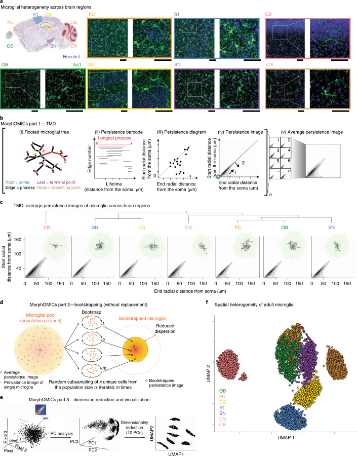
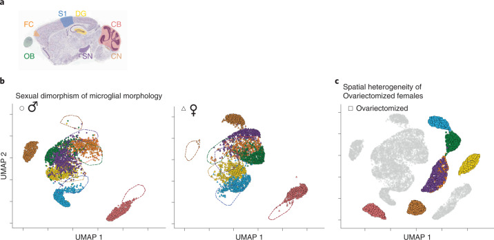
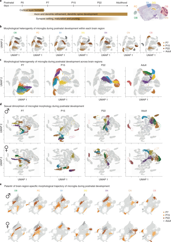
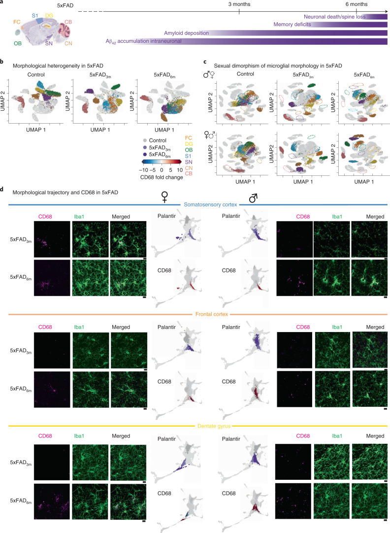
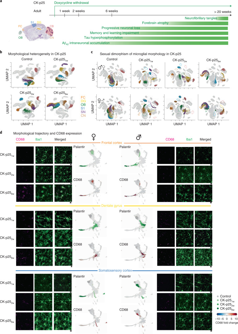
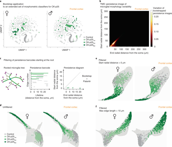
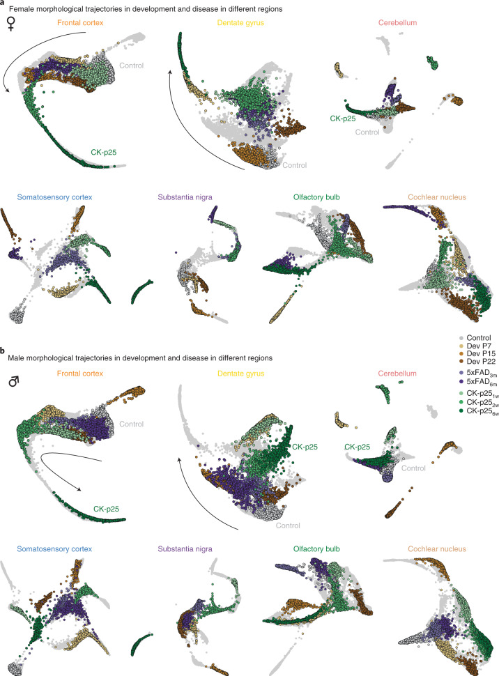
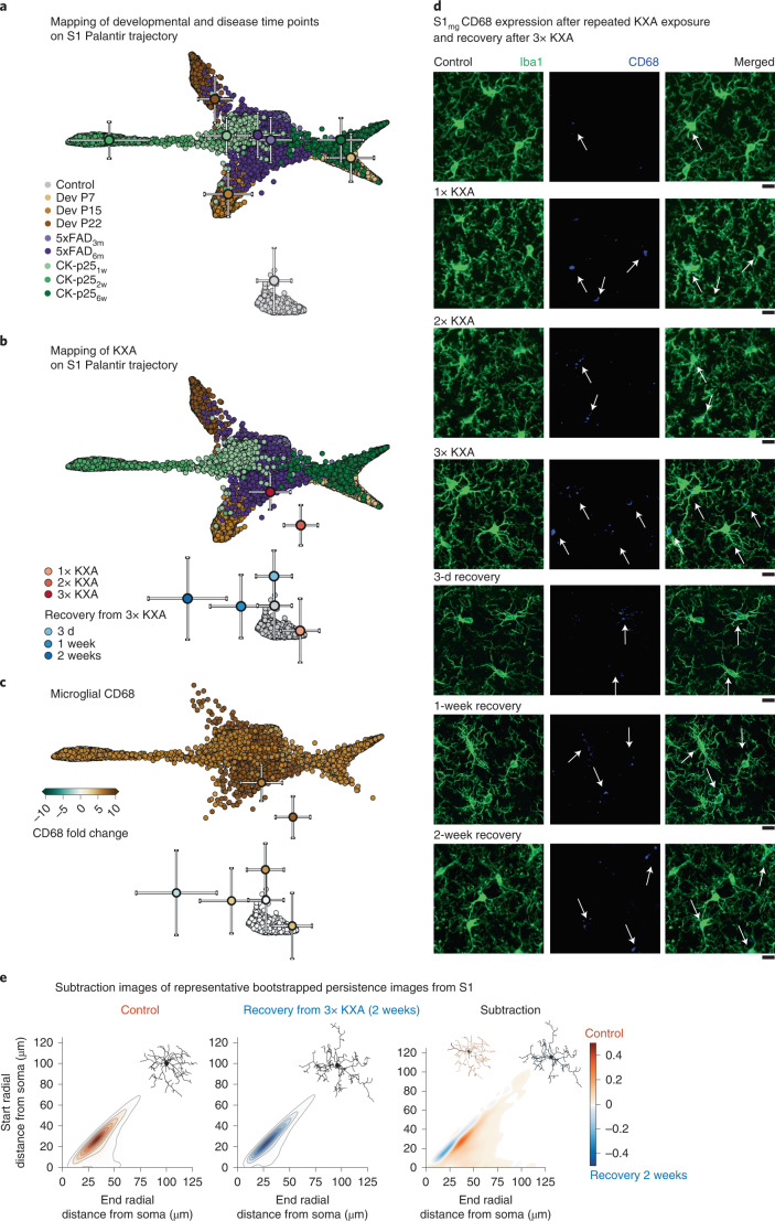
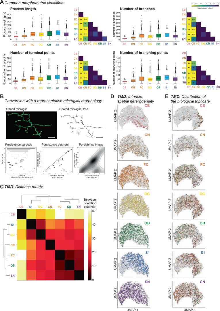
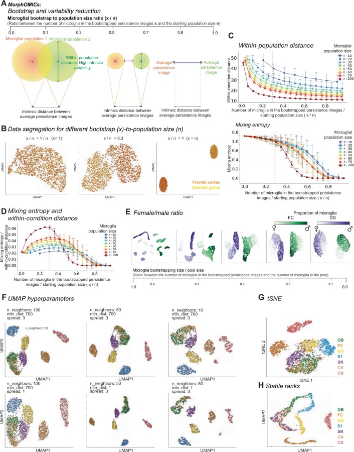
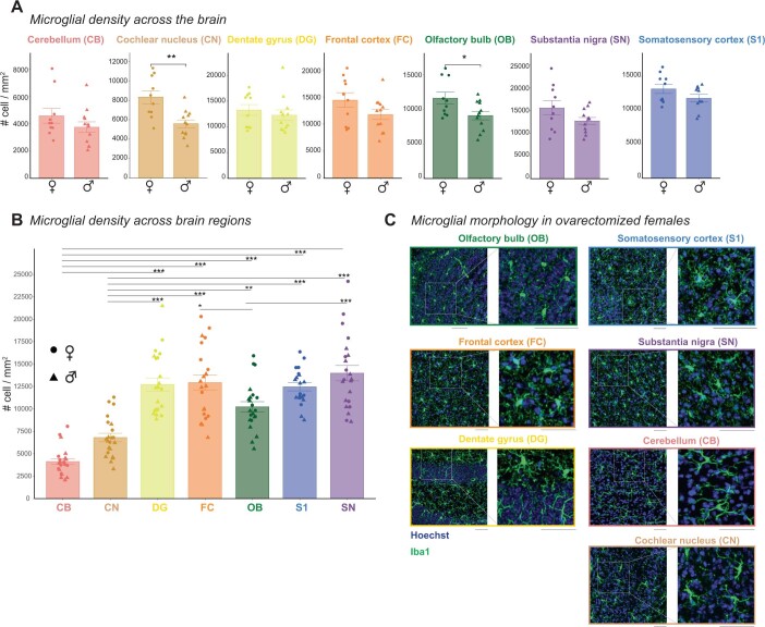
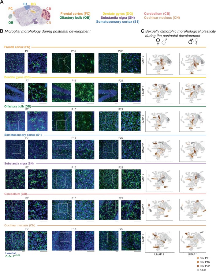
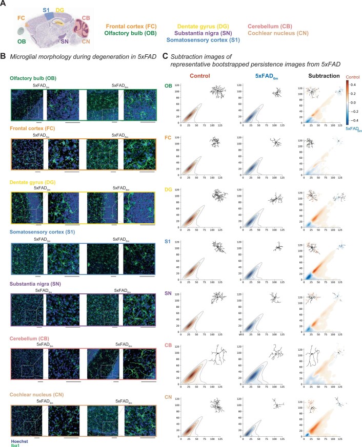
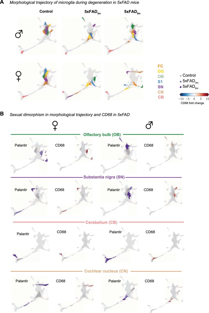
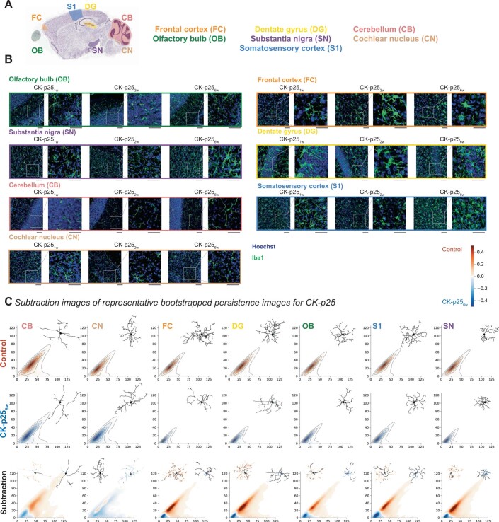
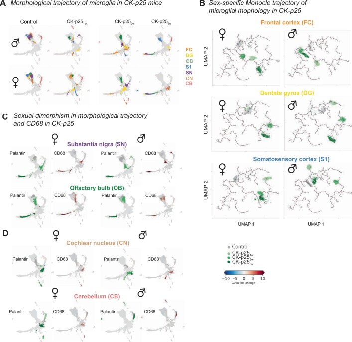
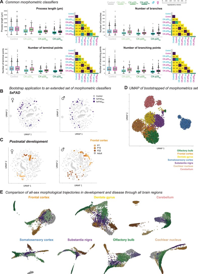
Environmental cues influence the highly dynamic morphology of microglia. Strategies to characterize these changes usually involve user-selected morphometric features, which preclude the identification of a spectrum of context-dependent morphological phenotypes. Here we develop MorphOMICs, a topological data analysis approach, which enables semiautomatic mapping of microglial morphology into an atlas of cue-dependent phenotypes and overcomes feature-selection biases and biological variability. We extract spatially heterogeneous and sexually dimorphic morphological phenotypes for seven adult mouse brain regions. This sex-specific phenotype declines with maturation but increases over the disease trajectories in two neurodegeneration mouse models, with females showing a faster morphological shift in affected brain regions. Remarkably, microglia morphologies reflect an adaptation upon repeated exposure to ketamine anesthesia and do not recover to control morphologies. Finally, we demonstrate that both long primary processes and short terminal processes provide distinct insights to morphological phenotypes. MorphOMICs opens a new perspective to characterize microglial morphology.
Overview
The claims and statistics of this paper haven’t been checked yet.
Run the check — free, no account needed. It reads the open-access text and checks the sources it cites.
Integrity signals
Registry
Cites
Statistics
Figures
Verification
Claims the paper makes
The claims and statistics of this paper haven’t been checked yet.
Run the check — free, no account needed. It reads the open-access text and checks the sources it cites.
Claims other papers make about this one
Each is checked against this paper’s own text: does it say what the citing paper says it does?
The source backs part of the claim, not all of it — or backs it for a different population, condition or comparison.
In the citing paper
“Cells portraying smaller cellular somas in addition to extensive and well-branched processes are usually linked to a surveillant state of microglia, whereas the shrinkage of processes and bigger somas are usually linked to reactive states [83–86].”
From Sex chromosomes and sex hormones differently shape microglial properties during normal physiological conditions in the adult mouse hippocampus (2025) · cites this paper as [84]
Experimental — accuracy not yet measured
Self-check
Percentages, sums and reported differences re-computed from the paper’s own numbers.
The self-check needs the paper’s full text.
Citations
Cites
UMAP: Uniform Manifold Approximation and Projection
McInnes, L., Healy, J., Saul, N., & Großberger, L. (2018) · Journal of Open Source Software · doi:10.21105/joss.00861 · Cited by 9,826
Shrestha, S., & Offer, S. M. (2016) · Medical Epigenetics · doi:10.1159/000444713 · Cited by 8,951
Trapnell, C., Cacchiarelli, D., Grimsby, J., Pokharel, P., Li, S., Morse, M., Lennon, N. J., Livak, K. J., Mikkelsen, T. S., & Rinn, J. L. (2014) · Nature Biotechnology · doi:10.1038/nbt.2859 · Cited by 7,314
UMAP: Uniform Manifold Approximation and Projection for Dimension Reduction
McInnes, L., Healy, J., & Melville, J. (2018) · arXiv · doi:10.48550/arxiv.1802.03426 · Cited by 7,101
Resting microglial cells are highly dynamic surveillants of brain parenchyma in vivo
Nimmerjahn, A., Kirchhoff, F., & Helmchen, F. (2005) · Science · doi:10.1126/science.1110647 · Cited by 5,827
Genome-wide atlas of gene expression in the adult mouse brain
Lein, E. S., Hawrylycz, M. J., Ao, N., Ayres, M., Bensinger, A., Bernard, A., Boe, A. F., Boguski, M. S., Brockway, K. S., Byrnes, E. J., et al. (2007) · Nature · doi:10.1038/nature05453 · Cited by 5,572
Fate Mapping Analysis Reveals That Adult Microglia Derive from Primitive Macrophages
Ginhoux, F., Greter, M., Leboeuf, M., Nandi, S., See, P., Gokhan, S., Mehler, M. F., Conway, S. J., Ng, L. G., Stanley, E. R., et al. (2010) · Science · doi:10.1126/science.1194637 · Cited by 5,175
The single-cell transcriptional landscape of mammalian organogenesis
Cao, J., Spielmann, M., Qiu, X., Huang, X., Ibrahim, D. M., Hill, A. J., Zhang, F., Mundlos, S., Christiansen, L., Steemers, F. J., et al. (2019) · Nature · doi:10.1038/s41586-019-0969-x · Cited by 4,490
Synaptic Pruning by Microglia Is Necessary for Normal Brain Development
Paolicelli, R. C., Bolasco, G., Pagani, F., Maggi, L., Scianni, M., Panzanelli, P., Giustetto, M., Ferreira, T. A., Guiducci, E., Dumas, L., et al. (2011) · Science · doi:10.1126/science.1202529 · Cited by 4,244
Host microbiota constantly control maturation and function of microglia in the CNS
Erny, D., Hrabě de Angelis, A. L., Jaitin, D., Wieghofer, P., Staszewski, O., David, E., Keren-Shaul, H., Mahlakoiv, T., Jakobshagen, K., Buch, T., et al. (2015) · Nature Neuroscience · doi:10.1038/nn.4030 · Cited by 3,569
Research works printed but not linked: 1
- Van Der Maaten, L. & Hinton, G. Visualizing data using t-SNE. J. Mach. Learn. Res. 9, 2579–2605 (2008).
Cited by
Sorted by citations
Microglia states and nomenclature: A field at its crossroads
Paolicelli, R. C., Sierra, A., Stevens, B., Tremblay, M.-E., Aguzzi, A., Ajami, B., Amit, I., Audinat, E., Bechmann, I., Bennett, M., et al. (2022) · Neuron · doi:10.1016/j.neuron.2022.10.020 · Cited by 1,973
Self-citationHuman microglial state dynamics in Alzheimer’s disease progression
Sun, N., Victor, M. B., Park, Y. P., Xiong, X., Scannail, A. N., Leary, N., Prosper, S., Viswanathan, S., Luna, X., Boix, C. A., et al. (2023) · Cell · doi:10.1016/j.cell.2023.08.037 · Cited by 462
Self-citationMicroglial morphometric analysis: so many options, so little consistency
Reddaway, J., Richardson, P., Bevan, R. J., Stoneman, J., & Palombo, M. (2023) · Frontiers in Neuroinformatics · doi:10.3389/fninf.2023.1211188 · Cited by 131
Quantifying microglial morphology: an insight into function
Green, T. R. F., & Rowe, R. K. (2024) · Clinical & Experimental Immunology · doi:10.1093/cei/uxae023 · Cited by 119
Müller, L., Benedetto, S. D., & Müller, V. (2025) · Cells · doi:10.3390/cells14010054 · Cited by 102
Kim, J. S., Pavlidis, P., & Ciernia, A. V. (2024) · eNeuro · doi:10.1523/eneuro.0014-24.2024 · Cited by 64
Johnson, N. R., Yuan, P., Castillo, E., Lopez, T. P., Yue, W., Bond, A., Rivera, B. M., Sullivan, M. C., Hirouchi, M., Giles, K., et al. (2023) · Nature Communications · doi:10.1038/s41467-022-35753-w · Cited by 46
Lewy body dementia promotion by air pollutants
Zhang, X., Liu, H., Wu, X., Jia, L., Gadhave, K., Wang, L., Zhang, K., Li, H., Chen, R., Kumbhar, R., et al. (2025) · Science · doi:10.1126/science.adu4132 · Cited by 48
Bobotis, B. C., Braniff, O., Gargus, M., Akinluyi, E. T., Awogbindin, I. O., & Tremblay, M.-È. (2023) · Brain Research Bulletin · doi:10.1016/j.brainresbull.2023.110752 · Cited by 40
The dual nature of neuroinflammation in networked brain
Müller, L., Benedetto, S. D., & Müller, V. (2025) · Frontiers in Immunology · doi:10.3389/fimmu.2025.1659947 · Cited by 43
Online conversion of reconstructed neural morphologies into standardized SWC format
Mehta, K., Ljungquist, B., Ogden, J., Nanda, S., Ascoli, R., Ng, L., & Ascoli, G. A. (2023) · Nature Communications · doi:10.1038/s41467-023-42931-x · Cited by 23
van Weering, H. R. J., Nijboer, T. W., Brummer, M. L., Boddeke, E., & Eggen, B. J. L. (2023) · Glia · doi:10.1002/glia.24427 · Cited by 26
Relationship between sex biases in gene expression and sex biases in autism and Alzheimer’s disease
Fass, S. B., Mulvey, B., Chase, R., Yang, W. C., Selmanovic, D., Chaturvedi, S. M., Tycksen, E., Weiss, L. A., & Dougherty, J. D. (2024) · Biology of Sex Differences · doi:10.1186/s13293-024-00622-2 · Cited by 25
Sheridan, S. D., Horng, J. E., Yeh, H., McCrea, L. T., Wang, J. P., Fu, T., & Perlis, R. H. (2022) · Biological Psychiatry · doi:10.1016/j.biopsych.2023.07.022 · Cited by 19
Type I interferon signalling and interferon‐responsive microglia in health and disease
López‐Atalaya, J. P., & Bhojwani-Cabrera, A. M. (2025) · FEBS Journal · doi:10.1111/febs.70126 · Cited by 23
Bobotis, B. C., Khakpour, M., Braniff, O., de Andrade, E. G., Gargus, M., Allen, M., Carrier, M., Baillargeon, J., Rangachari, M., & Tremblay, M.-È. (2025) · Journal of Neuroinflammation · doi:10.1186/s12974-025-03341-6 · Cited by 17
“This analysis provides insight into microglial function across contexts of health and disease [57, 84].”
Regulation of disease-associated microglia in the optic nerve by lipoxin B4 and ocular hypertension
Maurya, S., Lin, M., Karnam, S., Singh, T., Kumar, M., Ward, E., Sivak, J. M., Flanagan, J. G., & Gronert, K. (2024) · Molecular Neurodegeneration · doi:10.1186/s13024-024-00775-z · Cited by 17
Shi, L., Liu, S., Chen, J., Wang, H., & Wang, Z. (2024) · Neural Regeneration Research · doi:10.4103/nrr.nrr-d-24-00810 · Cited by 15
Le, L., Eliseeva, S. I., Plunk, E. C., Kara-Pabani, K., Li, H., Yarovinsky, F., & Majewska, A. K. (2025) · Cell Reports · doi:10.1016/j.celrep.2024.115176 · Cited by 13
Sanchez-Molina, P., Rosmus, D.-D., Brownell, D., Meral, M., Gohlich, C., Pratapa, A., Peymanfar, Y., Whitley, A., Hou, Y., Nikulina, N., et al. (2026) · Nature Neuroscience · doi:10.1038/s41593-026-02267-3 · Cited by 21
Dong, M., Qian, Y., Geng, Y., Wang, R., Wang, Y., Cai, J., Wang, X., Huang, Y., Liu, Y., Liu, W., et al. (2025) · Journal of Alzheimer s Disease · doi:10.1177/13872877251378007 · Cited by 12
Microglia contribute to cognitive decline in hypercholesterolemic LDLr −/− mice
Rodrigues, M. S., do Nascimento, N. B., Farias, H. R., Schons, T., Machado, A. G., Medeiros, E. B., Mesquita, A., Bast, R. K., Budni, J., Engblom, D., et al. (2023) · Journal of Neurochemistry · doi:10.1111/jnc.15952 · Cited by 13
Uweru, J. O., Okojie, A. K., Trivedi, A., Benderoth, J., Thomas, L. S., Davidson, G. K., Cox, K., & Eyo, U. B. (2024) · Journal of Neuroinflammation · doi:10.1186/s12974-024-03079-7 · Cited by 11
Maksour, S., & Ooi, L. (2023) · Journal of Neurochemistry · doi:10.1111/jnc.15885 · Cited by 12
Microglia role as the regulator of cognitive function
Sousa, R. A. L. D., & Cassilhas, R. C. (2023) · Revista da Associação Médica Brasileira · doi:10.1590/1806-9282.20230412 · Cited by 11
Cell morphologies in the nervous system: Glia steal the limelight
Ascoli, G. A. (2022) · The Journal of Comparative Neurology · doi:10.1002/cne.25429 · Cited by 9
Maes, M. E., Colombo, G., Uiterkamp, F. E. S., Sternberg, F., Venturino, A., Pohl, E. E., & Siegert, S. (2023) · iScience · doi:10.1016/j.isci.2023.107780 · Cited by 9
Self-citationContralateral Astrocyte Response to Acute Optic Nerve Damage Is Mitigated by PANX1 Channel Activity
Wurl, J. A., Nair, C. E. M., Dietz, J. A., Shestopalov, V. I., & Nickells, R. W. (2023) · International Journal of Molecular Sciences · doi:10.3390/ijms242115641 · Cited by 9
A model-based hierarchical Bayesian approach to Sholl analysis
VonKaenel, E. D., Feidler, A. M., Lowery, R. L., Andersh, K. M., Love, T., Majewska, A. K., & McCall, M. N. (2024) · Bioinformatics · doi:10.1093/bioinformatics/btae156 · Cited by 8
Zhang, Y., & Tang, C. (2024) · Aging and Disease · doi:10.14336/ad.2024.0326 · Cited by 7
Perochon, T., Krsnik, Ž., Massimo, M., Ruchiy, Y., Romero, A. L., Mohammadi, E., Li, X., Long, K. R., Parkkinen, L., Blomgren, K., et al. (2025) · Nature Communications · doi:10.1038/s41467-025-56560-z · Cited by 7
Ward, E., Benowitz, L. I., Brunner, T. M., Bu, G., Cayouette, M., Canto-Soler, V., Mesquita, S. D., Polo, A. D., DiAntonio, A., Duan, X., et al. (2025) · Molecular Neurodegeneration · doi:10.1186/s13024-025-00858-5 · Cited by 7
Microglial function interacts with the environment to affect sex-specific depression risk
Fitzgerald, E., Pokhvisneva, I., Patel, S. H., Chan, S. Y., Tan, A. P., Chen, H., Silveira, P. P., & Meaney, M. J. (2024) · Brain Behavior and Immunity · doi:10.1016/j.bbi.2024.04.030 · Cited by 5
Johnson, N. R., Yuan, P., Castillo, E., Lopez, T. P., Yue, W., Bond, A., Rivera, B. M., Sullivan, M. C., Hirouchi, M., Giles, K., et al. (2021) · bioRxiv (Cold Spring Harbor Laboratory) · doi:10.1101/2021.03.20.436288 · Cited by 5
Lessons from ex vivo and in vitro models in microglia research
Cserép, C., Berki, P., Mars, M., Pósfai, B., Pasterkamp, R. J., Benkő, S., & Dénes, Á. (2025) · Trends in Neurosciences · doi:10.1016/j.tins.2025.10.004 · Cited by 5
The cellular choreography of brain aging: a neuroimmune network perspective
Müller, L., Benedetto, S. D., & Müller, V. (2026) · Frontiers in Aging Neuroscience · doi:10.3389/fnagi.2026.1848642 · Cited by 5
Maya-Arteaga, J. P., Martínez-Orozco, H., & Díaz-Cintra, S. Y. (2024) · Frontiers in Cellular Neuroscience · doi:10.3389/fncel.2024.1505048 · Cited by 6
Birkle, T. J. Y., Willems, H. M. G., Skidmore, J., & Brown, G. C. (2024) · iScience · doi:10.1016/j.isci.2024.109454 · Cited by 4
Characterizing Microglial Morphology: Methodological Advances in Confocal Imaging and Analysis
Taborda-Bejarano, J. P., Nowak, D. B., Chaure, F. J., Allen, M. L., Blek, K. A., Walterhouse, S. H., Mantsch, J. R., & García‐Keller, C. (2025) · Cells · doi:10.3390/cells14171354 · Cited by 3
Benkeder, S., Dinh, S.-M., Marchal, P., Gea, P. D., Thoby‐Brisson, M., Hubert, V., Hristovska, I., Pitollat, G., Combet, K., Cardoit, L., et al. (2024) · eLife · doi:10.1101/2024.08.29.609024 · Cited by 3
Matoba, K., Kochi, T., Mreyoud, Y., Badrani, J. H., Patel, H., Tsujioka, H., Yamashita, T., Crossman, D. K., Niwa, M., & Kano, S. (2024) · bioRxiv (Cold Spring Harbor Laboratory) · doi:10.1101/2024.09.19.609743 · Cited by 3
Multi-gate Weighted Fusion Network for neuronal morphology classification
Sun, C., & Zhao, F. (2024) · Frontiers in Neuroscience · doi:10.3389/fnins.2024.1322623 · Cited by 2
3-dimensional morphological analysis of microglia in the sheep brain
Stock, V., Hadzajlic, E., Connelly, A. A., Bassi, J. K., Jufar, A. H., May, C. N., Lankadeva, Y. R., Yao, S. T., & Booth, L. C. (2025) · Journal of Neuroscience Methods · doi:10.1016/j.jneumeth.2025.110624 · Cited by 2
The Central Nervous System Source Modulates Microglia Function and Morphology In Vitro
Pinho, A. G., Monteiro, A., Fernandes, S., de Sousa, N., Salgado, A. J., Silva, N. A., & Monteiro, S. (2023) · International Journal of Molecular Sciences · doi:10.3390/ijms24097685 · Cited by 2
Jha, A., & Kumar, H. (2024) · Neuroglia · doi:10.3390/neuroglia5030017 · Cited by 3
Geertsema, J., Franssen, M. M. M., Barban, F., Sarauskyte, L., Giera, M. A., Kooij, G., & Kőrösi, A. (2024) · Prostaglandins Leukotrienes and Essential Fatty Acids · doi:10.1016/j.plefa.2024.102662 · Cited by 2
Exploring Graph and Digraph Persistence
Bergomi, M. G., & Ferri, M. (2023) · Algorithms · doi:10.3390/a16100465 · Cited by 2
AutoMorFi: Automated Whole-image Morphometry in Fiji/ImageJ for Diverse Analyses and DiscoveriesPreprint
Bouadi, O., Yao, C., Zeng, J., Beason, D., Inda, N., Malone, Z., Yoshihara, J., Manjally, A. V., Johnson, C., Cherry, J. D., et al. (2024) · bioRxiv (Cold Spring Harbor Laboratory) · doi:10.1101/2024.07.26.605357 · Cited by 2
Mcsweeney-Davis, A., Fang, C., Caruyer, E., Kerbrat, A., & Li, J. (2025) · Briefings in Bioinformatics · doi:10.1093/bib/bbaf258 · Cited by 1
Bonilla, M. I., Lee, H.-L., Foster, S., Kim, I.-J., & Rudenko, A. (2025) · Biological Psychiatry Global Open Science · doi:10.1016/j.bpsgos.2025.100591 · Cited by 1
Allopurinol treatment changes microglial characteristics in neonatal mice
Teruya, R.-I., Okajima, T., & Tsuruta, F. (2025) · PubMed Central · doi:10.17912/micropub.biology.001739 · Cited by 1
Venturino, A., Alamalhoda, M., Negrello, T., Jin, K., van Velthoven, C. T. J., Cubero, R. J., Yeung, J., Koppensteiner, P., Tasic, B., & Siegert, S. (2026) · Science Advances · doi:10.1126/sciadv.adz6517 · Cited by 1
Self-citationBoland, S. W., Coyner, G. N., Risen, S. J., Alsulami, A., McGrath, S., & Moreno, J. A. (2026) · Frontiers in Aging · doi:10.3389/fragi.2026.1829442 · Cited by 1
Du, J. T., Chartrand, T., Jayadev, S., Prater, K. E., & Lin, K. (2026) · Journal of Computational Biology · doi:10.1177/15578666261492321 · Cited by 2
Geometry based gene expression signatures detect cancer treatment responders in clinical trialsPreprint
Chachólski, W., & Ramanujam, R. (2024) · medRxiv · doi:10.1101/2024.07.01.24309803 · Cited by 1
Self-citationTemporal topology provides an interpretable framework for neuronal morphogenesisPreprint
Rigaux, K., Castro, A. F., & Kanari, L. (2025) · bioRxiv (Cold Spring Harbor Laboratory) · doi:10.1101/2025.06.02.657366 · Cited by 1
Self-citationSalter, E. W., Lackie, E. R. L., Georgiou, J., & Collingridge, G. L. (2026) · bioRxiv (Cold Spring Harbor Laboratory) · doi:10.64898/2026.02.04.703826 · Cited by 1
Uncovering the Basis of Human Connectome Complexity: The Role of Neuronal MorphologyPreprint
Barros-Zulaica, N., Santander, D. E., Kanari, L., Shi, Y., Perin, R., Pezzoli, M., Benavides‐Piccione, R., DeFelipe, J., de Kock, C. P. J., Segev, I., et al. (2026) · bioRxiv (Cold Spring Harbor Laboratory) · doi:10.64898/2026.02.12.705493 · Cited by 1
Self-citationAn organotypic neocortical slice culture for studying neuroglial interactionsPreprint
Higgins, K. P., Naqib, V. A. B. A., Mayo, P., Lodder, B., Masuda, T., Amann, L., Prinz, M., & Kole, M. H. P. (2026) · bioRxiv (Cold Spring Harbor Laboratory) · doi:10.64898/2026.05.15.725074 · Cited by 1
Maes, M. E., Colombo, G., Uiterkamp, F. E. S., Sternberg, F., Venturino, A., Pohl, E. E., & Siegert, S. (2022) · bioRxiv (Cold Spring Harbor Laboratory) · doi:10.1101/2022.08.22.504738
Self-citationSheridan, S. D., Horng, J. E., Yeh, H., McCrea, L. T., Fu, T., & Perlis, R. H. (2022) · bioRxiv (Cold Spring Harbor Laboratory) · doi:10.1101/2022.10.24.513576
Mapping tool reveals microglia’s shape-shifting secrets
Askham, A. V. (2022) · The Transmitter · doi:10.53053/vpzu2045
Fitzgerald, E., Pokhvisneva, I., Patel, S. H., Chan, S. Y., Tan, A. P., Chen, H., Silveira, P. P., & Meaney, M. J. (2023) · medRxiv · doi:10.1101/2023.02.18.23286124
Pruning vineyards: Updating barcodes and representative cycles by removing simplices
Giunti, B., & Lazovskis, J. (2026) · Foundations of Data Science · doi:10.3934/fods.2026007
Uweru, J. O., Okojie, K. A., Trivedi, A., Benderoth, J., Thomas, L. S., Davidson, G. K., Cox, K., & Eyo, U. B. (2024) · Research Square · doi:10.21203/rs.3.rs-3997803/v1
Maurya, S., Lin, M., Karnam, S., Singh, T., Kumar, M., Ward, E., Sivak, J. M., Flanagan, J. G., & Gronert, K. (2024) · bioRxiv (Cold Spring Harbor Laboratory) · doi:10.1101/2024.03.18.585452
Le, L., Eliseeva, S. I., Kara-Pabani, K., Li, H., Yarovinsky, F., & Majewska, A. K. (2024) · SSRN Electronic Journal · doi:10.2139/ssrn.4795163
Unsupervised deep learning enables blur-free super-resolution in two-photon microscopyPreprint
Morita, H., Hayashi, S., Tsuji, T., Kato, D., Wake, H., & Shimamura, T. (2024) · bioRxiv (Cold Spring Harbor Laboratory) · doi:10.1101/2024.04.30.591870
Benkeder, S., Dinh, S.-M., Marchal, P., Gea, P. D., Thoby‐Brisson, M., Hubert, V., Hristovska, I., Pitollat, G., Combet, K., Cardoit, L., et al. (2024) · eLife · doi:10.7554/elife.101630.1
Morphometric Identification of Parvalbumin-Positive Interneurons: A Data-Driven ApproachPreprint
Panday, M., Monteiro, L., Daudi, A., & Murphy, K. M. (2024) · bioRxiv (Cold Spring Harbor Laboratory) · doi:10.1101/2024.12.27.630528
Representation of conceptual affordances in eye movements and the entorhinal cortexPreprint
Eperon, A., Doeller, C. F., Theves, S., & Bottini, R. (2025) · bioRxiv (Cold Spring Harbor Laboratory) · doi:10.1101/2025.01.21.633955
Benkeder, S., Dinh, S.-M., Marchal, P., Gea, P. D., Thoby‐Brisson, M., Hubert, V., Hristovska, I., Pitollat, G., Combet, K., Cardoit, L., et al. (2025) · eLife · doi:10.7554/elife.101630.2
Topology across scales on heterogeneous cell data
Torras-Pérez, M., Yoon, I. H. R., Weeratunga, P. N., Ho, L., Byrne, H. M., Tillmann, U., & Harrington, H. A. (2025) · PLoS Computational Biology · doi:10.1371/journal.pcbi.1013460
Unsupervised deep learning enables blur-free resolution enhancement in two-photon microscopy
Morita, H., Hayashi, S., Tsuji, T., Kato, D., Wake, H., & Shimamura, T. (2026) · Cell Reports Methods · doi:10.1016/j.crmeth.2026.101476
Brain-wide changes in microglial morphology following peripheral nerve injury
Cazuza, R. A., Laoumtzis, M., Lacagnina, M. J., Morphett, J., Zagrai, S. M., Grieco, A. R., Hutchinson, M. R., & Grace, P. M. (2025) · Journal of Pain · doi:10.1016/j.jpain.2025.105582
Sex Dependent Effects of Minocycline on Contextual Fear MemoryPreprint
Lin, T. E., Hall, J., & Thomas, K. L. (2025) · bioRxiv (Cold Spring Harbor Laboratory) · doi:10.1101/2025.11.11.687783
Yuge, M., Koiwa, J., Shiromizu, T., Wakai, E., Mizoguchi, A., & Nishimura, Y. (2025) · Current Research in Toxicology · doi:10.1016/j.crtox.2025.100276
Uweru, J. O., Okojie, A. K., Trivedi, A., Benderoth, J., Thomas, L. S., Davidson, G. K., Cox, K., & Eyo, U. B. (2024) · Figshare · doi:10.6084/m9.figshare.26706966
Fass, S. B., Mulvey, B., Chase, R., Yang, W., Selmanovic, D., Chaturvedi, S. M., Tycksen, E., Weiss, L. A., & Dougherty, J. D. (2024) · Figshare · doi:10.6084/m9.figshare.26730706
Fass, S. B., Mulvey, B., Chase, R., Yang, W., Selmanovic, D., Chaturvedi, S. M., Tycksen, E., Weiss, L. A., & Dougherty, J. D. (2024) · Figshare · doi:10.6084/m9.figshare.26730724
Fass, S. B., Mulvey, B., Chase, R., Yang, W., Selmanovic, D., Chaturvedi, S. M., Tycksen, E., Weiss, L. A., & Dougherty, J. D. (2024) · Figshare · doi:10.6084/m9.figshare.26730709
Fass, S. B., Mulvey, B., Chase, R., Yang, W., Selmanovic, D., Chaturvedi, S. M., Tycksen, E., Weiss, L. A., & Dougherty, J. D. (2024) · Figshare · doi:10.6084/m9.figshare.26730715
Fass, S. B., Mulvey, B., Chase, R., Yang, W., Selmanovic, D., Chaturvedi, S. M., Tycksen, E., Weiss, L. A., & Dougherty, J. D. (2024) · Figshare · doi:10.6084/m9.figshare.26730730
Fass, S. B., Mulvey, B., Chase, R., Yang, W., Selmanovic, D., Chaturvedi, S. M., Tycksen, E., Weiss, L. A., & Dougherty, J. D. (2024) · Figshare · doi:10.6084/m9.figshare.26730718
Uweru, J. O., Okojie, A. K., Trivedi, A., Benderoth, J., Thomas, L. S., Davidson, G. K., Cox, K., & Eyo, U. B. (2024) · Figshare · doi:10.6084/m9.figshare.25608684
Fass, S. B., Mulvey, B., Chase, R., Yang, W., Selmanovic, D., Chaturvedi, S. M., Tycksen, E., Weiss, L. A., & Dougherty, J. D. (2024) · Figshare · doi:10.6084/m9.figshare.26730721
Uweru, J. O., Okojie, A. K., Trivedi, A., Benderoth, J., Thomas, L. S., Davidson, G. K., Cox, K., & Eyo, U. B. (2024) · Figshare · doi:10.6084/m9.figshare.25608855
Fass, S. B., Mulvey, B., Chase, R., Yang, W., Selmanovic, D., Chaturvedi, S. M., Tycksen, E., Weiss, L. A., & Dougherty, J. D. (2024) · Figshare · doi:10.6084/m9.figshare.26730727
Uweru, J. O., Okojie, A. K., Trivedi, A., Benderoth, J., Thomas, L. S., Davidson, G. K., Cox, K., & Eyo, U. B. (2024) · Figshare · doi:10.6084/m9.figshare.26706972
Uweru, J. O., Okojie, A. K., Trivedi, A., Benderoth, J., Thomas, L. S., Davidson, G. K., Cox, K., & Eyo, U. B. (2024) · Figshare · doi:10.6084/m9.figshare.26706969
Fass, S. B., Mulvey, B., Chase, R., Yang, W., Selmanovic, D., Chaturvedi, S. M., Tycksen, E., Weiss, L. A., & Dougherty, J. D. (2024) · Figshare · doi:10.6084/m9.figshare.26730712
Astrocyte morphology: complex or trivial?
Kriuchechnikova, A., Soldatova, A., Tiaglik, A. B., Tang, Y., & Brazhe, A. (2025) · Biophysical Reviews · doi:10.1007/s12551-025-01402-x
Subcellular orchestration of microglial aging
Schaible, P., & Erny, D. (2026) · Nature Aging · doi:10.1038/s43587-026-01109-y
Emissah, H., Tecuatl, C., & Ascoli, G. A. (2026) · bioRxiv (Cold Spring Harbor Laboratory) · doi:10.64898/2026.03.27.714818
Mesaoudi, A. E. (2024) · theses.fr (ABES) · doi:10.70675/f339157ez1688z4acaz87fcz0ecae062a7cc
Manifold learning for personalized and label-free detection of cardiac arrhythmias
Vazifeh, A. R., & Fleischer, J. W. (2026) · Informatics in Medicine Unlocked · doi:10.1016/j.imu.2026.101770
Chromatic topological mapping reveals organelle-specific spatial organization within microgliaPreprint
Agerberg, J., Kanari, L., Maes, M. E., Chachólski, W., Siegert, S., & Scolamiero, M. (2026) · bioRxiv (Cold Spring Harbor Laboratory) · doi:10.64898/2026.06.05.730307
Self-citationXia, Y., Yuan, P., Fei, G., & Cheng, X. (2026) · Brain medicine : · doi:10.61373/bm026y.0051
Sriram, A., Attili, S. M., & Seshaiyer, P. (2026) · Research Square · doi:10.21203/rs.3.rs-10233914/v1
Manifold Alignment for Label-Free Cell Phenotyping in Multimodal Microscopy
Vazifeh, A. R., & Fleischer, J. W. (2026) · Optics in the Life Sciences · doi:10.1364/microscopy.2026.mw1b.4
Metadata
- Authors
- Colombo, G., Cubero, R. J. A., Kanari, L., Venturino, A., Schulz, R., Scolamiero, M., Agerberg, J., Mathys, H., Tsai, L.-H., Chachólski, W., Hess, K., & Siegert, S.
- Affiliations
- Institute of Science and Technology Austria (AT)
- École Polytechnique Fédérale de Lausanne (CH)
- KTH Royal Institute of Technology (SE)
- Broad Institute (US)
- Corresponding author
- Siegert, Sandra
- ORCID
- Venue
- Nature Neuroscience
- Published
- 2022-09-30
- Volume
- 25
- Issue
- 10
- Pages
- 1379-1393
- ISSN
- 1097-6256 (ISSN-L)
- 1546-1726
- PMC
- PMC9534764
- OpenAlex
- W4298137907
- Cited by
- 100
- References
- 125
- Citations by year
- 2026: 25 · 2025: 23 · 2024: 31 · 2023: 15 · 2022: 5
- FWCI
- 7.20
- Type
- journal article
- Language
- en
- Open access
- hybrid
- doi.org
- Locations
- Nature Neuroscience (publishedVersion, cc-by)
- bioRxiv (Cold Spring Harbor Laboratory) (acceptedVersion, cc-by-nd)
- PubMed Central (publishedVersion, cc-by)
- PubMed (publishedVersion)
- DSpace@MIT (Massachusetts Institute of Technology) (submittedVersion, cc-by-nc)
- Infoscience (Ecole Polytechnique Fédérale de Lausanne) (submittedVersion, cc-by-nc-nd)
- SDL (submittedVersion)
- Funders
- JPB Foundation
- Institute of Science and Technology Austria (715571, 754411)
- European Commission (HORIZON2020, 754411)
- École Polytechnique Fédérale de Lausanne
- Bundesministerium für Wissenschaft, Forschung und Wirtschaft
- Board of the Swiss Federal Institutes of Technology
- National Institutes of Health
- National Institute of Neurological Disorders and Stroke (R37NS051874)
- H2020 Marie Skłodowska-Curie Actions (754411, HORIZON2020, 715571)
- Topics
- Life Sciences › Neuroscience › Neurology › Neuroinflammation and Neurodegeneration Mechanisms
- Life Sciences › Biochemistry, Genetics and Molecular Biology › Biophysics › Cell Image Analysis Techniques
- Life Sciences › Biochemistry, Genetics and Molecular Biology › Molecular Biology › Single-cell and spatial transcriptomics
- Keywords
- microglial morphology, morphomics, topological data analysis, sex differences, regional brain differences, microglial phenotype, neurodegeneration, ketamine anesthesia, microglial maturation, mouse brain
- MeSH terms
- Animals, Brain, Disease Models, Animal, Female, Ketamine, Male, Phenotype, Microglia, Mice
About these checks
Registry, retraction, Cites and statistics results are computed, not judged, with one exception: a printed reference that no index lists is read once by a model before it counts as a citation error. Claims are extracted by a model and kept only when their quote appears verbatim in the text; each cited source is then checked, with a model’s help, for whether it says what the claim relies on. Those verdicts can be wrong — read the claim’s quote and the cited source before relying on one.
Statistics and arithmetic are recomputed, not judged.
Confidence bands are shown only for fields calibrated on our annotated test set. None is calibrated yet, so no band is shown.
What counts as a claim ›How we measure confidence ›How we check this ›
Are you an author, or do you see a problem with this page? Request a review
