

The DOI matches its Crossref record; 60 of the 60 references it cites were found in a registry or an index.
The molecular structure of IFT-A and IFT-B in anterograde intraflagellar transport trains
Published version of a preprint
Abstract
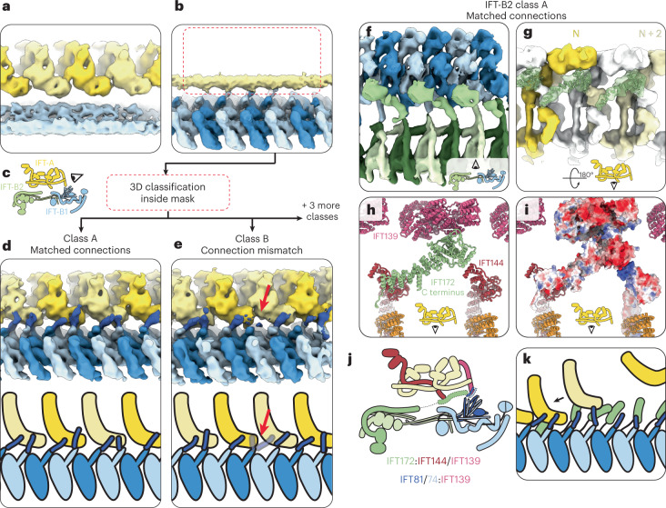
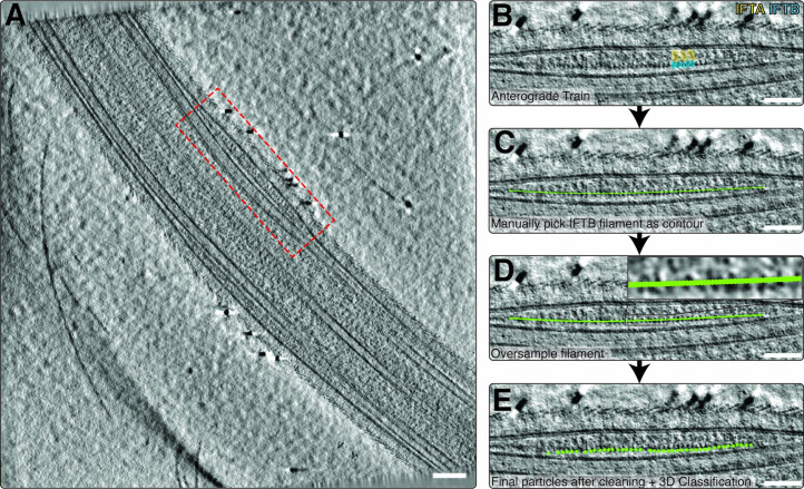
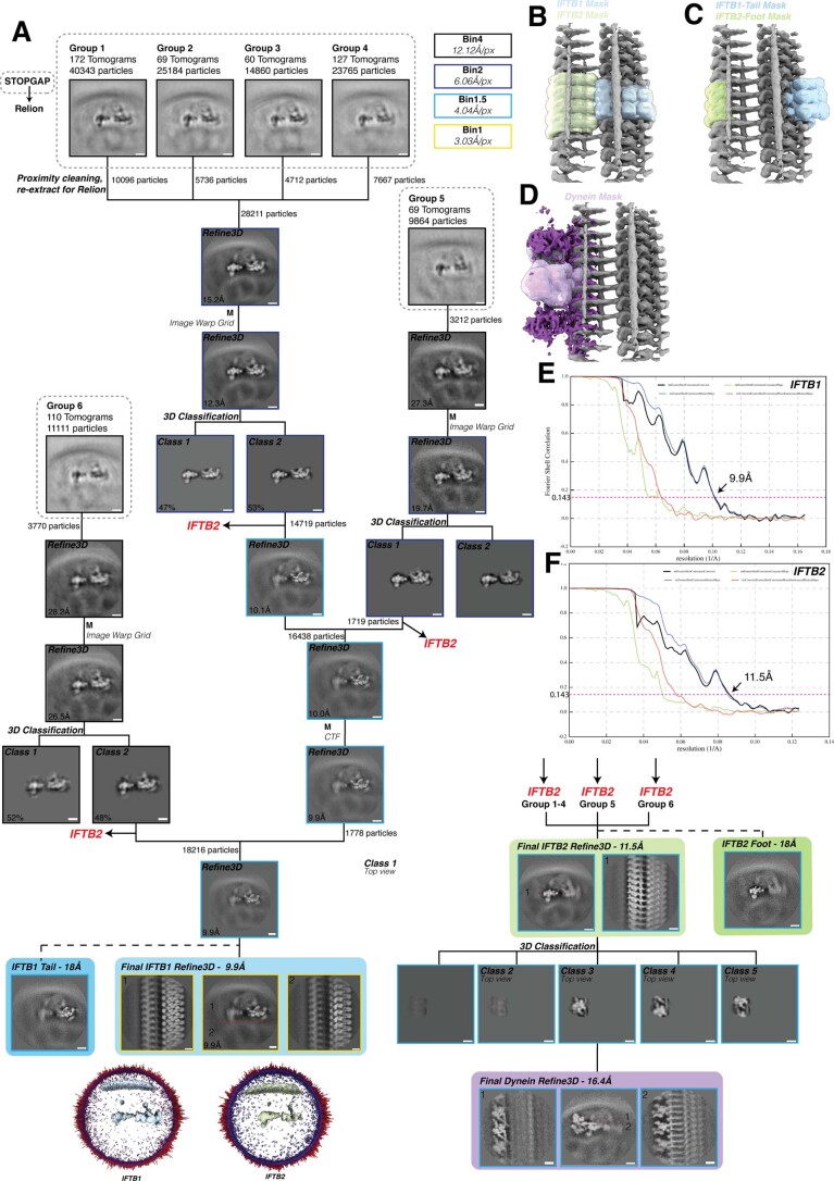
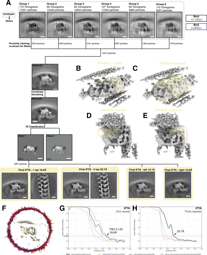
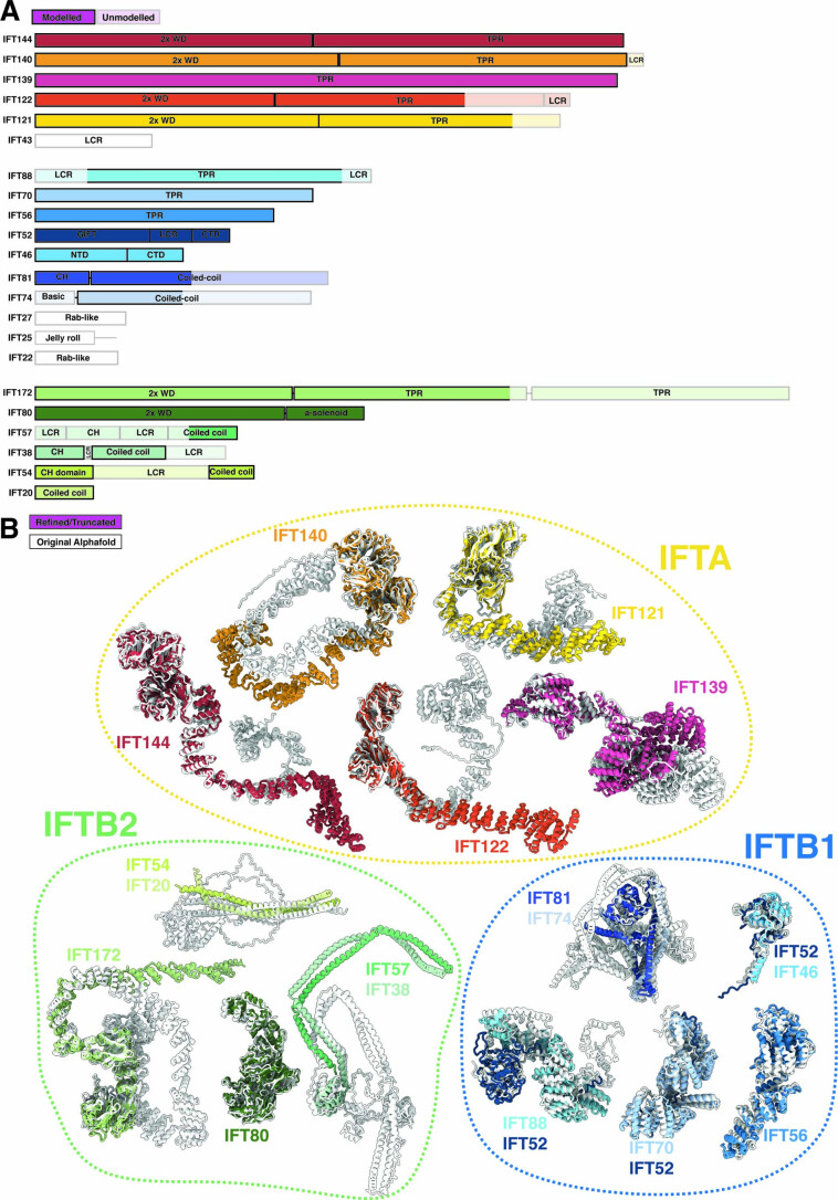
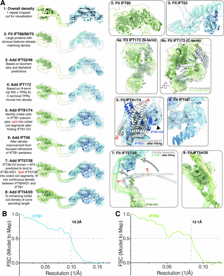
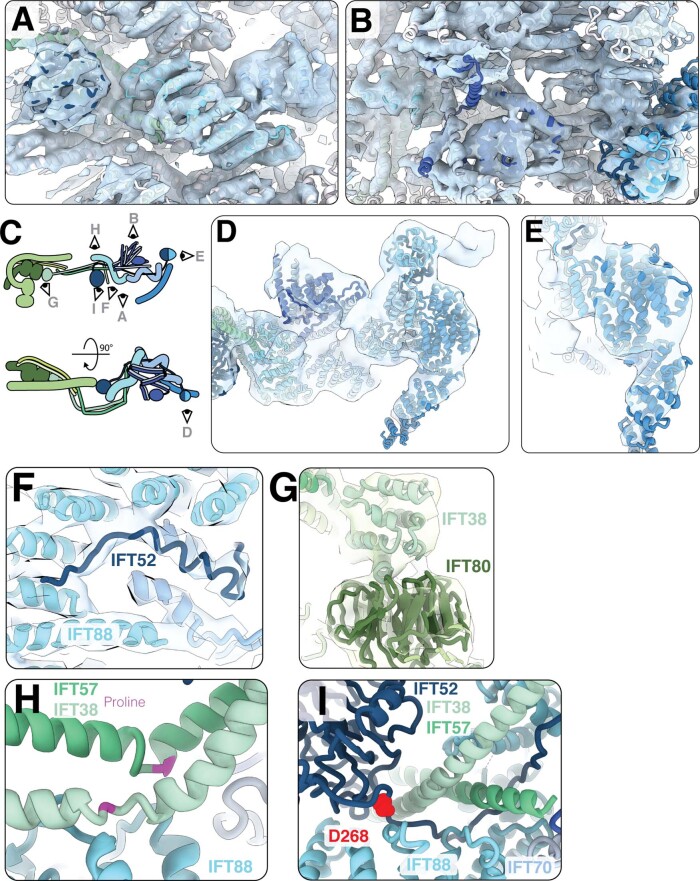
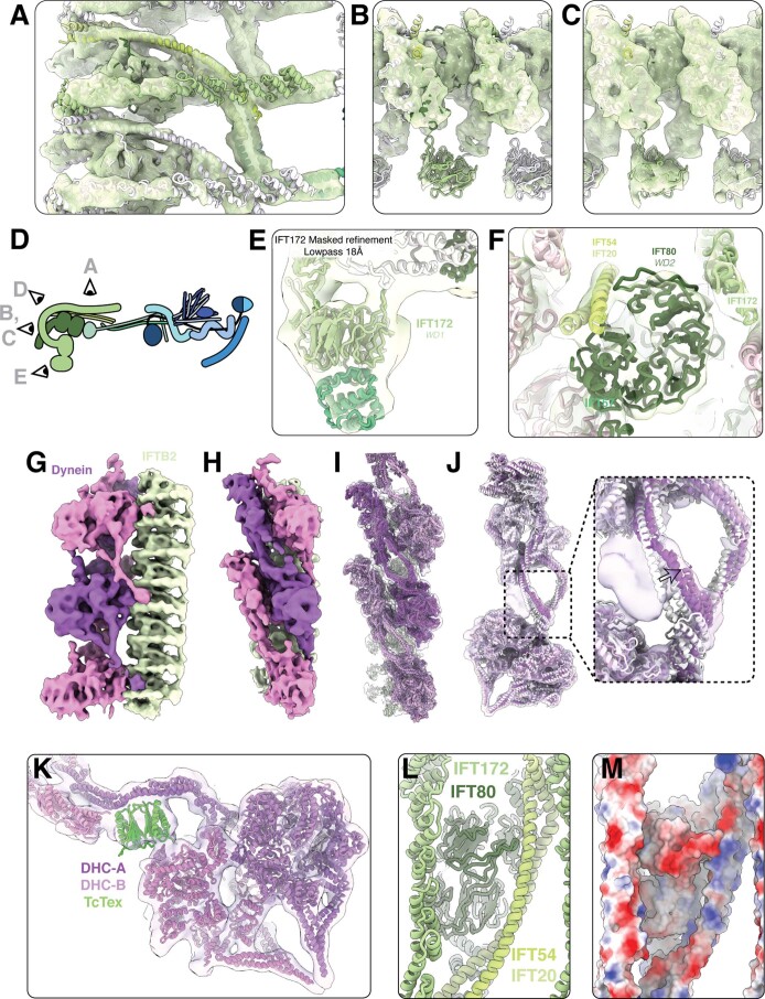
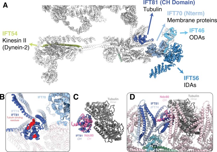
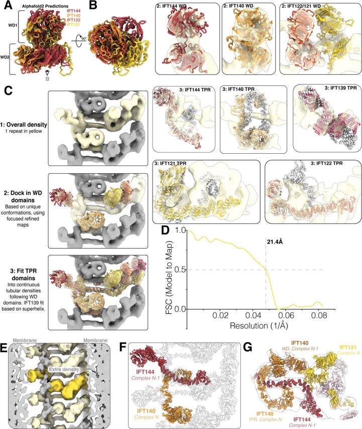
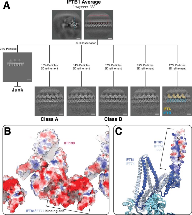
Anterograde intraflagellar transport (IFT) trains are essential for cilia assembly and maintenance. These trains are formed of 22 IFT-A and IFT-B proteins that link structural and signaling cargos to microtubule motors for import into cilia. It remains unknown how the IFT-A/-B proteins are arranged into complexes and how these complexes polymerize into functional trains. Here we use in situ cryo-electron tomography of Chlamydomonas reinhardtii cilia and AlphaFold2 protein structure predictions to generate a molecular model of the entire anterograde train. We show how the conformations of both IFT-A and IFT-B are dependent on lateral interactions with neighboring repeats, suggesting that polymerization is required to cooperatively stabilize the complexes. Following three-dimensional classification, we reveal how IFT-B extends two flexible tethers to maintain a connection with IFT-A that can withstand the mechanical stresses present in actively beating cilia. Overall, our findings provide a framework for understanding the fundamental processes that govern cilia assembly.
Overview
The claims and statistics of this paper haven’t been checked yet.
Run the check — free, no account needed. It reads the open-access text and checks the sources it cites.
Integrity signals
Registry
Cites
Statistics
Figures
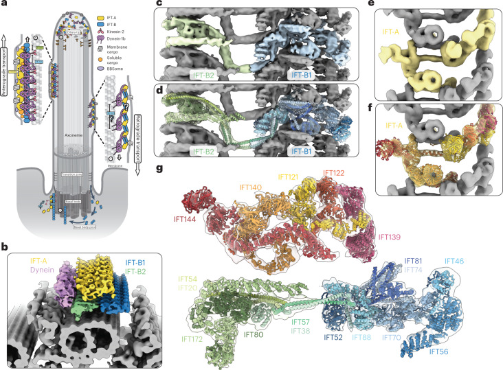
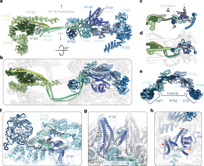
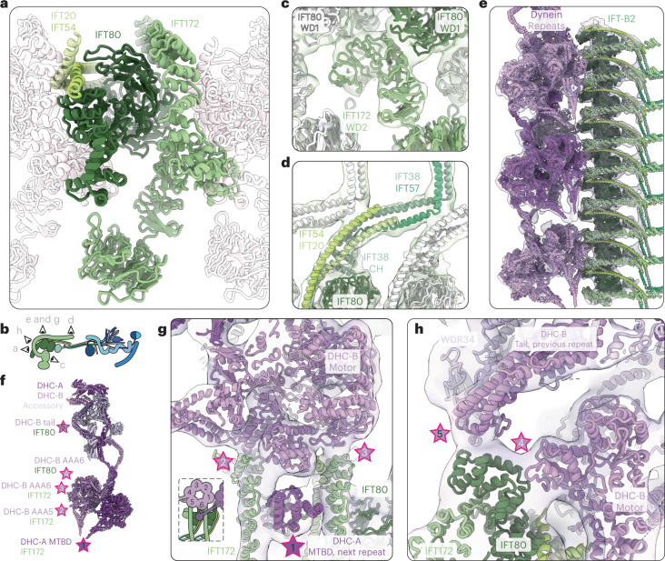
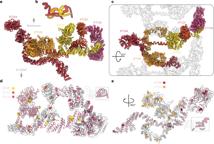
Verification
Claims the paper makes
The claims and statistics of this paper haven’t been checked yet.
Run the check — free, no account needed. It reads the open-access text and checks the sources it cites.
Claims other papers make about this one
Each is checked against this paper’s own text: does it say what the citing paper says it does?
In the citing paper
“Notably, the structure closely matches that predicted by AlphaFold2 multimer56,66 (Methods S1).”
From Structure of the endosomal Commander complex linked to Ritscher-Schinzel syndrome (2023) · cites this paper as [66]
In the citing paper
“The CCDC proteins share some structural similarity with other CH domain-containing and coiled-coil heterodimers,88 predominantly involved in regulating cytoskeletal interactions such as with ciliary microtubules,66,71,72 within dynein-adaptor assemblies,89,90,91 or at the kinetochore,92”
From Structure of the endosomal Commander complex linked to Ritscher-Schinzel syndrome (2023) · cites this paper as [66]
Self-check
Percentages, sums and reported differences re-computed from the paper’s own numbers.
The self-check needs the paper’s full text.
Citations
Cites
Coot : model-building tools for molecular graphics
Emsley, P., & Cowtan, K. (2004) · Acta Crystallographica Section D Biological Crystallography · doi:10.1107/s0907444904019158 · Cited by 30,571
UCSF ChimeraX: Structure visualization for researchers, educators, and developers
Pettersen, E. F., Goddard, T. D., Huang, C. C., Meng, E. C., Couch, G. S., Croll, T. I., Morris, J. H., & Ferrin, T. E. (2021) · Protein Science · doi:10.1002/pro.3943 · Cited by 9,514
Computer Visualization of Three-Dimensional Image Data Using IMOD
Kremer, J. R., Mastronarde, D. N., & McIntosh, J. R. (1996) · Journal of Structural Biology · doi:10.1006/jsbi.1996.0013 · Cited by 5,530
Automated electron microscope tomography using robust prediction of specimen movements
Mastronarde, D. N. (2005) · Journal of Structural Biology · doi:10.1016/j.jsb.2005.07.007 · Cited by 5,136
CTFFIND4: Fast and accurate defocus estimation from electron micrographs
Rohou, A., & Grigorieff, N. (2015) · Journal of Structural Biology · doi:10.1016/j.jsb.2015.08.008 · Cited by 4,913
New tools for automated high-resolution cryo-EM structure determination in RELION-3
Zivanov, J., Nakane, T., Forsberg, B. O., Kimanius, D., Hagen, W. J., Lindahl, E., & Scheres, S. H. (2018) · eLife · doi:10.7554/elife.42166 · Cited by 4,621
Protein complex prediction with AlphaFold-Multimer
Evans, R., O’Neill, M., Pritzel, A., Antropova, N., Senior, A., Green, T., Žídek, A., Bates, R., Blackwell, S., Yim, J., et al. (2021) · doi:10.1101/2021.10.04.463034 · Cited by 3,808
Genes and molecular pathways underpinning ciliopathies
Reiter, J. F., & Leroux, M. R. (2017) · Nature Reviews Molecular Cell Biology · doi:10.1038/nrm.2017.60 · Cited by 1,588
Real-time cryo-electron microscopy data preprocessing with Warp
Tegunov, D., & Cramer, P. (2019) · Nature Methods · doi:10.1038/s41592-019-0580-y · Cited by 1,332
Cellular signalling by primary cilia in development, organ function and disease
Anvarian, Z., Mykytyn, K., Mukhopadhyay, S., Pedersen, L. B., & Christensen, S. T. (2019) · Nature Reviews Nephrology · doi:10.1038/s41581-019-0116-9 · Cited by 845
Stenson, P. D., Mort, M., Ball, E. V., Chapman, M., Evans, K., Azevedo, L., Hayden, M., Heywood, S., Millar, D. S., Phillips, A. D., et al. (2020) · Human Genetics · doi:10.1007/s00439-020-02199-3 · Cited by 789
UCSF ChimeraX : Structure Visualization for Researchers, Educators, and Developers
Pettersen, E. F., Goddard, T. D., Huang, C. C., Meng, E. C., Couch, G. S., Croll, T. I., Morris, J. H., & Ferrin, T. E. (2020) · Apollo (University of Cambridge) · doi:10.17863/cam.57077 · Cited by 617
Multi-particle cryo-EM refinement with M visualizes ribosome-antibiotic complex at 3.5 Å in cells
Tegunov, D., Xue, L., Dienemann, C., Cramer, P., & Mahamid, J. (2021) · Nature Methods · doi:10.1038/s41592-020-01054-7 · Cited by 515
Kidmose, R. T., Juhl, J., Nissen, P., Boesen, T. P., Karlsen, J. L., & Pedersen, B. P. (2019) · IUCrJ · doi:10.1107/s2052252519007619 · Cited by 360
The Intraflagellar Transport Machinery
Taschner, M., & Lorentzen, E. (2016) · Cold Spring Harbor Perspectives in Biology · doi:10.1101/cshperspect.a028092 · Cited by 349
Lechtreck, K.-F., Johnson, E. C., Sakai, T., Cochran, D., Ballif, B. A., Rush, J., Pazour, G. J., Ikebe, M., & Witman, G. B. (2009) · Journal of Cell Biology · doi:10.1083/jcb.200909183 · Cited by 347
Intraflagellar transport (IFT) cargo
Qin, H., Diener, D. R., Geimer, S., Cole, D. G., & Rosenbaum, J. L. (2004) · The Journal of Cell Biology · doi:10.1083/jcb.200308132 · Cited by 342
Molecular Basis of Tubulin Transport Within the Cilium by IFT74 and IFT81
Bhogaraju, S., Cajanek, L., Fort, C., Blisnick, T., Weber, K., Taschner, M., Mizuno, N., Lamla, S., Bastin, P., Nigg, E. A., et al. (2013) · Science · doi:10.1126/science.1240985 · Cited by 317
The Ndc80 kinetochore complex forms oligomeric arrays along microtubules
Alushin, G. M., Ramey, V. H., Pasqualato, S., Ball, D. A., Grigorieff, N., Musacchio, A., & Nogales, E. (2010) · Nature · doi:10.1038/nature09423 · Cited by 303
When protein folding is simplified to protein coiling: the continuum of solenoid protein structures
Kobe, B., & Kajava, A. V. (2000) · Trends in Biochemical Sciences · doi:10.1016/s0968-0004(00)01667-4 · Cited by 281
An organelle-specific protein landscape identifies novel diseases and molecular mechanisms
Boldt, K., van Reeuwijk, J., Lu, Q., Koutroumpas, K., Nguyen, T.-M. T., Texier, Y., van Beersum, S. E. C., Horn, N., Willer, J. R., Mans, D. A., et al. (2016) · Nature Communications · doi:10.1038/ncomms11491 · Cited by 240
Electron-tomographic analysis of intraflagellar transport particle trains in situ
Pigino, G., Geimer, S., Lanzavecchia, S., Paccagnini, E., Cantele, F., Diener, D. R., Rosenbaum, J. L., & Lupetti, P. (2009) · The Journal of Cell Biology · doi:10.1083/jcb.200905103 · Cited by 210
Self-citationTaschner, M. J., Weber, K., Mourão, A., Vetter, M., Awasthi, M., Stiegler, M., Bhogaraju, S., & Lorentzen, E. (2016) · The EMBO Journal · doi:10.15252/embj.201593164 · Cited by 192
Jordan, M. A., Diener, D. R., Stepanek, L., & Pigino, G. (2018) · Nature Cell Biology · doi:10.1038/s41556-018-0213-1 · Cited by 186
Self-citationvan den Hoek, H., Klena, N., Jordan, M. A., Viar, G. A., Righetto, R. D., Schaffer, M., Erdmann, P. S., Wan, W., Geimer, S., Plitzko, J. M., et al. (2022) · Science · doi:10.1126/science.abm6704 · Cited by 148
Self-citationGuo, H., Franken, E., Deng, Y., Benlekbir, S., Lezcano, G. S., Janssen, B., Yu, L., Ripstein, Z. A., Tan, Y. Z., & Rubinstein, J. L. (2020) · IUCrJ · doi:10.1107/s205225252000929x · Cited by 140
Brazelton, W. J., Amundsen, C. D., Silflow, C. D., & Lefebvre, P. A. (2001) · Current Biology · doi:10.1016/s0960-9822(01)00485-7 · Cited by 136
Liu, P., & Lechtreck, K. F. (2018) · Proceedings of the National Academy of Sciences · doi:10.1073/pnas.1713226115 · Cited by 133
Taschner, M. J., Kotsis, F., Bräuer, P., Kuehn, E. W., & Lorentzen, E. (2014) · The Journal of Cell Biology · doi:10.1083/jcb.201408002 · Cited by 124
Structure of the dynein-2 complex and its assembly with intraflagellar transport trains
Toropova, K., Zalyte, R., Mukhopadhyay, A. G., Mladenov, M., Carter, A. P., & Roberts, A. J. (2019) · Nature Structural & Molecular Biology · doi:10.1038/s41594-019-0286-y · Cited by 120
Behal, R. H., Miller, M. S., Qin, H., Lucker, B. F., Jones, A., & Cole, D. G. (2012) · Journal of Biological Chemistry · doi:10.1074/jbc.m111.287102 · Cited by 119
Katoh, Y., Terada, M., Nishijima, Y., Takei, R., Nozaki, S., Hamada, H., & Nakayama, K. (2016) · Journal of Biological Chemistry · doi:10.1074/jbc.m116.713883 · Cited by 118
Crystal structure of the intraflagellar transport complex 25/27
Bhogaraju, S., Taschner, M. J., Morawetz, M., Minard‐Basquin, C., & Lorentzen, E. (2011) · The EMBO Journal · doi:10.1038/emboj.2011.110 · Cited by 107
Together, the IFT81 and IFT74 N-termini form the main module for intraflagellar transport of tubulin
Kubo, T., Brown, J., Bellve, K. D., Craige, B., Weghe, J. C. V. D., Fogarty, K. E., Lechtreck, K. F., & Witman, G. B. (2016) · Journal of Cell Science · doi:10.1242/jcs.187120 · Cited by 106
IFT-A structure reveals carriages for membrane protein transport into cilia
Hesketh, S. J., Mukhopadhyay, A. G., Nakamura, D., Toropova, K., & Roberts, A. J. (2022) · Cell · doi:10.1016/j.cell.2022.11.010 · Cited by 83
Cryo-EM structure of an active central apparatus
Han, L., Rao, Q., Yang, R., Wang, Y., Chai, P., Xiong, Y., & Zhang, K. (2022) · Nature Structural & Molecular Biology · doi:10.1038/s41594-022-00769-9 · Cited by 79
Ishikawa, H., Ide, T., Yagi, T., Jiang, X., Hirono, M., Sasaki, H., Yanagisawa, H., Wemmer, K. A., Stainier, D. Y. R., Qin, H., et al. (2014) · eLife · doi:10.7554/elife.01566 · Cited by 73
Kobayashi, T., Ishida, Y., Hirano, T., Katoh, Y., & Nakayama, K. (2020) · Molecular Biology of the Cell · doi:10.1091/mbc.e20-08-0556 · Cited by 64
Richey, E. A., & Qin, H. (2012) · PLoS ONE · doi:10.1371/journal.pone.0043118 · Cited by 58
Dai, J., Barbieri, F., Mitchell, D. R., & Lechtreck, K. F. (2018) · Molecular Biology of the Cell · doi:10.1091/mbc.e18-05-0291 · Cited by 57
Solution Structure of the Tctex1 Dimer Reveals a Mechanism for Dynein-Cargo Interactions
Wu, H., Maciejewski, M. W., Takebe, S., & King, S. M. (2005) · Structure · doi:10.1016/j.str.2004.11.013 · Cited by 54
Binding of IFT22 to the intraflagellar transport complex is essential for flagellum assembly
Wachter, S., Jung, J., Shafiq, S., Basquin, J., Fort, C., Bastin, P., & Lorentzen, E. (2019) · The EMBO Journal · doi:10.15252/embj.2018101251 · Cited by 53
Taschner, M. J., Mourão, A., Awasthi, M., Basquin, J., & Lorentzen, E. (2017) · Journal of Biological Chemistry · doi:10.1074/jbc.m117.780155 · Cited by 52
Biochemically validated structural model of the 15‐subunit intraflagellar transport complex IFT‐B
Petriman, N. A., Loureiro‐López, M., Taschner, M., Zacharia, N. K., Georgieva, M. M., Boegholm, N., Wang, J., Mourão, A., Russell, R. B., Andersen, J. S., et al. (2022) · The EMBO Journal · doi:10.15252/embj.2022112440 · Cited by 51
The structural basis of intraflagellar transport at a glance
Jordan, M. A., & Pigino, G. (2021) · Journal of Cell Science · doi:10.1242/jcs.247163 · Cited by 50
Self-citationHou, Y., & Witman, G. B. (2017) · Molecular Biology of the Cell · doi:10.1091/mbc.e17-03-0172 · Cited by 41
The IDA3 adapter, required for intraflagellar transport of I1 dynein, is regulated by ciliary length
Hunter, E. L., Lechtreck, K. F., Fu, G., Hwang, J., Lin, H., Gokhale, A., Alford, L. M., Lewis, B. L., Yamamoto, R., Kamiya, R., et al. (2018) · Molecular Biology of the Cell · doi:10.1091/mbc.e17-12-0729 · Cited by 41
McCafferty, C. L., Papoulas, O., Jordan, M. A., Hoogerbrugge, G., Nichols, C., Pigino, G., Taylor, D. W., Wallingford, J. B., & Marcotte, E. M. (2022) · eLife · doi:10.7554/elife.81977 · Cited by 40
Self-citationMembrane association and remodeling by intraflagellar transport protein IFT172
Wang, Q., Taschner, M. J., Ganzinger, K. A., Kelley, C. F., Villasenor, A., Heymann, M., Schwille, P., Lorentzen, E., & Mizuno, N. (2018) · Nature Communications · doi:10.1038/s41467-018-07037-9 · Cited by 38
Zhu, X., Wang, J., Li, S., Lechtreck, K. F., & Pan, J. (2020) · The EMBO Journal · doi:10.15252/embj.2020105781 · Cited by 38
Impaired cooperation between IFT74/BBS22–IFT81 and IFT25–IFT27/BBS19 causes Bardet-Biedl syndrome
Zhou, Z., Qiu, H., Castro-Araya, R.-F., Takei, R., Nakayama, K., & Katoh, Y. (2021) · Human Molecular Genetics · doi:10.1093/hmg/ddab354 · Cited by 35
Taschner, M. J., Lorentzen, A., Mourão, A., Collins, T., Freke, G., MOULDING, D. A., Basquin, J., Jenkins, D., & Lorentzen, E. (2018) · eLife · doi:10.7554/elife.33067 · Cited by 34
Wingfield, J. L., Mekonnen, B., Mengoni, I., Liu, P., Jordan, M. A., Diener, D. R., Pigino, G., & Lechtreck, K. F. (2021) · Journal of Cell Science · doi:10.1242/jcs.259010 · Cited by 32
Self-citationDupont, M. A., Humbert, C., Huber, C., Siour, Q., Guerrera, I. C., Jung, V. A., Christensen, A., Pouliet, A., Garfa‐Traoré, M., Nitschké, P., et al. (2019) · Human Molecular Genetics · doi:10.1093/hmg/ddz091 · Cited by 29
Dong, B., Wu, S., Wang, J., Liu, Y., Peng, Z., Meng, D., Huang, K., Wu, M., & Fan, Z. (2017) · Biology Open · doi:10.1242/bio.026278 · Cited by 27
Nievergelt, A. P., Zykov, I., Diener, D. R., Chhatre, A., Buchholz, T.-O., Delling, M., Diez, S., Jug, F., S̆tĕpánek, L., & Pigino, G. (2022) · Current Biology · doi:10.1016/j.cub.2022.07.033 · Cited by 23
Self-citationZhao, Q., Li, S., Shao, S., Wang, Z., & Pan, J. (2020) · PLoS Genetics · doi:10.1371/journal.pgen.1008561 · Cited by 19
Llabrés, S., Tsenkov, M. I., MacGowan, S. A., Barton, G. J., & Zachariae, U. (2019) · Journal of Structural Biology · doi:10.1016/j.jsb.2019.107405 · Cited by 9
Intraflagellar transport trains can turn around without the ciliary tip complexPreprint
Nievergelt, A. P., Zykov, I., Diener, D. R., Buchholz, T.-O., Delling, M., Diez, S., Jug, F., S̆tĕpánek, L., & Pigino, G. (2021) · bioRxiv (Cold Spring Harbor Laboratory) · doi:10.1101/2021.03.19.436138 · Cited by 9
Self-citationwan-lab-vanderbilt/STOPGAP: STOPGAP 0.7.3
Wan, W. (2020) · Zenodo (CERN European Organization for Nuclear Research) · doi:10.5281/zenodo.3973663 · Cited by 8
Cited by
Primary cilia as dynamic and diverse signalling hubs in development and disease
Mill, P., Christensen, S. T., & Pedersen, L. B. (2023) · Nature Reviews Genetics · doi:10.1038/s41576-023-00587-9 · Cited by 417
Structure of the endosomal Commander complex linked to Ritscher-Schinzel syndrome
Healy, M. D., McNally, K. E., Butkovič, R., Chilton, M., Kato, K., Sacharz, J., McConville, C., Moody, E. R. R., Shaw, S., Planelles-Herrero, V. J., et al. (2023) · Cell · doi:10.1016/j.cell.2023.04.003 · Cited by 91
IFT-A structure reveals carriages for membrane protein transport into cilia
Hesketh, S. J., Mukhopadhyay, A. G., Nakamura, D., Toropova, K., & Roberts, A. J. (2022) · Cell · doi:10.1016/j.cell.2022.11.010 · Cited by 83
McCafferty, C. L., Papoulas, O., Jordan, M. A., Hoogerbrugge, G., Nichols, C., Pigino, G., Taylor, D. W., Wallingford, J. B., & Marcotte, E. M. (2022) · eLife · doi:10.7554/elife.81977 · Cited by 40
Self-citationA conserved mechanism for the retrieval of polyubiquitinated proteins from cilia
Lange, S. M., Bennett, J. A., Eisert, R. J., & Brown, A. (2025) · Cell · doi:10.1016/j.cell.2025.07.043 · Cited by 11
Metadata
- Authors
- Lacey, S. E., Foster, H. E., & Pigino, G.
- ORCID
- Pigino, Gaia
- Venue
- Nature Structural & Molecular Biology
- Published
- 2023-01-02
- Volume
- 30
- Issue
- 5
- Pages
- 584-593
- ISSN
- 1545-9993
- 1545-9985
- PMC
- PMC10191852
- Cited by
- 119
- References
- 60
- Language
- en
- Licence
- creativecommons.org/licenses/by/4.0 (tdm)
- creativecommons.org/licenses/by/4.0 (vor)
About these checks
Registry, retraction, Cites and statistics results are computed, not judged, with one exception: a printed reference that no index lists is read once by a model before it counts as a citation error. Claims are extracted by a model and kept only when their quote appears verbatim in the text; each cited source is then checked, with a model’s help, for whether it says what the claim relies on. Those verdicts can be wrong — read the claim’s quote and the cited source before relying on one.
Statistics and arithmetic are recomputed, not judged.
Confidence bands are shown only for fields calibrated on our annotated test set. None is calibrated yet, so no band is shown.
What counts as a claim ›How we measure confidence ›How we check this ›
Are you an author, or do you see a problem with this page? Request a review
Data sources
Bibliographic data: Semantic Scholar, OpenAlex, Crossref.
Contains information from the Semantic Scholar Academic Graph Datasets, made available under the ODC Attribution License (ODC-BY 1.0).
Semantic Scholar’s data is described in: Kinney, R., et al. (2023). The Semantic Scholar Open Data Platform. arXiv:2301.10140.
