

The DOI matches its Crossref record; 3 of the 3 references it cites were found in a registry or an index.
Cdc6 contributes to abrogating the G1 checkpoint under hypoxic conditions in HPV E7 expressing cells
Abstract
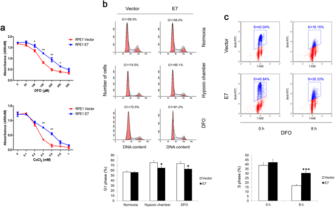
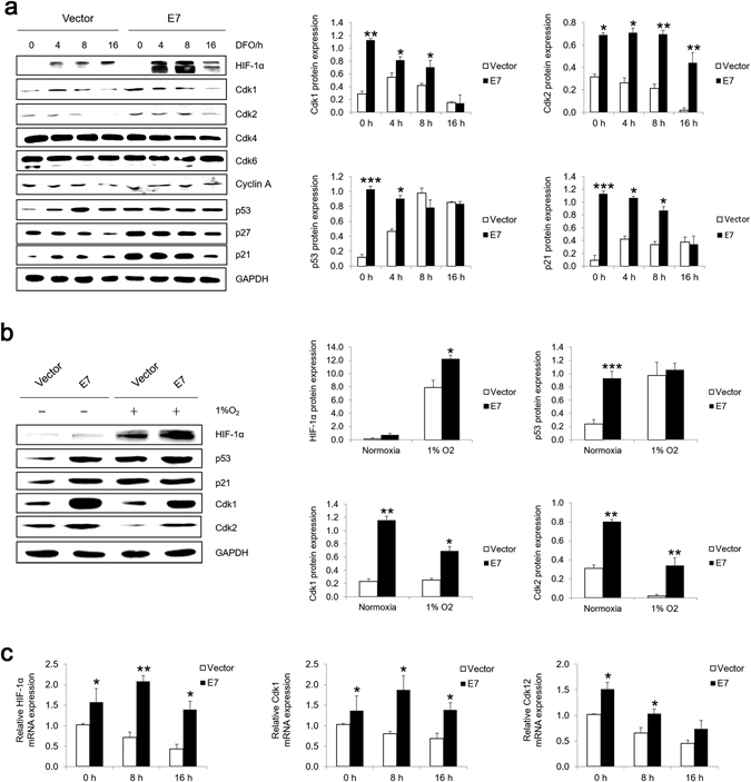
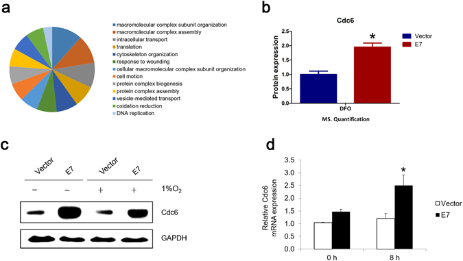
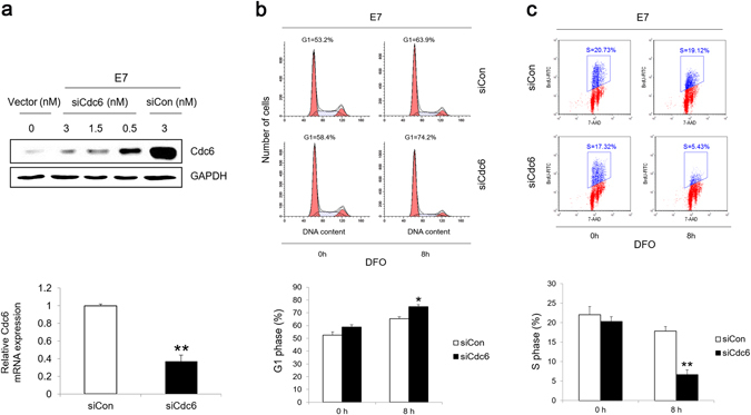
The human papillomavirus (HPV) plays a central role in cervical carcinogenesis and its oncogene E7 is essential in this process. We showed here that E7 abrogated the G1 cell cycle checkpoint under hypoxia and analyzed key cell cycle related proteins for their potential role in this process. To further explore the mechanism by which E7 bypasses hypoxia-induced G1 arrest, we applied a proteomic approach and used mass spectrometry to search for proteins that are differentially expressed in E7 expressing cells under hypoxia. Among differentially expressed proteins identified, Cdc6 is a DNA replication initiation factor and exhibits oncogenic activities when overexpressed. We have recently demonstrated that Cdc6 was required for E7-induced re-replication. Significantly, here we showed that Cdc6 played a role in E7-mediated G1 checkpoint abrogation under hypoxic condition, and the function could possibly be independent from its role in DNA replication initiation. This study uncovered a new function of Cdc6 in regulating cell cycle progression and has important implications in HPV-associated cancers.
Overview
The claims and statistics of this paper haven’t been checked yet.
Run the check — free, no account needed. It reads the open-access text and checks the sources it cites.
Integrity signals
Registry
Cites
Statistics
Figures
Verification
Claims the paper makes
The claims and statistics of this paper haven’t been checked yet.
Run the check — free, no account needed. It reads the open-access text and checks the sources it cites.
Claims other papers make about this one
Each is checked against this paper’s own text: does it say what the citing paper says it does?
In the citing paper
“this abrogation of p21Cip1 activity is less efficiently done by low-risk E7 [145,146]”
From The Not-So-Good, the Bad and the Ugly: HPV E5, E6 and E7 Oncoproteins in the Orchestration of Carcinogenesis (2021) · cites this paper as [146]
In the citing paper
“E7-expressing cells were shown to have upregulated cdc6 [143], which promotes cell cycle progression by activating cdk2 [144,145], and furthermore, cdc6 was shown to be important in the G1/S transition in E7-expressing cells under hypoxic conditions [146]”
From The Not-So-Good, the Bad and the Ugly: HPV E5, E6 and E7 Oncoproteins in the Orchestration of Carcinogenesis (2021) · cites this paper as [146]
Self-check
Percentages, sums and reported differences re-computed from the paper’s own numbers.
The self-check needs the paper’s full text.
Citations
Cites
The Regulation of INK4/ARF in Cancer and Aging
Kim, W. Y., & Sharpless, N. E. (2006) · Cell · doi:10.1016/j.cell.2006.10.003 · Cited by 1,009
HIF-1α induces cell cycle arrest by functionally counteracting Myc
Koshiji, M., Kageyama, Y., Pete, E. A., Horikawa, I., Barrett, J. C., & Huang, L. E. (2004) · The EMBO Journal · doi:10.1038/sj.emboj.7600196 · Cited by 652
A Nontranscriptional Role for HIF-1α as a Direct Inhibitor of DNA Replication
Hubbi, M. E., Kshitiz, Gilkes, D. M., Rey, S., Wong, C. C., Luo, W., Kim, D.-H., Dang, C. V., Levchenko, A., & Semenza, G. L. (2013) · Science Signaling · doi:10.1126/scisignal.2003417 · Cited by 116
Research works printed but not linked: 52
- zur Hausen, H. Papillomaviruses in the causation of human cancers - a brief historical account. Virology. 384, 260–265 (2009). · DOI · Paper page
- Baseman, J. G. & Koutsky, L. A. The epidemiology of human papillomavirus infections. Journal of clinical virology: the official publication of the Pan American Society for Clinical Virology. 32(Suppl 1), S16–24 (2005). · DOI
- Van Doorslaer, K. et al. The Papillomavirus Episteme: a central resource for papillomavirus sequence data and analysis. Nucleic acids research. 41, D571–578 (2013). · DOI
- de Villiers, E. M. Cross-roads in the classification of papillomaviruses. Virology. 445, 2–10 (2013). · DOI
- Burd, E. M. Human papillomavirus and cervical cancer. Clinical microbiology reviews. 16, 1–17 (2003). · DOI · Paper page
- Gao, G. et al. A novel RT-PCR method for quantification of human papillomavirus transcripts in archived tissues and its application in oropharyngeal cancer prognosis. International journal of cancer. 132, 882–890 (2013). · DOI
- Banerjee, N. S. et al. Conditionally activated E7 proteins of high-risk and low-risk human papillomaviruses induce S phase in postmitotic, differentiated human keratinocytes. Journal of virology. 80, 6517–6524 (2006). · DOI
- Chen, J. J., Reid, C. E., Band, V. & Androphy, E. J. Interaction of papillomavirus E6 oncoproteins with a putative calcium-binding protein. Science (New York, N.Y.). 269, 529–531 (1995). · DOI · Paper page
- Dyson, N., Howley, P. M., Munger, K. & Harlow, E. The human papilloma virus-16 E7 oncoprotein is able to bind to the retinoblastoma gene product. Science (New York, N.Y.). 243, 934–937 (1989). · DOI · Paper page
- Johnson, D. G., Schwarz, J. K., Cress, W. D. & Nevins, J. R. Expression of transcription factor E2F1 induces quiescent cells to enter S phase. Nature. 365, 349–352 (1993). · DOI
- Chen, J. J. Genomic Instability Induced By Human Papillomavirus Oncogenes. North American journal of medicine & science. 3, 43–47 (2010). · DOI
- Shin, M. K., Sage, J. & Lambert, P. F. Inactivating all three rb family pocket proteins is insufficient to initiate cervical cancer. Cancer research. 72, 5418–5427 (2012). · DOI
- Demers, G. W., Espling, E., Harry, J. B., Etscheid, B. G. & Galloway, D. A. Abrogation of growth arrest signals by human papillomavirus type 16 E7 is mediated by sequences required for transformation. Journal of virology. 70, 6862–6869 (1996). · DOI
- Fan, X. & Chen, J. J. Role of Cdk1 in DNA damage-induced G1 checkpoint abrogation by the human papillomavirus E7 oncogene. Cell cycle (Georgetown, Tex.). 13, 3249–3259 (2014). · DOI
- Zhou, Y. et al. Role of WDHD1 in Human Papillomavirus-Mediated Oncogenesis Identified by Transcriptional Profiling of E7-Expressing Cells. Journal of virology. 90, 6071–6084 (2016). · DOI
- Borlado, L. R. & Mendez, J. CDC6: from DNA replication to cell cycle checkpoints and oncogenesis. Carcinogenesis. 29, 237–243 (2008). · DOI
- Cocker, J. H., Piatti, S., Santocanale, C., Nasmyth, K. & Diffley, J. F. An essential role for the Cdc6 protein in forming the pre-replicative complexes of budding yeast. Nature. 379, 180–182 (1996). · DOI
- Kan, Q., Jinno, S., Yamamoto, H., Kobayashi, K. & Okayama, H. ATP-dependent activation of p21WAF1/CIP1-associated Cdk2 by Cdc6. Proceedings of the National Academy of Sciences of the United States of America. 105, 4757–4762 (2008). · DOI · Paper page
- Uranbileg, B. et al. Cdc6 protein activates p27KIP1-bound Cdk2 protein only after the bound p27 protein undergoes C-terminal phosphorylation. The Journal of biological chemistry. 287, 6275–6283 (2012). · DOI · Paper page
- Lau, E., Zhu, C., Abraham, R. T. & Jiang, W. The functional role of Cdc6 in S-G2/M in mammalian cells. EMBO reports. 7, 425–430 (2006). · DOI
- Hateboer, G. et al. Cell cycle-regulated expression of mammalian CDC6 is dependent on E2F. Molecular and cellular biology. 18, 6679–6697 (1998). · DOI
- Gonzalez, S. et al. Oncogenic activity of Cdc6 through repression of the INK4/ARF locus. Nature. 440, 702–706 (2006). · DOI
- Bonds, L., Baker, P., Gup, C. & Shroyer, K. R. Immunohistochemical localization of cdc6 in squamous and glandular neoplasia of the uterine cervix. Archives of pathology & laboratory medicine. 126, 1164–1168 (2002). · DOI
- Fan, X., Zhou, Y. & Chen, J. J. Role of Cdc6 in re-replication in cells expressing human papillomavirus E7 oncogene. Carcinogenesis. 37, 799–809 (2016). · DOI · Paper page
- Zhong, H. et al. Overexpression of hypoxia-inducible factor 1alpha in common human cancers and their metastases. Cancer research. 59, 5830–5835 (1999).
- Pugh, C. W. & Ratcliffe, P. J. Regulation of angiogenesis by hypoxia: role of the HIF system. Nature medicine. 9, 677–684 (2003). · DOI · Paper page
- Cho, Y. S. et al. HIF-1alpha controls keratinocyte proliferation by up-regulating p21(WAF1/Cip1). Biochimica et biophysica acta. 1783, 323–333 (2008). · DOI
- Gardner, L. B. et al. Hypoxia inhibits G1/S transition through regulation of p27 expression. The Journal of biological chemistry. 276, 7919–7926 (2001). · DOI
- Bodily, J. M., Mehta, K. P. & Laimins, L. A. Human papillomavirus E7 enhances hypoxia-inducible factor 1-mediated transcription by inhibiting binding of histone deacetylases. Cancer research. 71, 1187–1195 (2011). · DOI · Paper page
- Nakamura, M. et al. Hypoxia-specific stabilization of HIF-1alpha by human papillomaviruses. Virology. 387, 442–448 (2009). · DOI
- Rodolico, V. et al. Hypoxia inducible factor-1 alpha expression is increased in infected positive HPV16 DNA oral squamous cell carcinoma and positively associated with HPV16 E7 oncoprotein. Infectious agents and cancer. 6, 18 (2011). · DOI
- Moon, S. U. et al. The expression of human papillomavirus type 16 (HPV16 E7) induces cell cycle arrest and apoptosis in radiation and hypoxia resistant glioblastoma cells. Molecular medicine reports. 4, 1247–1253 (2011). · DOI
- Bagwell, C. B. et al. Optimizing flow cytometric DNA ploidy and S-phase fraction as independent prognostic markers for node-negative breast cancer specimens. Cytometry. 46, 121–135 (2001). · DOI
- Zhou, N., Yuan, S., Wang, R., Zhang, W. & Chen, J. J. Role of dual specificity tyrosine-phosphorylation-regulated kinase 1B (Dyrk1B) in S-phase entry of HPV E7 expressing cells from quiescence. Oncotarget. 6, 30745–30761 (2015). · DOI
- Gordan, J. D., Bertout, J. A., Hu, C. J., Diehl, J. A. & Simon, M. C. HIF-2alpha promotes hypoxic cell proliferation by enhancing c-myc transcriptional activity. Cancer cell. 11, 335–347 (2007). · DOI · Paper page
- Goda, N. et al. Hypoxia-inducible factor 1alpha is essential for cell cycle arrest during hypoxia. Molecular and cellular biology. 23, 359–369 (2003). · DOI
- Gordan, J. D., Thompson, C. B. & Simon, M. C. HIF and c-Myc: sibling rivals for control of cancer cell metabolism and proliferation. Cancer cell. 12, 108–113 (2007). · DOI
- Fan, X., Liu, Y., Heilman, S. A. & Chen, J. J. Human papillomavirus E7 induces rereplication in response to DNA damage. Journal of virology. 87, 1200–1210 (2013). · DOI
- Tang, S., Tao, M., McCoy, J. P. Jr. & Zheng, Z. M. The E7 oncoprotein is translated from spliced E6*I transcripts in high-risk human papillomavirus type 16- or type 18-positive cervical cancer cell lines via translation reinitiation. Journal of virology. 80, 4249–4263 (2006). · DOI
- Tang, S., Tao, M., McCoy, J. P. Jr. & Zheng, Z. M. Short-term induction and long-term suppression of HPV16 oncogene silencing by RNA interference in cervical cancer cells. Oncogene. 25, 2094–2104 (2006). · DOI
- Zhang, W. et al. The human papillomavirus type 58 E7 oncoprotein modulates cell cycle regulatory proteins and abrogates cell cycle checkpoints. Virology. 397, 139–144 (2010). · DOI
- Wu, R., Abramson, A. L., Symons, M. H. & Steinberg, B. M. Pak1 and Pak2 are activated in recurrent respiratory papillomas, contributing to one pathway of Rac1-mediated COX-2 expression. International journal of cancer. 127, 2230–2237 (2010). · DOI
- Yiu, G. K. et al. SPARC (secreted protein acidic and rich in cysteine) induces apoptosis in ovarian cancer cells. The American journal of pathology. 159, 609–622 (2001). · DOI
- Songock, W. K., Kim, S. M. & Bodily, J. M. The human papillomavirus E7 oncoprotein as a regulator of transcription. Virus research (2016). · DOI
- Hu, D. et al. HPV-16 E6/E7 promotes cell migration and invasion in cervical cancer via regulating cadherin switch in vitro and in vivo. Archives of gynecology and obstetrics. 292, 1345–1354 (2015). · DOI
- Roman, A. & Munger, K. The papillomavirus E7 proteins. Virology. 445, 138–168 (2013). · DOI
- Munger, K. et al. Mechanisms of human papillomavirus-induced oncogenesis. Journal of virology. 78, 11451–11460 (2004). · DOI · Paper page
- De Marco, F. Oxidative stress and HPV carcinogenesis. Viruses. 5, 708–731 (2013). · DOI
- Zerfass-Thome, K. et al. Inactivation of the cdk inhibitor p27KIP1 by the human papillomavirus type 16 E7 oncoprotein. Oncogene. 13, 2323–2330 (1996).
- Massimi, P. & Banks, L. Repression of p53 transcriptional activity by the HPV E7 proteins. Virology. 227, 255–259 (1997). · DOI
- Kelly, T. J., Nurse, P. & Forsburg, S. L. Coupling DNA replication to the cell cycle. Cold Spring Harbor symposia on quantitative biology. 58, 637–644 (1993). · DOI
- Tada, S. Cdt1 and geminin: role during cell cycle progression and DNA damage in higher eukaryotes. Frontiers in bioscience: a journal and virtual library. 12, 1629–1641 (2007). · DOI
Cited by
Basukala, O., & Banks, L. (2021) · Viruses · doi:10.3390/v13101892 · Cited by 137
Metadata
- Authors
- Chen, H., Zhang, Q., Qiao, L., Fan, X., Zhang, W., Zhao, W., & Chen, J. J.
- Venue
- Scientific Reports
- Published
- 2017-06-07
- Volume
- 7
- Issue
- 1
- ISSN
- 2045-2322
- PMC
- PMC5462782
- Cited by
- 10
- References
- 3
- Language
- en
About these checks
Registry, retraction, Cites and statistics results are computed, not judged, with one exception: a printed reference that no index lists is read once by a model before it counts as a citation error. Claims are extracted by a model and kept only when their quote appears verbatim in the text; each cited source is then checked, with a model’s help, for whether it says what the claim relies on. Those verdicts can be wrong — read the claim’s quote and the cited source before relying on one.
Statistics and arithmetic are recomputed, not judged.
Confidence bands are shown only for fields calibrated on our annotated test set. None is calibrated yet, so no band is shown.
What counts as a claim ›How we measure confidence ›How we check this ›
Are you an author, or do you see a problem with this page? Request a review
