

The DOI matches its Crossref record; 51 of the 51 references it cites were found in a registry or an index.
Activation-induced cytidine deaminase an antibody diversification enzyme interacts with chromatin modifier UBN1 in B-cells
Open accessPublished version of a preprint
Abstract
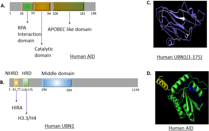
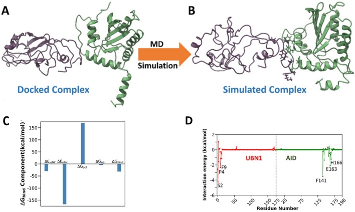
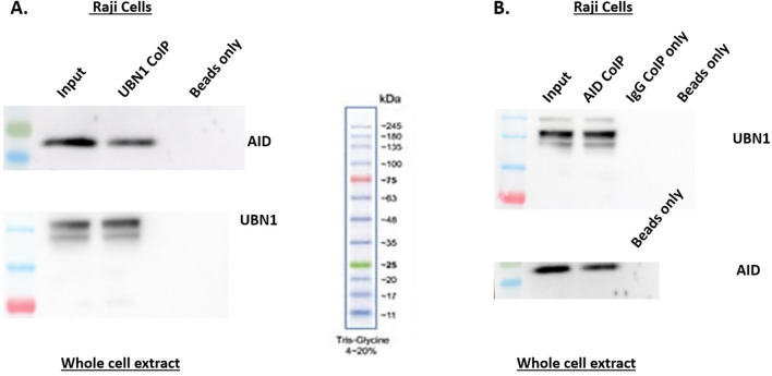
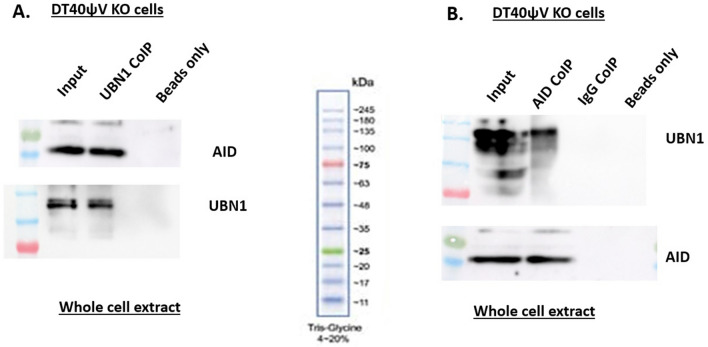
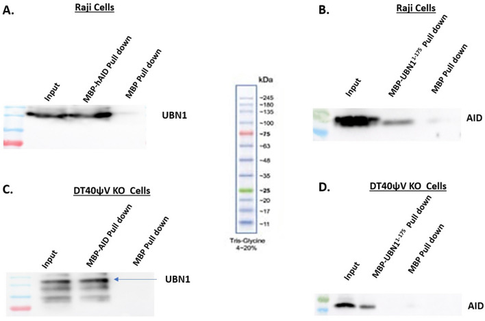
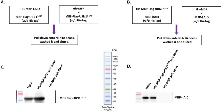
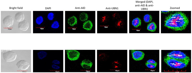
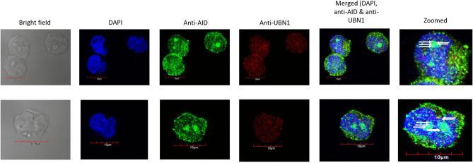
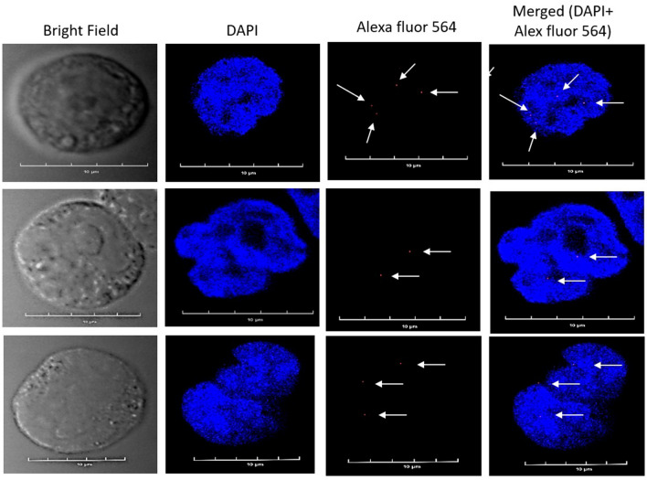
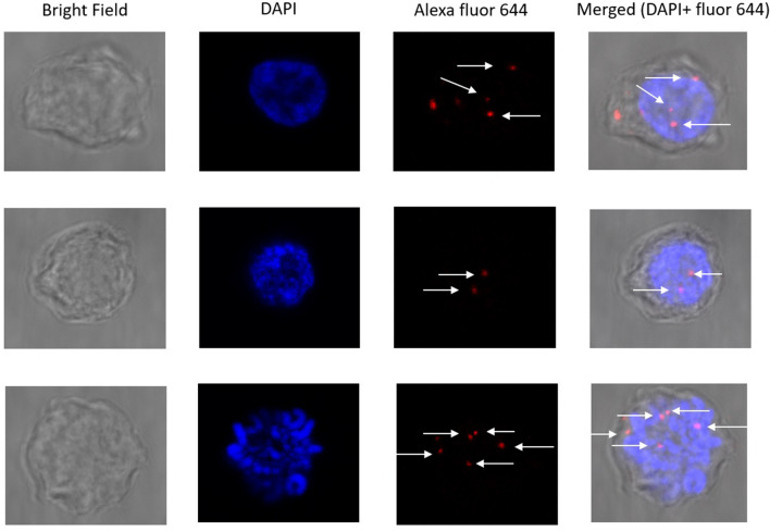
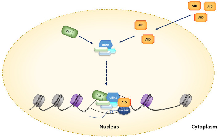
Activation-induced cytidine deaminase (AID) is the key mediator of antibody diversification in activated B-cells by the process of somatic hypermutation (SHM) and class switch recombination (CSR). Targeting AID to the Ig genes requires transcription (initiation and elongation), enhancers, and its interaction with numerous factors. Furthermore, the HIRA chaperon complex, a regulator of chromatin architecture, is indispensable for SHM. The HIRA chaperon complex consists of UBN1, ASF1a, HIRA, and CABIN1 that deposit H3.3 onto the DNA, the SHM hallmark. We explored whether UBN1 interacts with AID using computational and in-vitro experiments. Interestingly, our in-silico studies, such as molecular docking and molecular dynamics simulation results, predict that AID interacts with UBN1. Subsequently, co-immunoprecipitation and pull-down experiments established interactions between UBN1 and AID inside B-cells. Additionally, a double immunofluorescence assay confirmed that AID and UBN1 were co-localized in the human and chicken B-cell lines. Moreover, proximity ligation assay studies validated that AID interacts with UBN1. Ours is the first report on the interaction of genome mutator enzyme AID with UBN1. Nevertheless, the fate of interaction between UBN1 and AID is yet to be explored in the context of SHM or CSR.
Overview
The claims and statistics of this paper haven’t been checked yet.
Run the check — free, no account needed. It reads the open-access text and checks the sources it cites.
Integrity signals
Registry
Cites
Statistics
Figures
Verification
Claims the paper makes
The claims and statistics of this paper haven’t been checked yet.
Run the check — free, no account needed. It reads the open-access text and checks the sources it cites.
Self-check
Percentages, sums and reported differences re-computed from the paper’s own numbers.
The self-check needs the paper’s full text.
Citations
Cites
Highly accurate protein structure prediction with AlphaFold
Jumper, J., Evans, R., Pritzel, A., Green, T., Figurnov, M., Ronneberger, O., Tunyasuvunakool, K., Bates, R., Žídek, A., Potapenko, A., et al. (2021) · Nature · doi:10.1038/s41586-021-03819-2 · Cited by 43,764
Particle mesh Ewald: An N ⋅log( N ) method for Ewald sums in large systems
Darden, T., York, D., & Pedersen, L. (1993) · The Journal of Chemical Physics · doi:10.1063/1.464397 · Cited by 30,546
ff14SB: Improving the Accuracy of Protein Side Chain and Backbone Parameters from ff99SB
Maier, J. A., Martinez, C., Kasavajhala, K., Wickstrom, L., Hauser, K. E., & Simmerling, C. (2015) · Journal of Chemical Theory and Computation · doi:10.1021/acs.jctc.5b00255 · Cited by 10,830
The Amber biomolecular simulation programs
Case, D. A., Cheatham, T. E., Darden, T., Gohlke, H., Luo, R., Merz, K. M., Onufriev, A., Simmerling, C., Wang, B., & Woods, R. J. (2005) · Journal of Computational Chemistry · doi:10.1002/jcc.20290 · Cited by 9,535
PTRAJ and CPPTRAJ: Software for Processing and Analysis of Molecular Dynamics Trajectory Data
Roe, D. R., & Cheatham, T. E. (2013) · Journal of Chemical Theory and Computation · doi:10.1021/ct400341p · Cited by 7,382
I-TASSER: a unified platform for automated protein structure and function prediction
Roy, A., Kucukural, A., & Zhang, Y. (2010) · Nature Protocols · doi:10.1038/nprot.2010.5 · Cited by 6,170
The I-TASSER Suite: protein structure and function prediction
Yang, J., Yan, R., Roy, A., Xu, D., Poisson, J., & Zhang, Y. (2015) · Nature Methods · doi:10.1038/nmeth.3213 · Cited by 5,596
Muramatsu, M., Kinoshita, K., Fagarasan, S., Yamada, S., Shinkai, Y., & Honjo, T. (2000) · Cell · doi:10.1016/s0092-8674(00)00078-7 · Cited by 3,474
Free energy calculations: Applications to chemical and biochemical phenomena
Kollman, P. (1993) · Chemical Reviews · doi:10.1021/cr00023a004 · Cited by 2,101
A modified TIP3P water potential for simulation with Ewald summation
Price, D. J., & Brooks, C. L. (2004) · The Journal of Chemical Physics · doi:10.1063/1.1808117 · Cited by 1,676
Revy, P., Muto, T., Levy, Y., Geissmann, F., Plebani, A., Sanal, O., Catalan, N., Forveille, M., Dufourcq-Lagelouse, R., Gennery, A., et al. (2000) · Cell · doi:10.1016/s0092-8674(00)00079-9 · Cited by 1,647
Distinct Factors Control Histone Variant H3.3 Localization at Specific Genomic Regions
Goldberg, A. D., Banaszynski, L. A., Noh, K.-M., Lewis, P. W., Elsaesser, S. J., Stadler, S., Dewell, S., Law, M., Guo, X., Li, X., et al. (2010) · Cell · doi:10.1016/j.cell.2010.01.003 · Cited by 1,280
Gohlke, H., Kiel, C., & Case, D. A. (2003) · Journal of Molecular Biology · doi:10.1016/s0022-2836(03)00610-7 · Cited by 1,192
The Histone Variant H3.3 Marks Active Chromatin by Replication-Independent Nucleosome Assembly
Ahmad, K., & Henikoff, S. (2002) · Molecular Cell · doi:10.1016/s1097-2765(02)00542-7 · Cited by 1,112
An analysis of the accuracy of Langevin and molecular dynamics algorithms
Pastor, R. W., Brooks, B. R., & Szabo, A. (1988) · Molecular Physics · doi:10.1080/00268978800101881 · Cited by 1,043
Béguelin, W., Popovic, R., Teater, M., Jiang, Y., Bunting, K. L., Rosen, M., Shen, H., Yang, S. N., Wang, L., Ezponda, T., et al. (2013) · Cancer Cell · doi:10.1016/j.ccr.2013.04.011 · Cited by 846
The RosettaDock server for local protein-protein docking
Lyskov, S., & Gray, J. J. (2008) · Nucleic Acids Research · doi:10.1093/nar/gkn216 · Cited by 592
Altering the pathway of immunoglobulin hypermutation by inhibiting uracil-DNA glycosylase
Di Noia, J., & Neuberger, M. S. (2002) · Nature · doi:10.1038/nature00981 · Cited by 587
Lyskov, S., Chou, F.-C., Conchúir, S. Ó., Der, B. S., Drew, K., Kuroda, D., Xu, J., Weitzner, B. D., Renfrew, P. D., Sripakdeevong, P., et al. (2013) · PLoS ONE · doi:10.1371/journal.pone.0063906 · Cited by 431
Somatic Hypermutation of Immunoglobulin Genes Is Linked to Transcription Initiation
Peters, A., & Storb, U. (1996) · Immunity · doi:10.1016/s1074-7613(00)80298-8 · Cited by 426
HIRA Is Critical for a Nucleosome Assembly Pathway Independent of DNA Synthesis
Ray-Gallet, D., Quivy, J.-P., Scamps, C., Martini, E. M.-D., Lipinski, M., & Almouzni, G. (2002) · Molecular Cell · doi:10.1016/s1097-2765(02)00526-9 · Cited by 425
Ray-Gallet, D., Woolfe, A., Vassias, I., Pellentz, C., Lacoste, N., Puri, A., Schultz, D. C., Pchelintsev, N. A., Adams, P. D., Jansen, L. E. T., et al. (2011) · Molecular Cell · doi:10.1016/j.molcel.2011.12.006 · Cited by 387
Betz, A. G., Milstein, C., González-Fernández, A., Pannell, R., Larson, T., & Neuberger, M. S. (1994) · Cell · doi:10.1016/0092-8674(94)90316-6 · Cited by 379
Pavri, R., Gazumyan, A., Jankovic, M., Di Virgilio, M., Klein, I., Ansarah-Sobrinho, C., Resch, W., Yamane, A., San-Martin, B. R., Barreto, V., et al. (2010) · Cell · doi:10.1016/j.cell.2010.09.017 · Cited by 378
Replication protein A interacts with AID to promote deamination of somatic hypermutation targets
Chaudhuri, J., Khuong, C., & Alt, F. W. (2004) · Nature · doi:10.1038/nature02821 · Cited by 378
Ito, S., Nagaoka, H., Shinkura, R., Begum, N., Muramatsu, M., Nakata, M., & Honjo, T. (2004) · Proceedings of the National Academy of Sciences · doi:10.1073/pnas.0307335101 · Cited by 304
Separate domains of AID are required for somatic hypermutation and class-switch recombination
Shinkura, R., Ito, S., Begum, N. A., et al. (2004) · Nature Immunology · doi:10.1038/ni1086 · Cited by 233
Rogozin, I. B., & Diaz, M. (2004) · The Journal of Immunology · doi:10.4049/jimmunol.172.6.3382 · Cited by 214
Gowrishankar, B., Somaiah, N., Zhang, R., Tang, Y., Hoffmann, J., Andrake, M., Ceulemans, H., Schultz, D., Marmorstein, R., & Adams, P. D. (2008) · Molecular and Cellular Biology · doi:10.1128/mcb.01047-08 · Cited by 156
Sherwood, P. W., Tsang, S., & Osley, M. A. (1993) · Molecular and Cellular Biology · doi:10.1128/mcb.13.1.28 · Cited by 153
Bransteitter, R., Pham, P., Calabrese, P. P., & Goodman, M. F. (2004) · Journal of Biological Chemistry · doi:10.1074/jbc.m408135200 · Cited by 151
Interaction between Antibody-Diversification Enzyme AID and Spliceosome-Associated Factor CTNNBL1
Conticello, S. G., Ganesh, K., Xue, K., Lu, M., Rada, C. A., & Neuberger, M. S. (2008) · Molecular Cell · doi:10.1016/j.molcel.2008.07.009 · Cited by 145
Nowak, U. M., Matthews, A. J., Zheng, S., & Chaudhuri, J. (2010) · Nature Immunology · doi:10.1038/ni.1977 · Cited by 131
DNA Methylation Dynamics of Germinal Center B Cells Are Mediated by AID
Domínguez, P. M., Teater, M. R., Chambwe, N., Kormáksson, M., Redmond, D., Ishii, J. K., Vuong, B. Q., Chaudhuri, J., Melnick, A. M., Vasanthakumar, A., et al. (2015) · Cell Reports · doi:10.1016/j.celrep.2015.08.036 · Cited by 117
RPA Interacts with HIRA and Regulates H3.3 Deposition at Gene Regulatory Elements in Mammalian Cells
Zhang, H., Gan, H., Wang, Z., Lee, J., Zhou, H., Ördög, T., Wold, M. S., Ljungman, M. E., & Zhang, Z. (2017) · Molecular Cell · doi:10.1016/j.molcel.2016.11.030 · Cited by 101
Xiong, C., Wen, Z., Yu, J., Chen, J., Liu, C.-P., Zhang, X., Chen, P., Xu, R.-M., & Li, G. (2018) · BMC Biology · doi:10.1186/s12915-018-0573-9 · Cited by 56
Aida, M., Hamad, N., Stanlie, A., et al. (2013) · Proceedings of the National Academy of Sciences · doi:10.1073/pnas.1305859110 · Cited by 49
Changes in RNA polymerase II progression influence somatic hypermutation of Ig-related genes by AID
Kodgire, P., Mukkawar, P., Ratnam, S., Martin, T. E., & Storb, U. (2013) · The Journal of Experimental Medicine · doi:10.1084/jem.20121523 · Cited by 48
Self-citationNagaoka, H., Tran, T. H., Kobayashi, M., Aida, M., & Honjo, T. (2010) · International Immunology · doi:10.1093/intimm/dxq023 · Cited by 44
Quinlan, E. M., King, J. J., Amemiya, C. T., Hsu, E., & Larijani, M. (2017) · Molecular and Cellular Biology · doi:10.1128/mcb.00077-17 · Cited by 31
Kanehiro, Y., Todo, K., 根岸, 美., Fukuoka, J., Gan, W., Hikasa, T., Kaga, Y., Takemoto, M., Magari, M., Li, X., et al. (2012) · Proceedings of the National Academy of Sciences · doi:10.1073/pnas.1120368109 · Cited by 30
Romanello, M. B., Schiavone, D., Frey, A., & Sale, J. E. (2016) · The EMBO Journal · doi:10.15252/embj.201693958 · Cited by 28
Roy, R., Mishra, A., Poddar, S., Nayak, D., & Kar, P. (2020) · Journal of Biomolecular Structure and Dynamics · doi:10.1080/07391102.2020.1838327 · Cited by 26
Self-citationNucleosome Stability Dramatically Impacts the Targeting of Somatic Hypermutation
Kodgire, P., Mukkawar, P., North, J. A., Poirier, M. G., & Storb, U. (2012) · Molecular and Cellular Biology · doi:10.1128/mcb.06722-11 · Cited by 25
Self-citationYu, G., Zhang, Y., Gupta, V., Zhang, J., MacCarthy, T., Duan, Z., & Scharff, M. D. (2021) · Proceedings of the National Academy of Sciences · doi:10.1073/pnas.2114743118 · Cited by 21
Chelico, L., Pham, P., Petruska, J. A., et al. (2009) · Journal of Biological Chemistry · doi:10.1074/jbc.r109.052449 · Cited by 18
Pham, P., Afif, S. A., Shimoda, M., Maeda, K., Sakaguchi, N., Pedersen, L. C., & Goodman, M. F. (2017) · DNA repair · doi:10.1016/j.dnarep.2017.03.007 · Cited by 16
Singh, A. K., Tamrakar, A., Jaiswal, A. K., Kanayama, N., & Kodgire, P. (2020) · Molecular Immunology · doi:10.1016/j.molimm.2020.01.005 · Cited by 13
Self-citationSingh, A. K., Tamrakar, A., Jaiswal, A. K., Kanayama, N., Agarwal, A., Tripathi, P., & Kodgire, P. (2019) · Molecular Immunology · doi:10.1016/j.molimm.2019.10.002 · Cited by 11
Self-citationRoy, R., Jonniya, N. A., Poddar, S., Sk, M. F., & Kar, P. (2021) · Journal of Chemical Information and Modeling · doi:10.1021/acs.jcim.1c00918 · Cited by 11
Self-citationAID preferentially targets the top strand in nucleosome sequences
Singh, A. K., Jaiswal, A. K., & Kodgire, P. (2019) · Molecular Immunology · doi:10.1016/j.molimm.2019.05.015 · Cited by 5
Self-citation
Research works printed but not linked: 1
- Kräutler, V., van Gunsteren, W. F. & Hünenberger, P. H. A fast SHAKE algorithm to solve distance constraint equations for small molecules in molecular dynamics simulations. J. Comput. Chem. 22(5), 501–508. https://doi.org/10.1002/1096-987X(20010415)22:5%3C501::AID-JCC1021%3E3.0.CO (2001). · DOI
Metadata
- Authors
- Jaiswal, A., Roy, R., Tamrakar, A., Singh, A. K., Kar, P., & Kodgire, P.
- Affiliations
- Indian Institute of Technology Indore (IN)
- Corresponding author
- Kodgire, Prashant
- Venue
- Scientific Reports
- Published
- 2023-11-10
- Volume
- 13
- Issue
- 1
- Pages
- 19615-19615
- ISSN
- 2045-2322 (ISSN-L)
- PMC
- PMC10638239
- OpenAlex
- W4388573268
- Cited by
- 3
- References
- 51
- Citations by year
- 2026: 2 · 2025: 1
- FWCI
- 0.27
- Type
- journal article
- Language
- en
- Open access
- gold
- doi.org
- Licence
- cc-by
- creativecommons.org/licenses/by/4.0 (tdm)
- creativecommons.org/licenses/by/4.0 (vor)
- Locations
- Scientific Reports (publishedVersion, cc-by)
- Research Square (acceptedVersion, cc-by)
- PubMed Central (publishedVersion, cc-by)
- PubMed (publishedVersion)
- DOAJ (DOAJ: Directory of Open Access Journals) (submittedVersion, cc-by-sa)
- Funders
- Science and Engineering Research Board (CRG/2020/358)
- Indian Council of Medical Research (OMI/17/2020-ECD-1)
- Topics
- Life Sciences › Biochemistry, Genetics and Molecular Biology › Molecular Biology › Genomics and Chromatin Dynamics
- Life Sciences › Biochemistry, Genetics and Molecular Biology › Molecular Biology › DNA Repair Mechanisms
- Life Sciences › Immunology and Microbiology › Immunology › T-cell and B-cell Immunology
- Keywords
- activation-induced cytidine deaminase, antibody diversification, somatic hypermutation, class switch recombination, B cells, protein-protein interaction, chromatin modification, immunoglobulin genes
- MeSH terms
- AICDA (Activation-Induced Cytidine Deaminase), Chromatin, Cytidine Deaminase, Humans, Immunoglobulins, Nuclear Proteins, Transcription Factors, Immunoglobulin Class Switching, Molecular Chaperones, Somatic Hypermutation, Immunoglobulin, Molecular Docking Simulation
About these checks
Registry, retraction, Cites and statistics results are computed, not judged, with one exception: a printed reference that no index lists is read once by a model before it counts as a citation error. Claims are extracted by a model and kept only when their quote appears verbatim in the text; each cited source is then checked, with a model’s help, for whether it says what the claim relies on. Those verdicts can be wrong — read the claim’s quote and the cited source before relying on one.
Statistics and arithmetic are recomputed, not judged.
Confidence bands are shown only for fields calibrated on our annotated test set. None is calibrated yet, so no band is shown.
What counts as a claim ›How we measure confidence ›How we check this ›
Are you an author, or do you see a problem with this page? Request a review
Data sources
Bibliographic data: Semantic Scholar, OpenAlex, Crossref.
Contains information from the Semantic Scholar Academic Graph Datasets, made available under the ODC Attribution License (ODC-BY 1.0).
Semantic Scholar’s data is described in: Kinney, R., et al. (2023). The Semantic Scholar Open Data Platform. arXiv:2301.10140.
