

The DOI matches its Crossref record; 24 of the 25 references it cites were found in a registry or an index; none of the 34 sources its claims cite is checked for support yet.
Evaluation of the genotoxic and transformation potential induced by asbestos compared to cleavage fragments
Open accessAbstract
The World Health Organization has confirmed that asbestos fibres are carcinogenic, claiming that asbestos-related diseases should be eradicated worldwide. Actinolite, amosite, anthophyllite, chrysotile, crocidolite, and tremolite are regulated asbestiform mineral phases. However, in nature, asbestos minerals occur either in a fibrous and asbestiform (original morphology characterized by high length-to-width ratio and provided of high tensile strength and flexibility) or fibrous but not asbestiform appearance. This study used human epithelial cancer cells (A549) and a mouse fibroblast cell line (Balb/c 3T3) to compare the genotoxic and carcinogenic effects of a sample of amphibole asbestos with samples of fibrous not asbestiform named cleavage fragments (CV) obtained by grinding non-asbestiform amphiboles. The results showed that exposure of alveolar lung cells to asbestos and elongated mineral particles, in the habit of cleavage fragments (CF) derived from the grinding of non-asbestiform amphiboles and serpentines, causes cytotoxic effects, oxidative stress and genotoxic damage. Moreover, CF obtained from an actinolite schist induces a transformation effect in the Balb/c 3T3 model. Together, these findings highlight the importance of considering CF as a potential threat to human health since it can cause genotoxic damage by triggering cellular transformation processes that overlap with the mechanisms involved in the carcinogenesis processes of asbestos.
Overview
AI snapshot
Written from the paper’s full text.
TL;DR
This in vitro study compared amphibole asbestos (F3) with non-asbestiform cleavage fragments (A1, A3, A4, A5) in A549 and Balb/c 3T3 cells, finding cytotoxicity, oxidative stress, DNA damage, and transformation effects for some CFs. The authors conclude that CFs may pose human health risks overlapping with asbestos carcinogenesis mechanisms.
Key findings
- Grinding non-asbestiform amphiboles produced cleavage fragments without asbestiform phases; A1, A3, A4, and A5 had average length-to-width ratios of 9, 18, 23, and 13, respectively, while asbestiform F3 had an average ratio of 36. Only 2 of 500 measured elongated mineral particles had a ratio < 3. Short fibers (<5 μm) were 28% in A1, 45% in A3, 14% in A4, and 2% in A5.
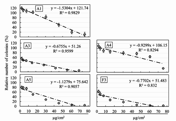
- All samples reduced clonogenic capacity with increasing concentration. IC50 values were 1.90 µg/cm² for F3, 46.88 for A1, 1.90 for A3, 60.38 for A4, and 22.73 for A5; no-effects doses were 0.40, 14.20, 0.40, 6.61, and 1.90 µg/cm², respectively.
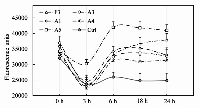
- In A549 cells, ROS increased from 3 to 6 h and then plateaued; at 6 h all samples reached P < 0.01. At 24 h, F3 and A5 showed further positive trends (P < 0.001), while A3, A1, and A4 showed declines or maintenance (P < 0.05 to P < 0.01).
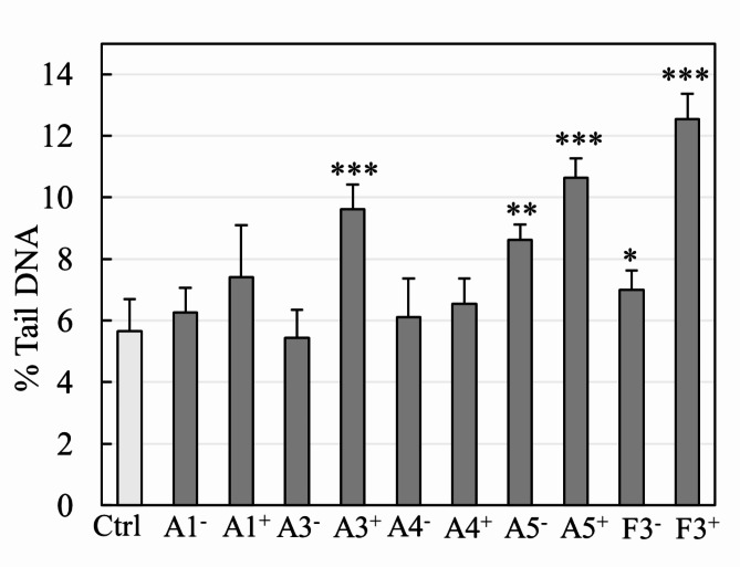
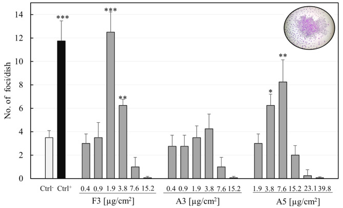
- Significant DNA damage occurred only with A3, A5, and F3. A3 induced damage at IC50 (P < 0.001); A5 induced damage at both no-effects dose (P < 0.01) and IC50 (P < 0.001); F3 followed the same trend as A5 with higher damage (F3 vs. A5 P < 0.01). In transformation assays, F3 was transforming at 1.9 µg/cm² (P < 0.001) and 3.8 µg/cm² (P < 0.01); A5 transformed at 3.8 µg/cm² (P < 0.05) and peaked at 7.6 µg/cm² (P < 0.01); A3 showed no transforming effect.
Methods
- Mineral samples included non-asbestiform amphiboles A1, A3, A4, and A5 and asbestiform amphibole F3; A2 is listed in Table 4 but not tested in the reported assays. Characterization used optical microscopy, SEM-EDS, µ-Raman, and ζ-potential. CFs were prepared by grinding, sieving through 63 μm, micronizing, freeze-drying, sterilizing, and sonicating before cell exposure.
- A549 human epithelial cancer cells were used for DCF-DA redox status and alkaline comet assays; Balb/c 3T3 cells were used for clonogenic and cell transformation assays. Cells were exposed to varying concentrations and times, with untreated cells as negative controls and F3 or 3-methylcholanthrene as positive controls where applicable.
- Statistical analysis used JMP version 17, one-way ANOVA with Bonferroni post hoc testing, and P < 0.05 as significant.
Limitations
- The paper states that further in vivo studies are needed to better understand EMP actions and clarify etiopathological mechanisms induced by inhalation, and that reassessment of current legislation may eventually be necessary.
- Evident from the text: all findings are in vitro using A549 and Balb/c 3T3 cell lines, with no in vivo or human exposure data.
- Evident from the text: transformation tests were performed only on F3, A3, and A5, not on A1 or A4, and sample A2 appears in the sample table but is not included in the reported biological results.
Model-written summary. Check it against the paper before relying on it.
Integrity signals
Registry
Cites
Statistics
Figures
Verification
Claims the paper makes
52 Own result · 9 Check pending · 8 Own interpretation · 6 No full text of the sources · 6 Own method · 5 No basis given · 4 Sources not found · 4 Not checked yet
A result the paper reports from its own data — it cites no other source for it, so there is none to check. The quote shows it is in the paper.
In the paper
“The ROS production in cultured cells treated with asbestos or CFs highlights the capacity to trigger oxidative stress by contact with the cell epithelium.”
§ Evaluation of the genotoxic and transformation potential induced by asbestos compared to cleavage fragments > Discussion and conclusions
plainstates a cause- Asserted: passes — The authors put it forward as true.
- Checkable: passes — It can be checked against evidence.
- Load-bearing: passes — The paper’s conclusions depend on it.
- Based: passes — The paper gives a reason why it holds.
A result the paper reports from its own data — it cites no other source for it, so there is none to check. The quote shows it is in the paper.
In the paper
“Only two Elongated Mineral Particles on 500 measured have L: D ratio < 3 (from samples A4 and A5).”
§ Evaluation of the genotoxic and transformation potential induced by asbestos compared to cleavage fragments > Results > Fibres morphology
plain- Asserted: passes — The authors put it forward as true.
- Checkable: passes — It can be checked against evidence.
- Load-bearing: passes — The paper’s conclusions depend on it.
- Based: passes — The paper gives a reason why it holds.
A result the paper reports from its own data — it cites no other source for it, so there is none to check. The quote shows it is in the paper.
In the paper
“In this regard, the genotoxic damage after A5 exposure reaches values like those reported for cells exposed to asbestiform amphiboles.”
§ Evaluation of the genotoxic and transformation potential induced by asbestos compared to cleavage fragments > Discussion and conclusions
plain- Asserted: passes — The authors put it forward as true.
- Checkable: passes — It can be checked against evidence.
- Load-bearing: passes — The paper’s conclusions depend on it.
- Based: passes — The paper gives a reason why it holds.
A result the paper reports from its own data — it cites no other source for it, so there is none to check. The quote shows it is in the paper.
In the paper
“The results show a significant increase in DNA damage only in the cells treated with A3, A5, and F3 samples.”
§ Evaluation of the genotoxic and transformation potential induced by asbestos compared to cleavage fragments > Results > Genotoxic damage
plainstates a cause- Asserted: passes — The authors put it forward as true.
- Checkable: passes — It can be checked against evidence.
- Load-bearing: passes — The paper’s conclusions depend on it.
- Based: passes — The paper gives a reason why it holds.
A result the paper reports from its own data — it cites no other source for it, so there is none to check. The quote shows it is in the paper.
In the paper
“The results showed that in vitro exposure of alveolar cells to asbestos, besides causing cytotoxic effects, oxidative stress, and genotoxic damage, induced a transforming effect in the Balb/c 3T3 model.”
§ Evaluation of the genotoxic and transformation potential induced by asbestos compared to cleavage fragments > Discussion and conclusions
plainstates a cause- Asserted: passes — The authors put it forward as true.
- Checkable: passes — It can be checked against evidence.
- Load-bearing: passes — The paper’s conclusions depend on it.
- Based: passes — The paper gives a reason why it holds.
A result the paper reports from its own data — it cites no other source for it, so there is none to check. The quote shows it is in the paper.
In the paper
“Briefly, it is possible to appreciate a clonogenic capacity decrease per all samples tested, consequently correlating to the concentration of CF to which the cell cultures are exposed.”
§ Evaluation of the genotoxic and transformation potential induced by asbestos compared to cleavage fragments > Results > Clonogenic assay
hedgedstates a cause- Asserted: passes — The authors put it forward as true.
- Checkable: passes — It can be checked against evidence.
- Load-bearing: unclear — It is unclear whether the paper’s conclusions depend on it.
- Based: passes — The paper gives a reason why it holds.
A result the paper reports from its own data — it cites no other source for it, so there is none to check. The quote shows it is in the paper.
In the paper
“However, EMPs obtained by grinding prismatic amphiboles significantly increase genotoxic damage and the transformation potentiality, highlighting typical asbestos mechanisms of the carcinogenesis process.”
§ Evaluation of the genotoxic and transformation potential induced by asbestos compared to cleavage fragments > Discussion and conclusions
plainstates a cause- Asserted: passes — The authors put it forward as true.
- Checkable: passes — It can be checked against evidence.
- Load-bearing: passes — The paper’s conclusions depend on it.
- Based: passes — The paper gives a reason why it holds.
A result the paper reports from its own data — it cites no other source for it, so there is none to check. The quote shows it is in the paper.
In the paper
“In parallel, when samples capable of triggering genotoxic damage were tested by transformation assay performed in Balb/c 3T3 cells, it was possible to observe a carcinogenic potential in both asbestiform and non-asbestiform amphibole.”
§ Evaluation of the genotoxic and transformation potential induced by asbestos compared to cleavage fragments > Discussion and conclusions
hedgedstates a cause- Asserted: passes — The authors put it forward as true.
- Checkable: passes — It can be checked against evidence.
- Load-bearing: passes — The paper’s conclusions depend on it.
- Based: passes — The paper gives a reason why it holds.
- Rests on an assumption: The process of in vitro cell transformation follows a multistage process closer to the model stages of in vivo carcinogenesis.
A result the paper reports from its own data — it cites no other source for it, so there is none to check. The quote shows it is in the paper.
In the paper
“In general, A549 cells exposed to sample A5 were more susceptible to redox alterations than other samples.”
§ Evaluation of the genotoxic and transformation potential induced by asbestos compared to cleavage fragments > Results > Redox status
plainstates an association- Asserted: passes — The authors put it forward as true.
- Checkable: passes — It can be checked against evidence.
- Load-bearing: unclear — It is unclear whether the paper’s conclusions depend on it.
- Based: passes — The paper gives a reason why it holds.
A result the paper reports from its own data — it cites no other source for it, so there is none to check. The quote shows it is in the paper.
In the paper
“After 18 h, the ROS levels maintained almost equal values for all the samples except F3, where a positive trend was observed (P < 0.001).”
§ Evaluation of the genotoxic and transformation potential induced by asbestos compared to cleavage fragments > Results > Redox status
plain- Asserted: passes — The authors put it forward as true.
- Checkable: passes — It can be checked against evidence.
- Load-bearing: unclear — It is unclear whether the paper’s conclusions depend on it.
- Based: passes — The paper gives a reason why it holds.
- Probably not a substantial claim. 26
- It may state two separate claims as one. 29
The paper states this without a source or data behind it here, so there is nothing to check it against. The quote shows it is in the paper.
In the paper
“The tested minerals (Table 4) represent non-asbestiform fibrous amphiboles from different host rocks, showing heterogeneous mineralogical and petrographic characteristics.”
§ Evaluation of the genotoxic and transformation potential induced by asbestos compared to cleavage fragments > Materials and methods > Minerals samples
plain- Asserted: passes — The authors put it forward as true.
- Checkable: passes — It can be checked against evidence.
- Load-bearing: fails — The paper’s conclusions may not depend on it.
- Based: passes — The paper gives a reason why it holds.
Probably not a substantial claim. The paper’s conclusions may not depend on it. It may state two separate claims as one.
A result the paper reports from its own data — it cites no other source for it, so there is none to check. The quote shows it is in the paper.
In the paper
“The regression coefficients were highly significant for samples A1, A3 and A5 (P < 0.001), with R2 values of 0.982, 0.959, and 0.905, respectively. Otherwise, samples A4 and F3 reported a P-value < 0.01 with R2 values of 0.829 and 0.832.”
§ Evaluation of the genotoxic and transformation potential induced by asbestos compared to cleavage fragments > Results > Clonogenic assay
plainstates an association- Asserted: passes — The authors put it forward as true.
- Checkable: passes — It can be checked against evidence.
- Load-bearing: unclear — It is unclear whether the paper’s conclusions depend on it.
- Based: passes — The paper gives a reason why it holds.
It may state two separate claims as one.
A result the paper reports from its own data — it cites no other source for it, so there is none to check. The quote shows it is in the paper.
In the paper
“The most negative measure is reached by sample F3, which shows − 30.25 ± 0.99 mV at 37 °C.”
§ Evaluation of the genotoxic and transformation potential induced by asbestos compared to cleavage fragments > Results > ζ-potential
plain- Asserted: passes — The authors put it forward as true.
- Checkable: passes — It can be checked against evidence.
- Load-bearing: fails — The paper’s conclusions may not depend on it.
- Based: passes — The paper gives a reason why it holds.
Probably not a substantial claim. The paper’s conclusions may not depend on it.
A result the paper reports from its own data — it cites no other source for it, so there is none to check. The quote shows it is in the paper.
In the paper
“Noteworthy, the genotoxic damage induced by F3 sample, corresponding to amphibole, followed the same trend as the A5 sample, albeit with higher damage levels (F3 vs. A5 P < 0.01).”
§ Evaluation of the genotoxic and transformation potential induced by asbestos compared to cleavage fragments > Results > Genotoxic damage
plainstates a cause- Asserted: passes — The authors put it forward as true.
- Checkable: passes — It can be checked against evidence.
- Load-bearing: unclear — It is unclear whether the paper’s conclusions depend on it.
- Based: passes — The paper gives a reason why it holds.
It may state two separate claims as one.
The paper states this without a source or data behind it here, so there is nothing to check it against. The quote shows it is in the paper.
In the paper
“However, the mineral chemistry of amphibole in A5 is balanced by assessing Fe2+ to C and B sites.”
§ Evaluation of the genotoxic and transformation potential induced by asbestos compared to cleavage fragments > Discussion and conclusions
plain- Asserted: passes — The authors put it forward as true.
- Checkable: passes — It can be checked against evidence.
- Load-bearing: fails — The paper’s conclusions may not depend on it.
- Based: fails — The paper may give no reason why it holds.
Probably not a substantial claim. The paper’s conclusions may not depend on it. The paper may give no reason why it holds.
A result the paper reports from its own data — it cites no other source for it, so there is none to check. The quote shows it is in the paper.
In the paper
“Sample A4, on the other hand, has the most negligible negative value of −20.37 ± 0.43 mV.”
§ Evaluation of the genotoxic and transformation potential induced by asbestos compared to cleavage fragments > Results > ζ-potential
plain- Asserted: passes — The authors put it forward as true.
- Checkable: passes — It can be checked against evidence.
- Load-bearing: fails — The paper’s conclusions may not depend on it.
- Based: passes — The paper gives a reason why it holds.
Probably not a substantial claim. The paper’s conclusions may not depend on it.
A result the paper reports from its own data — it cites no other source for it, so there is none to check. The quote shows it is in the paper.
In the paper
“The results obtained at 24 h indicate a slight negative decline for samples A3 (P < 0.05) and A1 (P < 0.01), maintenance of the observed ROS values for samples A4 (P < 0.05) and A5 (P < 0.001) and a further positive trend for F3 (P < 0.001).”
§ Evaluation of the genotoxic and transformation potential induced by asbestos compared to cleavage fragments > Results > Redox status
hedgedstates a cause- Asserted: passes — The authors put it forward as true.
- Checkable: passes — It can be checked against evidence.
- Load-bearing: unclear — It is unclear whether the paper’s conclusions depend on it.
- Based: passes — The paper gives a reason why it holds.
It may state two separate claims as one.
A result the paper reports from its own data — it cites no other source for it, so there is none to check. The quote shows it is in the paper.
In the paper
“Sample F3 is an asbestiform amphibole, with L: D between 6 and 139.”
§ Evaluation of the genotoxic and transformation potential induced by asbestos compared to cleavage fragments > Results > Fibres morphology
plainSingle case- Asserted: passes — The authors put it forward as true.
- Checkable: passes — It can be checked against evidence.
- Load-bearing: passes — The paper’s conclusions depend on it.
- Based: passes — The paper gives a reason why it holds.
It may state two separate claims as one.
A result the paper reports from its own data — it cites no other source for it, so there is none to check. The quote shows it is in the paper.
In the paper
“Noteworthy, the IC50 in the tested samples is achieved at different CF concentrations except the F3 and A3 samples, where no-effects dose and IC50 values overlap.”
§ Evaluation of the genotoxic and transformation potential induced by asbestos compared to cleavage fragments > Results > Clonogenic assay
plain- Asserted: passes — The authors put it forward as true.
- Checkable: passes — It can be checked against evidence.
- Load-bearing: unclear — It is unclear whether the paper’s conclusions depend on it.
- Based: passes — The paper gives a reason why it holds.
It may state two separate claims as one.
A claim is checked only against the cited paper’s full text, never its abstract, and none is available for its sources.
In the paper
“This analysis confirms the belonging of the amphibole class to the subgroup of calcium amphiboles, as reported by the IMA classification17. Furthermore, according to the classification reported by Leake et al.18, the calcium amphiboles themselves of the analyzed samples belong to the tremolite-actinolite series.”
§ Evaluation of the genotoxic and transformation potential induced by asbestos compared to cleavage fragments > Results > Mineral chemistry
What the sources say
- Not checked — no full text of the sourcecites [17] Hawthorne, F. C. et al. Welch M.D. nomenclature of the amphibole supergroup. Am. Mineral. 97, 2031–2048 (2012). source
- Not checked — no full text of the sourcecites [18] Leake, B. E. et al. Nomenclature of amphiboles; Report of the subcommittee on amphiboles of the international mineralogical association commission on new minerals and mineral names. Mineral. Mag. 61, 295–310. https://doi.org/10.1180/minmag.1997.061.405.13 (1997). source
boosted- Asserted: passes — The authors put it forward as true.
- Checkable: passes — It can be checked against evidence.
- Load-bearing: unclear — It is unclear whether the paper’s conclusions depend on it.
- Based: passes — The paper gives a reason why it holds.
It may state two separate claims as one.
A result the paper reports from its own data — it cites no other source for it, so there is none to check. The quote shows it is in the paper.
In the paper
“For sample A5, the transforming effect is seen at concentrations of 3.8 µg/cm2 (P < 0.05), reaching the peak transforming effect at concentrations of 7.6 µg/cm2 (P < 0.01).”
§ Evaluation of the genotoxic and transformation potential induced by asbestos compared to cleavage fragments > Results > Transforming effect of CFs
plainstates a cause- Asserted: passes — The authors put it forward as true.
- Checkable: passes — It can be checked against evidence.
- Load-bearing: unclear — It is unclear whether the paper’s conclusions depend on it.
- Based: passes — The paper gives a reason why it holds.
It may state two separate claims as one.
A result the paper reports from its own data — it cites no other source for it, so there is none to check. The quote shows it is in the paper.
In the paper
“No transforming effect is observable at doses below 1.9 µg/cm2, while at concentrations above 3.8 µg/cm2, a toxic action is observed that also inhibits the formation of a homogeneous cell mat in the plates in which the cells were cultured.”
§ Evaluation of the genotoxic and transformation potential induced by asbestos compared to cleavage fragments > Results > Transforming effect of CFs
plain- Asserted: passes — The authors put it forward as true.
- Checkable: passes — It can be checked against evidence.
- Load-bearing: passes — The paper’s conclusions depend on it.
- Based: passes — The paper gives a reason why it holds.
It may state two separate claims as one.
A result the paper reports from its own data — it cites no other source for it, so there is none to check. The quote shows it is in the paper.
In the paper
“At the same time, sample A5 induced DNA damage both at the lowest tested concentrations, corresponding to no-effects dose and signed as – (P < 0.01), and at the highest concentrations corresponding to IC50 (P < 0.001).”
§ Evaluation of the genotoxic and transformation potential induced by asbestos compared to cleavage fragments > Results > Genotoxic damage
plainstates a cause- Asserted: passes — The authors put it forward as true.
- Checkable: passes — It can be checked against evidence.
- Load-bearing: passes — The paper’s conclusions depend on it.
- Based: passes — The paper gives a reason why it holds.
It may state two separate claims as one.
A result the paper reports from its own data — it cites no other source for it, so there is none to check. The quote shows it is in the paper.
In the paper
“Noteworthy are the potential values of samples A3 (−25.76 ± 0.37 mV) and A5 (−23.58 ± 1.36 mV), which are lower than F3 but higher than samples A1 (-21.63 ± 0.27 mV) and A4.”
§ Evaluation of the genotoxic and transformation potential induced by asbestos compared to cleavage fragments > Results > ζ-potential
plain- Asserted: passes — The authors put it forward as true.
- Checkable: passes — It can be checked against evidence.
- Load-bearing: fails — The paper’s conclusions may not depend on it.
- Based: passes — The paper gives a reason why it holds.
Probably not a substantial claim. The paper’s conclusions may not depend on it. It may state two separate claims as one.
A result the paper reports from its own data — it cites no other source for it, so there is none to check. The quote shows it is in the paper.
In the paper
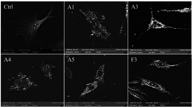
“Photomicrographs A1 and A3 show a scarcely fibrous appearance that seems to adhere to the surface of the cell membrane, englobing its entire structure.”
§ Evaluation of the genotoxic and transformation potential induced by asbestos compared to cleavage fragments > Results > Genotoxic damage
hedged- Asserted: passes — The authors put it forward as true.
- Checkable: passes — It can be checked against evidence.
- Load-bearing: unclear — It is unclear whether the paper’s conclusions depend on it.
- Based: passes — The paper gives a reason why it holds.
It may state two separate claims as one.
The paper states this without a source or data behind it here, so there is nothing to check it against. The quote shows it is in the paper.
In the paper
“Sample F3 identifies an asbestiform amphibole, while samples A1, A3, A4, and A5 contain non-asbestiform amphiboles.”
§ Evaluation of the genotoxic and transformation potential induced by asbestos compared to cleavage fragments > Materials and methods > Minerals samples
plain- Asserted: passes — The authors put it forward as true.
- Checkable: passes — It can be checked against evidence.
- Load-bearing: passes — The paper’s conclusions depend on it.
- Based: unclear — It is unclear why the paper says it holds.
It may state two separate claims as one.
A result the paper reports from its own data — it cites no other source for it, so there is none to check. The quote shows it is in the paper.
In the paper
“In particular, the curves show a first phase corresponding to the third hour, in which there is a slight decrease in ROS. From the third to the 6th hour, an increase in reactive oxygen species (ROS) is possible, a time beyond which a plateau is observed.”
§ Evaluation of the genotoxic and transformation potential induced by asbestos compared to cleavage fragments > Results > Redox status
hedgedstates a cause- Asserted: passes — The authors put it forward as true.
- Checkable: passes — It can be checked against evidence.
- Load-bearing: unclear — It is unclear whether the paper’s conclusions depend on it.
- Based: passes — The paper gives a reason why it holds.
It may state two separate claims as one.
A result the paper reports from its own data — it cites no other source for it, so there is none to check. The quote shows it is in the paper.
In the paper
“Furthermore, cell contact with sample F3 at doses between 0.4 and 15.2 µg/cm2 shows a highly transforming effect at doses of 1.9 µg/cm2 (P < 0.001) corresponding to the IC50 and 3.8 µg/cm2, although the latter less significantly (P < 0.01).”
§ Evaluation of the genotoxic and transformation potential induced by asbestos compared to cleavage fragments > Results > Transforming effect of CFs
plainstates a cause- Asserted: passes — The authors put it forward as true.
- Checkable: passes — It can be checked against evidence.
- Load-bearing: passes — The paper’s conclusions depend on it.
- Based: passes — The paper gives a reason why it holds.
It may state two separate claims as one.
A result the paper reports from its own data — it cites no other source for it, so there is none to check. The quote shows it is in the paper.
In the paper
“| F3 | 0.40 | 1.90 |”
- Also stated in: § Clonogenic assay · plain
“| A1 | 14.20 | 46.88 |”
- Also stated in: § Clonogenic assay · plain
“| A3 | 0.40 | 1.90 |”
§ Evaluation of the genotoxic and transformation potential induced by asbestos compared to cleavage fragments > Results > Clonogenic assay
plain- Asserted: passes — The authors put it forward as true.
- Checkable: passes — It can be checked against evidence.
- Load-bearing: passes — The paper’s conclusions depend on it.
- Based: passes — The paper gives a reason why it holds.
It may state two separate claims as one.
- Also stated in: § Clonogenic assay · plain
In the paper
“Actinolite, amosite, anthophyllite, chrysotile, crocidolite, and tremolite are regulated asbestiform mineral phases.”
§ Evaluation of the genotoxic and transformation potential induced by asbestos compared to cleavage fragments > Abstract
What the sources say
- Not matchedcites [7] IARC Working Group on the Evaluation of Carcinogenic Risks to Humans. Lyon (FR): International Agency for Research on Cancer (2012).
Couldn’t be matched to a paper in Crossref, so it can’t be checked.
plain- Asserted: passes — The authors put it forward as true.
- Checkable: passes — It can be checked against evidence.
- Load-bearing: fails — The paper’s conclusions may not depend on it.
- Based: passes — The paper gives a reason why it holds.
Probably not a substantial claim. The paper’s conclusions may not depend on it.
- Not matched
What the paper did — it cites no source for this, so there is none to check. The quote shows it is in the paper.
In the paper
“Only unequivocally positive foci, greater than 1 mm in diameter and intensely basophilic, were counted, consisting of randomly arranged, multilayered cells with an infiltrating phenotype toward the surrounding monolayer exhibiting contact inhibition.”
§ Evaluation of the genotoxic and transformation potential induced by asbestos compared to cleavage fragments > Materials and methods > Cells transformation assay
plain- Asserted: passes — The authors put it forward as true.
- Checkable: passes — It can be checked against evidence.
- Load-bearing: passes — The paper’s conclusions depend on it.
- Based: passes — The paper gives a reason why it holds.
Probably not a substantial claim. It may state two separate claims as one.
In the paper
“The World Health Organization has confirmed that asbestos fibres are carcinogenic, claiming that asbestos-related diseases should be eradicated worldwide.”
- Also stated in: § Introduction · plain
“In 2012, the International Agency for Research on Cancer (IARC) confirmed that chrysotile, crocidolite, amosite, tremolite, actinolite, and anthophyllite are cancerogenic to humans (Group 1)1.”
§ Evaluation of the genotoxic and transformation potential induced by asbestos compared to cleavage fragments > Abstract
What the sources say
- Not checked yetcites [1] A review of human carcinogens. Part C: Arsenic, metals, dusts, and fibres. Lyons, International Agency for Research on Cancer, 2009 (IARC monographs on the evaluation of carcinogenic risks to humans, Vol. 100 C). http://monographs.iarc.fr/ENG/Monographs/vol100C/mono100C.pdf (Accessed 13 April 2013) (2009).
plainstates a cause- Asserted: passes — The authors put it forward as true.
- Checkable: passes — It can be checked against evidence.
- Load-bearing: fails — The paper’s conclusions may not depend on it.
- Based: passes — The paper gives a reason why it holds.
Probably not a substantial claim. The paper’s conclusions may not depend on it.
- Also stated in: § Introduction · plain
What the paper did — it cites no source for this, so there is none to check. The quote shows it is in the paper.
In the paper
“A viability of at least 80% was a prerequisite to proceed.”
§ Evaluation of the genotoxic and transformation potential induced by asbestos compared to cleavage fragments > Materials and methods > Alkaline comet assay
plain- Asserted: passes — The authors put it forward as true.
- Checkable: passes — It can be checked against evidence.
- Load-bearing: fails — The paper’s conclusions may not depend on it.
- Based: passes — The paper gives a reason why it holds.
Probably not a substantial claim. The paper’s conclusions may not depend on it.
In the paper
“The surfaces of negatively charged CFs promote HO° radical formation through reaction with peroxide, Ca2+ sequestration that impairs the mitochondrial apoptotic pathway, and activation of agglomeration processes responsible for chronic tumourigenic inflammation23.”
§ Evaluation of the genotoxic and transformation potential induced by asbestos compared to cleavage fragments > Discussion and conclusions
What the sources say
- Check pendingcites [23] Pollastri, S. et al. The zeta potential of mineral fibres. J. Hazard. Mater. 15 469–79. https://doi.org/10.1016/j.jhazmat.2014.05.060 (2014). source
plainstates a cause- Asserted: passes — The authors put it forward as true.
- Checkable: passes — It can be checked against evidence.
- Load-bearing: passes — The paper’s conclusions depend on it.
- Based: passes — The paper gives a reason why it holds.
It may state two separate claims as one.
In the paper
“According to WHO, “breathable fibre” particles showed the following parameters: length > 5 μm, width < 3 μm or have an Aspect Ratio (length of the particles divided by the width, A/R) of at least 3:19.”
§ Evaluation of the genotoxic and transformation potential induced by asbestos compared to cleavage fragments > Introduction
What the sources say
- Not matchedcites [9] WHO. Determination of airborne fibre number concentrations. A recommended method, by phase contrast optical microscopy (Membrane Filter Method), World Health Organization, Geneva. (1996).
Couldn’t be matched to a paper in Crossref, so it can’t be checked.
plain- Asserted: passes — The authors put it forward as true.
- Checkable: passes — It can be checked against evidence.
- Load-bearing: fails — The paper’s conclusions may not depend on it.
- Based: passes — The paper gives a reason why it holds.
Probably not a substantial claim. The paper’s conclusions may not depend on it. It may state two separate claims as one.
- Not matched
In the paper
“The regulated definition of asbestos fibre excludes several other fibrous minerals that may possess an asbestiform habitus but do not exhibit all of the properties of asbestos, such as sepiolite, erionite and nemolite6.”
§ Evaluation of the genotoxic and transformation potential induced by asbestos compared to cleavage fragments > Introduction
What the sources say
- Not checked — no full text of the sourcecites [6] Ross, M., Kuntze, R. A. & Clifton, R. A. in B. Levadie, ed., Definition for Asbestos and Other Health Related Silicates, ASTMSTP 834, American Society for Testing and Materials, Philadelphia, Pa.139–147 (1984). source
plain- Asserted: passes — The authors put it forward as true.
- Checkable: passes — It can be checked against evidence.
- Load-bearing: fails — The paper’s conclusions may not depend on it.
- Based: passes — The paper gives a reason why it holds.
Probably not a substantial claim. The paper’s conclusions may not depend on it.
What the paper did — it cites no source for this, so there is none to check. The quote shows it is in the paper.
In the paper
“All tests were conducted by exposing cells to CF obtained after grinding non-asbestiform amphiboles (samples A1-A5) and using a positive control (F3). Untreated cells were considered as a negative control.”
§ Evaluation of the genotoxic and transformation potential induced by asbestos compared to cleavage fragments > Materials and methods > Cell cultures and treatment conditions
plain- Asserted: passes — The authors put it forward as true.
- Checkable: passes — It can be checked against evidence.
- Load-bearing: fails — The paper’s conclusions may not depend on it.
- Based: passes — The paper gives a reason why it holds.
Probably not a substantial claim. The paper’s conclusions may not depend on it. It may state two separate claims as one.
The paper states this without a source or data behind it here, so there is nothing to check it against. The quote shows it is in the paper.
In the paper
“Although CFs are not asbestiform, they have the same composition as asbestos minerals and aspect ratios falling in the definition of fibers.”
§ Evaluation of the genotoxic and transformation potential induced by asbestos compared to cleavage fragments > Introduction
plain- Asserted: passes — The authors put it forward as true.
- Checkable: passes — It can be checked against evidence.
- Load-bearing: passes — The paper’s conclusions depend on it.
- Based: passes — The paper gives a reason why it holds.
It may state two separate claims as one.
A conclusion the paper draws from its own results — it cites no other source for it, so there is none to check. The quote shows it is in the paper.
In the paper
“Together, these findings highlight the importance of considering CF as a potential threat to human health since it can cause genotoxic damage by triggering cellular transformation processes that overlap with the mechanisms involved in the carcinogenesis processes of asbestos.”
§ Evaluation of the genotoxic and transformation potential induced by asbestos compared to cleavage fragments > Abstract
plainstates a cause- Asserted: passes — The authors put it forward as true.
- Checkable: passes — It can be checked against evidence.
- Load-bearing: passes — The paper’s conclusions depend on it.
- Based: passes — The paper gives a reason why it holds.
It may state two separate claims as one.
- Rests on an assumption: In vitro results in A549 and Balb/c 3T3 cell lines are predictive of human carcinogenic potential and health threat.
- Rests on an assumption: The Balb/c 3T3 cell transformation assay foci indicate carcinogenic transformation.
- Rests on an assumption: Carcinogenesis requires mutational events that ROS and genotoxic damage could trigger.
- Rests on an assumption: Cleavage fragments produced by laboratory grinding are representative of CFs generated by mechanical action in environmental or occupational settings.
What the paper did — it cites no source for this, so there is none to check. The quote shows it is in the paper.
In the paper
“The results regarding multiple individual experiments were expressed as means ± SD, and data were analysed by one-way analysis of variance (ANOVA) with post hoc testing using the Bonferroni test. A P value of < 0.05 was considered statistically significant.”
§ Evaluation of the genotoxic and transformation potential induced by asbestos compared to cleavage fragments > Materials and methods > Statistical analyses
plain- Asserted: passes — The authors put it forward as true.
- Checkable: passes — It can be checked against evidence.
- Load-bearing: passes — The paper’s conclusions depend on it.
- Based: passes — The paper gives a reason why it holds.
Probably not a substantial claim. It may state two separate claims as one.
A conclusion the paper draws from its own results — it cites no other source for it, so there is none to check. The quote shows it is in the paper.
In the paper
“Additionally, the differences in reactive oxygen species (ROS) and other toxic effects observed in samples A1 and A5 could be attributed to the size of the fibers or their ratio, which was more significant for sample A5.”
§ Evaluation of the genotoxic and transformation potential induced by asbestos compared to cleavage fragments > Discussion and conclusions
hedgedstates a possible cause- Asserted: passes — The authors put it forward as true.
- Checkable: passes — It can be checked against evidence.
- Load-bearing: fails — The paper’s conclusions may not depend on it.
- Based: fails — The paper may give no reason why it holds.
Probably not a substantial claim. The paper’s conclusions may not depend on it. The paper may give no reason why it holds.
A claim is checked only against the cited paper’s full text, never its abstract, and none is available for its sources.
In the paper
“Different authors reported that asbestos induces DNA damage by directly or indirectly breaking it by triggering ROS production28–30.”
§ Evaluation of the genotoxic and transformation potential induced by asbestos compared to cleavage fragments > Discussion and conclusions
What the sources say
- Not checked — no full text of the sourcecites [28] Walker, C., Everitt, J. & Barrett, J. C. Possible cellular and molecular mechanisms for asbestos carcinogenicity. Am. J. Ind. Med. 21, 253–273. https://doi.org/10.1002/ajim.4700210214 (1992). source
- Not checked — no full text of the sourcecites [29] Kamp, D. W., Graceffa, P., Pryor, W. A. & Weitzman, S. A. The role of free radicals in asbestos-induced diseases. Free Radic Biol. Med. 12, 293–315. https://doi.org/10.1016/0891-5849(92)90117-y (1992). source
- Not checked — no full text of the sourcecites [30] Upadhyay, D. & Kamp, D. W. Asbestos-induced pulmonary toxicity: role of DNA damage and apoptosis. Exp. Biol. Med. 228, 650–659. https://doi.org/10.1177/153537020322800602 (2023). source
plainstates a cause- Asserted: passes — The authors put it forward as true.
- Checkable: passes — It can be checked against evidence.
- Load-bearing: fails — The paper’s conclusions may not depend on it.
- Based: passes — The paper gives a reason why it holds.
Probably not a substantial claim. The paper’s conclusions may not depend on it.
In the paper
“However, increasing evidence for a comprehensive approach to risk factors models points to a direct correlation between asbestiform amphibole and variance in mesothelioma potency16, considering the impact of cleavage fragments is not significant.”
§ Evaluation of the genotoxic and transformation potential induced by asbestos compared to cleavage fragments > Introduction
What the sources say
- Not checked — no full text of the sourcecites [16] Wylie, A. G., Korchevskiy, A., Segrave, A. M. & Duane, A. Modeling mesothelioma risk factors from amphibole fiber dimensionality: mineralogical and epidemiological perspective. J. Appl. Toxicol. 40, 515–524. https://doi.org/10.1002/jat.3923 (2020). source
hedgedstates an association- Asserted: passes — The authors put it forward as true.
- Checkable: passes — It can be checked against evidence.
- Load-bearing: passes — The paper’s conclusions depend on it.
- Based: passes — The paper gives a reason why it holds.
It may state two separate claims as one.
What the paper did — it cites no source for this, so there is none to check. The quote shows it is in the paper.
In the paper
“The results (Fig. 5) show a highly significant effect induced by 3-methylchloranthene (P < 0.001) when compared with the control sample (Ctrl-) as a sign of the high sensitivity of the system.”
§ Evaluation of the genotoxic and transformation potential induced by asbestos compared to cleavage fragments > Results > Transforming effect of CFs
plainstates a cause- Asserted: passes — The authors put it forward as true.
- Checkable: passes — It can be checked against evidence.
- Load-bearing: passes — The paper’s conclusions depend on it.
- Based: passes — The paper gives a reason why it holds.
It may state two separate claims as one.
In the paper
“It is well known that the length of the fibers significantly impacts the genesis of stressogenic processes in the cellular comparts due mainly to frustrated phagocytosis25.”
§ Evaluation of the genotoxic and transformation potential induced by asbestos compared to cleavage fragments > Discussion and conclusions
What the sources say
- Check pendingcites [25] Kuroda, A. Recent progress and perspectives on the mechanisms underlying asbestos toxicity. Genes Environ. 12 (43), 46. https://doi.org/10.1186/s41021-021-00215-0 (2021). source
boostedstates a cause- Asserted: passes — The authors put it forward as true.
- Checkable: passes — It can be checked against evidence.
- Load-bearing: fails — The paper’s conclusions may not depend on it.
- Based: passes — The paper gives a reason why it holds.
Probably not a substantial claim. The paper’s conclusions may not depend on it.
In the paper
“According to previous studies, DNA damage was evaluated by single-cell gel electrophoresis20.”
§ Evaluation of the genotoxic and transformation potential induced by asbestos compared to cleavage fragments > Materials and methods > Alkaline comet assay
What the sources say
- Check pendingcites [20] La Maestra, S., Micale, R. T., Ferretti, M., Izzotti, A. & Gaggero, L. Attenuation of oxidative stress and chromosomal aberrations in cultured macrophages and pulmonary cells following self-sustained high temperature synthesis of asbestos. Sci. Rep. 10, 8581. https://doi.org/10.1038/s41598-020-65620-x (2020). source
plain- Asserted: passes — The authors put it forward as true.
- Checkable: passes — It can be checked against evidence.
- Load-bearing: passes — The paper’s conclusions depend on it.
- Based: passes — The paper gives a reason why it holds.
Probably not a substantial claim.
In the paper
“All these factors have been linked to the formation of RNOs, the release of cytokines and growth factors promoting chronic inflammation, leukocyte invasion, and DNA damage20–22.”
§ Evaluation of the genotoxic and transformation potential induced by asbestos compared to cleavage fragments > Discussion and conclusions
What the sources say
- Check pendingcites [20] La Maestra, S., Micale, R. T., Ferretti, M., Izzotti, A. & Gaggero, L. Attenuation of oxidative stress and chromosomal aberrations in cultured macrophages and pulmonary cells following self-sustained high temperature synthesis of asbestos. Sci. Rep. 10, 8581. https://doi.org/10.1038/s41598-020-65620-x (2020). source
- Not checked yetcites [21] Huang, S. X., Jaurand, M. C., Kamp, D. W., Whysner, J. & Hei, T. K. Role of mutagenicity in asbestos fiber-induced carcinogenicity and other diseases. J. Toxicol. Environ. Health B Crit. Rev. 14, 179–245. https://doi.org/10.1080/10937404.2011.556051 (2011). source
- Not checked — no full text of the sourcecites [22] Kim, H. N. et al. Changes in DNA 8-hydroxyguanine levels, 8-hydroxyguanine repair activity, and hOGG1 and hMTH1 mRNA expression in human lung alveolar epithelial cells induced by crocidolite asbestos. Carcinogenesis 22, 265–269. https://doi.org/10.1093/carcin/22.2.265 (2001). source
plainstates an association- Asserted: passes — The authors put it forward as true.
- Checkable: passes — It can be checked against evidence.
- Load-bearing: unclear — It is unclear whether the paper’s conclusions depend on it.
- Based: passes — The paper gives a reason why it holds.
It may state two separate claims as one.
In the paper
“The elongated mineral particles include both asbestiform and non-asbestiform minerals.”
§ Evaluation of the genotoxic and transformation potential induced by asbestos compared to cleavage fragments > Introduction
What the sources say
- Check pendingcites [5] Militello, G. M., Gaggero, L. & La Maestra, S. Asbestiform amphiboles and cleavage fragments analogues: overview of critical dimensions, aspect ratios. Exposure Health Eff. Min. 11, 525. https://doi.org/10.3390/min11050525 (2021). source
plain- Asserted: passes — The authors put it forward as true.
- Checkable: passes — It can be checked against evidence.
- Load-bearing: fails — The paper’s conclusions may not depend on it.
- Based: passes — The paper gives a reason why it holds.
Probably not a substantial claim. The paper’s conclusions may not depend on it.
A conclusion the paper draws from its own results — it cites no other source for it, so there is none to check. The quote shows it is in the paper.
In the paper
“Noteworthy, the highest levels of ROS highlighted for sample A5 could be referred to as the almost total absence of fibers with a length less than 5 μm (short fibers), which, unlike the other samples, are at values close to zero as for sample F3.”
§ Evaluation of the genotoxic and transformation potential induced by asbestos compared to cleavage fragments > Discussion and conclusions
hedgedstates a possible cause- Asserted: passes — The authors put it forward as true.
- Checkable: passes — It can be checked against evidence.
- Load-bearing: unclear — It is unclear whether the paper’s conclusions depend on it.
- Based: passes — The paper gives a reason why it holds.
It may state two separate claims as one.
In the paper
“The National Research Council (1984) reported that the crystal structure defines mineral asbestiform and non-asbestiform habitus10.”
§ Evaluation of the genotoxic and transformation potential induced by asbestos compared to cleavage fragments > Introduction
What the sources say
- Not matchedcites [10] National Research Council (US) Committee on Nonoccupational Health Risks of Asbestiform Fibers. Asbestiform Fibers: Nonoccupational Health Risks (National Academies Press (US), 1984).
Couldn’t be matched to a paper in Crossref, so it can’t be checked.
plainstates a cause- Asserted: passes — The authors put it forward as true.
- Checkable: passes — It can be checked against evidence.
- Load-bearing: fails — The paper’s conclusions may not depend on it.
- Based: passes — The paper gives a reason why it holds.
Probably not a substantial claim. The paper’s conclusions may not depend on it.
- Not matched
In the paper
“Asbestos carcinogenesis depends on numerous factors, including inhaled dose, dimension, physical durability, surface features, crystallinity, and chemical composition.”
§ Evaluation of the genotoxic and transformation potential induced by asbestos compared to cleavage fragments > Discussion and conclusions
What the sources say
- Check pendingcites [20] La Maestra, S., Micale, R. T., Ferretti, M., Izzotti, A. & Gaggero, L. Attenuation of oxidative stress and chromosomal aberrations in cultured macrophages and pulmonary cells following self-sustained high temperature synthesis of asbestos. Sci. Rep. 10, 8581. https://doi.org/10.1038/s41598-020-65620-x (2020). source
- Not checked yetcites [21] Huang, S. X., Jaurand, M. C., Kamp, D. W., Whysner, J. & Hei, T. K. Role of mutagenicity in asbestos fiber-induced carcinogenicity and other diseases. J. Toxicol. Environ. Health B Crit. Rev. 14, 179–245. https://doi.org/10.1080/10937404.2011.556051 (2011). source
- Not checked — no full text of the sourcecites [22] Kim, H. N. et al. Changes in DNA 8-hydroxyguanine levels, 8-hydroxyguanine repair activity, and hOGG1 and hMTH1 mRNA expression in human lung alveolar epithelial cells induced by crocidolite asbestos. Carcinogenesis 22, 265–269. https://doi.org/10.1093/carcin/22.2.265 (2001). source
plainstates a cause- Asserted: passes — The authors put it forward as true.
- Checkable: passes — It can be checked against evidence.
- Load-bearing: fails — The paper’s conclusions may not depend on it.
- Based: passes — The paper gives a reason why it holds.
Probably not a substantial claim. The paper’s conclusions may not depend on it.
What the paper did — it cites no source for this, so there is none to check. The quote shows it is in the paper.
In the paper
“One hundred randomly selected cell nuclei were taken per sample.”
§ Evaluation of the genotoxic and transformation potential induced by asbestos compared to cleavage fragments > Materials and methods > Alkaline comet assay
plain- Asserted: passes — The authors put it forward as true.
- Checkable: passes — It can be checked against evidence.
- Load-bearing: fails — The paper’s conclusions may not depend on it.
- Based: passes — The paper gives a reason why it holds.
Probably not a substantial claim. The paper’s conclusions may not depend on it.
In the paper
“The toxicity of asbestos fibres is mainly due to dimension, shape, and bio-persistence8.”
§ Evaluation of the genotoxic and transformation potential induced by asbestos compared to cleavage fragments > Introduction
What the sources say
- Not checked yetcites [8] Aust, A., Cook, P. & Dodson, R. Morphological and chemical mechanisms of elongated mineral particle toxicities. J. Toxicol. Environ. Health B Crit. Rev. 14, 40–75. https://doi.org/10.1080/10937404.2011.556046 (2011). source
plainstates a cause- Asserted: passes — The authors put it forward as true.
- Checkable: passes — It can be checked against evidence.
- Load-bearing: passes — The paper’s conclusions depend on it.
- Based: passes — The paper gives a reason why it holds.
Probably not a substantial claim.
None of the sources cited for this claim could be matched to a paper in Crossref, so there is nothing to check it against.
In the paper
“CTA was conducted according to published guidelines (IARC/NCI Working Group, 1985) as amended by Hayashi31.”
§ Evaluation of the genotoxic and transformation potential induced by asbestos compared to cleavage fragments > Materials and methods > Cells transformation assay
What the sources say
- Not matchedcites IARC/NCI Working Group, 1985
Couldn’t be matched to a paper in Crossref, so it can’t be checked.
- Not matchedcites Hayashi31
Couldn’t be matched to a paper in Crossref, so it can’t be checked.
plain- Asserted: passes — The authors put it forward as true.
- Checkable: passes — It can be checked against evidence.
- Load-bearing: passes — The paper’s conclusions depend on it.
- Based: passes — The paper gives a reason why it holds.
Probably not a substantial claim.
- Not matched
In the paper
“The Occupational Safety and Health Administration (OSHA) in 1994 indicated that fibres with not-asbestiform aspects are “cleavage fragments (CF)”, referring to amphibole or serpentine that split into fragments with dimensions similar to asbestos fibers14.”
§ Evaluation of the genotoxic and transformation potential induced by asbestos compared to cleavage fragments > Introduction
What the sources say
- Not checked yetcites [14] Occupational Safety and Health Administration. Rules and regulations, Department of Labor: 29 CFR Parts 1910, 1915 and 1926, 59 FR 40964, RIN: 1218-AB25; occupational exposure to asbestos; August -final rule; appendix B of 1910.1001 (1), (1994).
hedged- Asserted: passes — The authors put it forward as true.
- Checkable: passes — It can be checked against evidence.
- Load-bearing: passes — The paper’s conclusions depend on it.
- Based: passes — The paper gives a reason why it holds.
Probably not a substantial claim.
In the paper
“The non-asbestiform minerals are elongated particles that lack the mechanical properties of the asbestiform analogue. They originate with massive, prismatic, needle-like habitus and might produce cleavage fragments by grinding5.”
§ Evaluation of the genotoxic and transformation potential induced by asbestos compared to cleavage fragments > Introduction
What the sources say
- Check pendingcites [5] Militello, G. M., Gaggero, L. & La Maestra, S. Asbestiform amphiboles and cleavage fragments analogues: overview of critical dimensions, aspect ratios. Exposure Health Eff. Min. 11, 525. https://doi.org/10.3390/min11050525 (2021). source
hedgedstates a possible cause- Asserted: passes — The authors put it forward as true.
- Checkable: passes — It can be checked against evidence.
- Load-bearing: fails — The paper’s conclusions may not depend on it.
- Based: passes — The paper gives a reason why it holds.
Probably not a substantial claim. The paper’s conclusions may not depend on it. It may state two separate claims as one.
In the paper
“Several studies have demonstrated the prominent role of ROS as a second messenger in asbestos toxicity mediation26, mainly in the pulmonary mesothelium27.”
§ Evaluation of the genotoxic and transformation potential induced by asbestos compared to cleavage fragments > Discussion and conclusions
What the sources say
- Not checked — no full text of the sourcecites [26] Chew, S. H. & Toyokuni, S. Malignant mesothelioma as an oxidative stress-induced cancer: an update. Free Radic. Bio Med. 86, 166–178. https://doi.org/10.1016/j.freeradbiomed.2015.05.002 (2015). source
- Check pendingcites [27] Gào, X. & Schöttker, B. Reduction oxidation pathways involved in cancer development: a systematic review of literature reviews. Oncotarget 8, 51888–51906. https://doi.org/10.18632/oncotarget.17128 (2017). source
boostedstates a cause- Asserted: passes — The authors put it forward as true.
- Checkable: passes — It can be checked against evidence.
- Load-bearing: fails — The paper’s conclusions may not depend on it.
- Based: passes — The paper gives a reason why it holds.
Probably not a substantial claim. The paper’s conclusions may not depend on it.
A conclusion the paper draws from its own results — it cites no other source for it, so there is none to check. The quote shows it is in the paper.
In the paper
“It is worth noting that the redox imbalance determined by the treatment of cells with sample A5 cannot be related to the dimensions of the fibres themselves or their ratio, which does not differ from the other samples tested except sample F3.”
§ Evaluation of the genotoxic and transformation potential induced by asbestos compared to cleavage fragments > Discussion and conclusions
plain- Asserted: passes — The authors put it forward as true.
- Checkable: passes — It can be checked against evidence.
- Load-bearing: passes — The paper’s conclusions depend on it.
- Based: passes — The paper gives a reason why it holds.
It may state two separate claims as one.
The paper relies on this without support.
In vitro results in A549 and Balb/c 3T3 cell lines are predictive of human carcinogenic potential and health threat.
“This study used human epithelial cancer cells (A549) and a mouse fibroblast cell line (Balb/c 3T3) to compare the genotoxic and carcinogenic effects of a sample of amphibole asbestos with samples of fibrous not asbestiform named cleavage fragments (CV) obtained by grinding non-asbestiform amphiboles.”
The paper relies on this without support.
The Balb/c 3T3 cell transformation assay foci indicate carcinogenic transformation.
“The possible transforming activity of CFs was investigated using the in vitro transformation model of Balb/c 3T3 cells.”
The paper relies on this without support.
Carcinogenesis requires mutational events that ROS and genotoxic damage could trigger.
“This choice arises from the awareness that the carcinogenesis process requires mutational events that ROS and genotoxic damage could trigger.”
The paper relies on this without support.
Cleavage fragments produced by laboratory grinding are representative of CFs generated by mechanical action in environmental or occupational settings.
“CF used in this research were obtained by grinding different non-asbestiform amphiboles derived from different rocks.”
The paper relies on this without support.
Oxidative stress can lead to chronic inflammation, which can trigger chronic diseases like cancer.
“Moreover, oxidative stress can lead to chronic inflammation, triggering chronic diseases like cancer.”
The paper relies on this without support.
Carcinogenesis is a long and complex multi-step process requiring different DNA mutations.
“Carcinogenesis is a long and complex multi-step process requiring different DNA mutations, transforming a healthy cell into a cancer cell.”
The paper relies on this without support.
The process of in vitro cell transformation follows a multistage process closer to the model stages of in vivo carcinogenesis.
“Different studies have shown that the process follows a multistage process closer to the model stages of in vivo carcinogenesis.”
Self-check
Percentages, sums and reported differences re-computed from the paper’s own numbers.
Each contradiction between two passages listed here was confirmed by an independent check.
Rated minor: it would not touch a result — a value no result rests on, a p-value that stays on the same side of the significance level, or a conflict that is clerical, resolved elsewhere in the paper, or outside its own findings.
In the paper
§ Evaluation of the genotoxic and transformation potential induced by asbestos compared to cleavage fragments > Discussion and conclusions
“the CFs obtained by grinding the actinolite schist A3”
§ Evaluation of the genotoxic and transformation potential induced by asbestos compared to cleavage fragments > Materials and methods > Minerals samples
“| A3 | Serpentinised peridotite | Piedmont (Italy) |”
Rated minor: it would not touch a result — a value no result rests on, a p-value that stays on the same side of the significance level, or a conflict that is clerical, resolved elsewhere in the paper, or outside its own findings.
In the paper
§ Evaluation of the genotoxic and transformation potential induced by asbestos compared to cleavage fragments > Discussion and conclusions
“the redox imbalance determined by the treatment of cells with sample A5 cannot be related to the dimensions of the fibres themselves or their ratio, which does not differ from the other samples tested except sample F3.”
§ Evaluation of the genotoxic and transformation potential induced by asbestos compared to cleavage fragments > Discussion and conclusions
“Additionally, the differences in reactive oxygen species (ROS) and other toxic effects observed in samples A1 and A5 could be attributed to the size of the fibers or their ratio, which was more significant for sample A5.”
Citations
Cites
Nomenclature of the amphibole supergroup
Hawthorne, F. C., Oberti, R., Harlow, G. E., Maresch, W. V., Martin, R. F., Schumacher, J. C., & Welch, M. D. (2012) · American Mineralogist · doi:10.2138/am.2012.4276 · Cited by 1,216
Leake, B. E., Woolley, A. R., Arps, C. E. S., Birch, W. D., Gilbert, M. C., Grice, J. D., Hawthorne, F. C., Kato, A., Kisch, H. J., Krivovichev, V. G., et al. (1997) · Mineralogical Magazine · doi:10.1180/minmag.1997.061.405.13 · Cited by 975
The role of free radicals in asbestos-induced diseases
Kamp, D. W., Graceffa, P., Pryor, W. A., & Weitzman, S. A. (1992) · Free Radical Biology and Medicine · doi:10.1016/0891-5849(92)90117-y · Cited by 428
Contribution of Angiogenesis to Inflammation and Cancer
Aguilar‐Cázares, D., Chavez-Dominguez, R., Carlos‐Reyes, Á., López‐Camarillo, C., la Cruz, O. N. H.-D., & López-González, J. S. (2019) · Frontiers in Oncology · doi:10.3389/fonc.2019.01399 · Cited by 330
The Composition and Morphology of Amphiboles from the Rainy Creek Complex, Near Libby, Montana
Meeker, G. P., Bern, A. M., Brownfield, I. K., Lowers, H. A., Sutley, S. J., Hoefen, T. M., & Vance, J. S. (2003) · American Mineralogist · doi:10.2138/am-2003-11-1239 · Cited by 171
Role of Mutagenicity in Asbestos Fiber-Induced Carcinogenicity and Other Diseases
Huang, S. X. L., Jaurand, M.-C., Kamp, D. W., Whysner, J., & Hei, T. K. (2011) · Journal of Toxicology and Environmental Health, Part B · doi:10.1080/10937404.2011.556051 · Cited by 163
Morphological and Chemical Mechanisms of Elongated Mineral Particle Toxicities
Aust, A. E., Cook, P. M., & Dodson, R. F. (2011) · Journal of Toxicology and Environmental Health, Part B · doi:10.1080/10937404.2011.556046 · Cited by 155
Gào, X., & Schöttker, B. (2017) · Oncotarget · doi:10.18632/oncotarget.17128 · Cited by 147
Malignant mesothelioma as an oxidative stress-induced cancer: An update
Chew, S. H., & Toyokuni, S. (2015) · Free Radical Biology and Medicine · doi:10.1016/j.freeradbiomed.2015.05.002 · Cited by 102
Asbestos-Induced Pulmonary Toxicity: Role of DNA Damage and Apoptosis
Upadhyay, D., & Kamp, D. W. (2003) · Experimental Biology and Medicine · doi:10.1177/153537020322800602 · Cited by 83
The zeta potential of mineral fibres
Pollastri, S., Gualtieri, A. F., Gualtieri, M. L., Hanuskova, M., Cavallo, A., & Gaudino, G. (2014) · Journal of Hazardous Materials · doi:10.1016/j.jhazmat.2014.05.060 · Cited by 83
Naturally Occurring Asbestos: Potential for Human Exposure, Southern Nevada, USA
Buck, B. J., Goossens, D., Metcalf, R. V., McLaurin, B., Ren, M., & Freudenberger, F. (2013) · Soil Science Society of America Journal · doi:10.2136/sssaj2013.05.0183 · Cited by 75
Kim, H.-N., Morimoto, Y., Tsuda, T., Ootsuyama, Y., Hirohashi, M., Hirano, T., Tanaka, I., Lim, Y., Yun, I.-G., & Kasai, H. (2001) · Carcinogenesis · doi:10.1093/carcin/22.2.265 · Cited by 65
Possible cellular and molecular mechanisms for asbestos carcinogenicity
Walker, C., Everitt, J., & Barrett, J. C. (1992) · American Journal of Industrial Medicine · doi:10.1002/ajim.4700210214 · Cited by 57
Gualtieri, A. F., Andreozzi, G. B., Tomatis, M., & Turci, F. (2019) · Free Radical Biology and Medicine · doi:10.1016/j.freeradbiomed.2018.07.023 · Cited by 45
Recent progress and perspectives on the mechanisms underlying Asbestos toxicity
Kuroda, A. (2021) · Genes and Environment · doi:10.1186/s41021-021-00215-0 · Cited by 38
Wylie, A. G., Korchevskiy, A., Segrave, A. M., & Duane, A. (2020) · Journal of Applied Toxicology · doi:10.1002/jat.3923 · Cited by 32
Militello, G. M., Gaggero, L., & La Maestra, S. (2021) · Minerals · doi:10.3390/min11050525 · Cited by 29
Self-citationLanger, A. M., Nolan, R. P., & Addison, J. (1991) · Mechanisms in Fibre Carcinogenesis · doi:10.1007/978-1-4684-1363-2_22 · Cited by 25
Ross, M., Kuntze, R., & Clifton, R. (1984) · Definitions for Asbestos and Other Health-Related Silicates · doi:10.1520/stp39141s · Cited by 17
A STUDY OF THE EFFECT OF CHRYSOTILE FIBER SURFACE COMPOSITION ON GENOTOXICITY IN VITRO
Michael J. Keane, J. W. S. (1999) · Journal of Toxicology and Environmental Health, Part A · doi:10.1080/009841099157494 · Cited by 14
La Maestra, S., Micale, R. T., Ferretti, M., Izzotti, A., & Gaggero, L. (2020) · Scientific Reports · doi:10.1038/s41598-020-65620-x · Cited by 14
Self-citationHayashi, K., Sasaki, K., Asada, S., Tsuchiya, T., Hayashi, M., Yoshimura, I., Tanaka, N., & Umeda, M. (2008) · Alternatives to Laboratory Animals · doi:10.1177/026119290803600609 · Cited by 13
Non-asbestiform elongate mineral particles and mesothelioma risk: Human and experimental evidence
Goodman, J. E., Becich, M. J., Bernstein, D. M., Case, B. W., Mandel, J. H., Nel, A. E., Nolan, R., Odo, N. U., Smith, S. R., Taioli, E., et al. (2023) · Environmental Research · doi:10.1016/j.envres.2022.114578 · Cited by 13
Research works printed but not linked: 2
- A review of human carcinogens. Part C: Arsenic, metals, dusts, and fibres. Lyons, International Agency for Research on Cancer, 2009 (IARC monographs on the evaluation of carcinogenic risks to humans, Vol. 100 C). http://monographs.iarc.fr/ENG/Monographs/vol100C/mono100C.pdf (Accessed 13 April 2013) (2009).
- National Research Council (US) Committee on Nonoccupational Health Risks of Asbestiform Fibers. Asbestiform Fibers: Nonoccupational Health Risks (National Academies Press (US), 1984).
Cited by
Sorted by citations
Korchevskiy, A. A., & Wylie, A. G. (2025) · Inhalation Toxicology · doi:10.1080/08958378.2025.2486087 · Cited by 6
Vijayaram, S., Kaarmukhilnilavan, R. S., Mahendran, K., Sandoval‐Hevia, G., Naman, S., & Baldi, A. (2026) · BioNanoScience · doi:10.1007/s12668-025-02363-x · Cited by 2
Elangasinghe, A., Patel, H., Dirks, K. N., Fan, W., de Zoysa, A., Yi, C. Z., Young, P. G., Hamilton, A. R., Talbot, N., Brook, M., et al. (2026) · Chemosphere · doi:10.1016/j.chemosphere.2026.144916 · Cited by 1
UVB-aged polystyrene microplastics induce enhanced stress responses in human proximal tubular cells
Maestra, S. L., Ferrea, L., Rumeo, N., Passalacqua, M., Esposito, P. B., Mancianti, N., Porta, E. L., & Verzola, D. (2026) · Toxicology · doi:10.1016/j.tox.2026.154585 · Cited by 1
Wylie, A. G., Mossman, B. T., & Korchevskiy, A. A. (2025) · Archives of Toxicology · doi:10.1007/s00204-025-04039-7
Metadata
- Authors
- La Maestra, S., Militello, G. M., Alberti, S., Benvenuti, M., & Gaggero, L.
- Affiliations
- University of Genoa (IT)
- Venue
- Scientific Reports
- Published
- 2025-01-29
- Volume
- 15
- Issue
- 1
- Pages
- 3613-3613
- ISSN
- 2045-2322 (ISSN-L)
- PMC
- PMC11775320
- OpenAlex
- W4406891208
- Semantic Scholar
- 082d8f3c408f302c77671e8d9a7847539e6fc062
- Cited by
- 10
- References
- 25
- Citations by year
- 2026: 8 · 2025: 2
- FWCI
- 5.68
- Type
- journal article
- Language
- en
- Open access
- gold
- doi.org
- Licence
- Locations
- Scientific Reports (publishedVersion, cc-by-nc-nd)
- PubMed Central (publishedVersion, cc-by-nc-nd)
- PubMed (publishedVersion)
- DOAJ (DOAJ: Directory of Open Access Journals) (submittedVersion, cc-by-sa)
- CINECA IRIS Institutial Research Information System (University of Genoa) (submittedVersion)
- Funders
- POligEnetic Mélanges: anatomy, significance and societal impacts (CUP 1 D33C22000350001)
- Topics
- Health Sciences › Medicine › Pulmonary and Respiratory Medicine › Occupational and environmental lung diseases
- Life Sciences › Biochemistry, Genetics and Molecular Biology › Cancer Research › Carcinogens and Genotoxicity Assessment
- Social Sciences › Health › Occupational Health and Global Justice
- Keywords
- asbestos, cleavage fragments, amphibole asbestos, asbestiform minerals, genotoxicity, carcinogenicity, cell transformation, oxidative stress, cytotoxicity, A549 cells, 3T3 fibroblasts, actinolite, serpentine, asbestos-related diseases
- MeSH terms
- A549 Cells, Animals, Asbestos, Cell Transformation, Neoplastic, DNA Damage, Humans, Asbestos, Amphibole, Oxidative Stress, BALB 3T3 Cells, Mice
About these checks
Registry, retraction, Cites and statistics results are computed, not judged, with one exception: a printed reference that no index lists is read once by a model before it counts as a citation error. Claims are extracted by a model and kept only when their quote appears verbatim in the text; each cited source is then checked, with a model’s help, for whether it says what the claim relies on. Those verdicts can be wrong — read the claim’s quote and the cited source before relying on one.
A language model drew the claims from the paper on 2026-11-11, under version 3 of the claim definition (model deepseek-v4.1-flash, prompt extract-v3).
A second model labelled each claim on 2026-11-11 (model jev-1.13-20260917, definition revision gold-guideline-v0.2).
Statistics and arithmetic are recomputed, not judged.
Confidence bands are shown only for fields calibrated on our annotated test set. None is calibrated yet, so no band is shown.
What counts as a claim ›How we measure confidence ›How we check this ›
Are you an author, or do you see a problem with this page? Request a review
