

The DOI matches its Crossref record; 7 of the 7 references it cites were found in a registry or an index.
Recent progress and perspectives on the mechanisms underlying Asbestos toxicity
Abstract
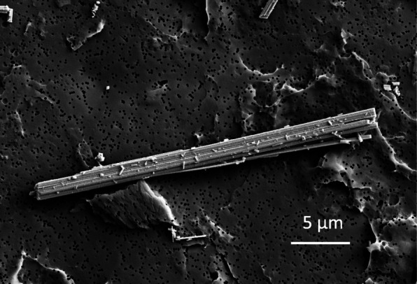
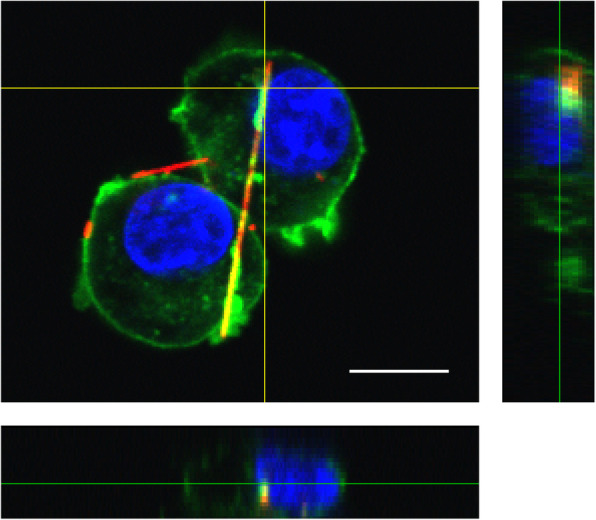
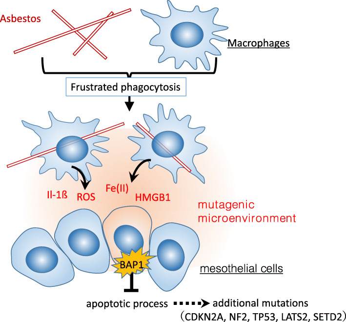
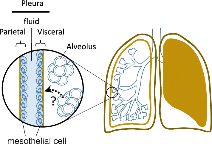
Most cases of mesothelioma are known to result from exposure to asbestos fibers in the environment or occupational ambient air. The following questions regarding asbestos toxicity remain partially unanswered: (i) why asbestos entering the alveoli during respiration exerts toxicity in the pleura; and (ii) how asbestos causes mesothelioma, even though human mesothelial cells are easily killed upon exposure to asbestos. As for the latter question, it is now thought that the frustrated phagocytosis of asbestos fibers by macrophages prolongs inflammatory responses and gives rise to a “mutagenic microenvironment” around mesothelial cells, resulting in their malignant transformation. Based on epidemiological and genetic studies, a carcinogenic model has been proposed in which BRCA1-associated protein 1 mutations are able to suppress cell death in mesothelial cells and increase genomic instability in the mutagenic microenvironment. This leads to additional mutations, such as CDKN2A [p16], NF2, TP53, LATS2, and SETD2, which are associated with mesothelioma carcinogenesis. Regarding the former question, the receptors involved in the intracellular uptake of asbestos and the mechanism of transfer of inhaled asbestos from the alveoli to the pleura are yet to be elucidated. Further studies using live-cell imaging techniques will be critical to fully understanding the mechanisms underlying asbestos toxicity.
Overview
The claims and statistics of this paper haven’t been checked yet.
Run the check — free, no account needed. It reads the open-access text and checks the sources it cites.
Integrity signals
Registry
Cites
Statistics
Figures
Verification
Claims the paper makes
The claims and statistics of this paper haven’t been checked yet.
Run the check — free, no account needed. It reads the open-access text and checks the sources it cites.
Claims other papers make about this one
Each is checked against this paper’s own text: does it say what the citing paper says it does?
In the citing paper
“It is well known that the length of the fibers significantly impacts the genesis of stressogenic processes in the cellular comparts due mainly to frustrated phagocytosis25.”
From Evaluation of the genotoxic and transformation potential induced by asbestos compared to cleavage fragments (2025) · cites this paper as [25]
Self-check
Percentages, sums and reported differences re-computed from the paper’s own numbers.
The self-check needs the paper’s full text.
Citations
Cites
Cholesterol, inflammation and innate immunity.
Tall, A. R., & Yvan-Charvet, L. (2015) · Nature Reviews Immunology · doi:10.1038/nri3793 · Cited by 1,479
Valavanidis, A., Vlachogianni, T., Fiotakis, K., & Loridas, S. (2013) · International Journal of Environmental Research and Public Health · doi:10.3390/ijerph10093886 · Cited by 910
Molecular pathways: targeting mechanisms of asbestos and erionite carcinogenesis in mesothelioma
Carbone, M., & Yang, H. (2012) · Clinical Cancer Research · doi:10.1158/1078-0432.ccr-11-2259 · Cited by 229
Toyokuni, S. (2019) · Free Radical Biology and Medicine · doi:10.1016/j.freeradbiomed.2018.10.401 · Cited by 96 · cited 1×
“Therefore, asbestos toxicity is associated with iron [20, 47].”
BackgroundHomozygous deletion of CDKN2A/2B is a hallmark of iron-induced high-grade rat mesothelioma
Hu, Q., Akatsuka, S., Yamashita, Y., Ohara, H., Nagai, H., Okazaki, Y., Takahashi, T., & Toyokuni, S. (2010) · Laboratory Investigation · doi:10.1038/labinvest.2009.140 · Cited by 62
Ito, F., Yanatori, I., Maeda, Y., Nimura, K., Ito, S., Hirayama, T., Nagasawa, H., Kohyama, N., Okazaki, Y., Akatsuka, S., et al. (2020) · Redox Biology · doi:10.1016/j.redox.2020.101616 · Cited by 45 · cited 1×
“proposed that macrophage necrosis occurs repeatedly along with lysosomal cell death [50], and ferroptosis [51] could establish an Fe (II)-rich mutagenic microenvironment [52], although they need to elucidate mutations induced under these conditions.”
BackgroundNagai, H., Okazaki, Y., Chew, S. H., Misawa, N., Yasui, H., & Toyokuni, S. (2013) · Cancer Prevention Research · doi:10.1158/1940-6207.capr-13-0244 · Cited by 39
Research works printed but not linked: 74
- Davis JM, Beckett ST, Bolton RE, Collings P, Middleton AP. Mass and number of fibres in the pathogenesis of asbestos-related lung disease in rats. Br J Cancer. 1978;37(5):673–88. https://doi.org/10.1038/bjc.1978.105. · DOI
- Suzuki Y, Yuen SR, Ashley R. Short, thin asbestos fibers contribute to the development of human malignant mesothelioma: pathological evidence. Int J Hyg Environ Health. 2005;208(3):201–10. https://doi.org/10.1016/j.ijheh.2005.01.015. · DOI · Paper page
- Kanarek MS. Mesothelioma from chrysotile asbestos: update. Ann Epidemiol. 2011;21(9):688–97. https://doi.org/10.1016/j.annepidem.2011.05.010. · DOI
- Wagner JC, Sleggs CA, Marchand P. Diffuse pleural mesothelioma and asbestos exposure in the North Western Cape Province. Occup Environ Med. 1960;17(4):260–71. https://doi.org/10.1136/oem.17.4.260. · DOI · Paper page
- Park EK, Takahashi K, Jiang Y, Movahed M, Kameda T. Elimination of asbestos use and asbestos-related diseases: an unfinished story. Cancer Sci. 2012;103(10):1751–5. https://doi.org/10.1111/j.1349-7006.2012.02366.x. · DOI
- Gordon RE, Fitzgerald S, Millette J. Asbestos in commercial cosmetic talcum powder as a cause of mesothelioma in women. Int J Occup Environ Health. 2014;20(4):318–32. https://doi.org/10.1179/2049396714Y.0000000081. · DOI
- Moline J, Bevilacqua K, Alexandri M, Gordon RE. Mesothelioma associated with the use of cosmetic talc. J Occup Environ Med. 2020;62(1):11–7. https://doi.org/10.1097/JOM.0000000000001723. · DOI
- Furuya S, Chimed-Ochir O, Takahashi K, David A, Takala J. Global Asbestos Disaster. Int J Environ Res Public Health. 2018;15(5):1000. https://doi.org/10.3390/ijerph15051000. · DOI
- Bariş B, Demir AU, Shehu V, Karakoca Y, Kisacik G, Bariş YI. Environmental fibrous zeolite (erionite) exposure and malignant tumors other than mesothelioma. J Environ Pathol Toxicol Oncol. 1996;15:183–9.
- Carbone M, Baris YI, Bertino P, Brass B, Comertpay S, Dogan AU, et al. Erionite exposure in North Dakota and Turkish villages with mesothelioma. Proc Natl Acad Sci U S A. 2011;108(33):13618–23. https://doi.org/10.1073/pnas.1105887108. · DOI · Paper page
- Carbone M, Emri S, Dogan AU, Steele I, Tuncer M, Pass HI, et al. A mesothelioma epidemic in Cappadocia: scientific developments and unexpected social outcomes. Nat Rev Cancer. 2007;7(2):147–54. https://doi.org/10.1038/nrc2068. · DOI
- Larson D, Powers A, Ambrosi JP, Tanji M, Napolitano A, Flores EG, et al. Investigating palygorskite's role in the development of mesothelioma in southern Nevada: insights into fiber-induced carcinogenicity. J Toxicol Environ Health, Part B. 2016;19(5-6):213–30. https://doi.org/10.1080/10937404.2016.1195321. · DOI
- Sanchez VC, Pietruska JR, Miselis NR, Hurt RH, Kane AB. Biopersistence and potential adverse health impacts of fibrous nanomaterials: what have we learned from asbestos? Wiley Interdiscip Rev: Nanomed Nanobiotechnol. 2009;1(5):511–29. https://doi.org/10.1002/wnan.41. · DOI · Paper page
- Olofsson K, Mark J. Specificity of asbestos-induced chromosomal aberrations in short-term cultured human mesothelial cells. Cancer Genet Cytogenet. 1989;41(1):33–9. https://doi.org/10.1016/0165-4608(89)90105-2. · DOI
- Yang H, Bocchetta M, Kroczynska B, Elmishad AG, Chen Y, Liu Z, et al. TNF-α inhibits asbestos-induced cytotoxicity via a NF-κB-dependent pathway, a possible mechanism for asbestos-induced oncogenesis. Proc Natl Acad Sci U S A. 2006;103(27):10397–402. https://doi.org/10.1073/pnas.0604008103. · DOI · Paper page
- Wang Y, Faux SP, Hallden G, Kirn DH, Houghton CE, Lemoine NR, et al. Interleukin-1β and tumour necrosis factor-alpha promote the transformation of human immortalised mesothelial cells by erionite. Int J Oncol. 2004;25(1):173–8.
- Xu A, Wu LJ, Santella RM, Hei TK. Role of oxyradicals in mutagenicity and DNA damage induced by crocidolite asbestos in mammalian cells. Cancer Res. 1999;59(23):5922–6.
- Qi F, Okimoto G, Jube S, Napolitano A, Pass HI, Laczko R, et al. Continuous exposure to chrysotile asbestos can cause transformation of human mesothelial cells via HMGB1 and TNF-α signaling. Am J Pathol. 2013;183(5):1654–66. https://doi.org/10.1016/j.ajpath.2013.07.029. · DOI
- Lehnert BE. Defense mechanisms against inhaled particles and associated particle-cell interactions. In: Guthrie Jr GD, Mossman BT, editors. Health effects of mineral dusts. Berlin, Boston: De Gruyter; 2018. p. 427–70. https://doi.org/10.1515/9781501509711-017. · DOI
- Oberdörster G. Macrophage-associated responses to chrysotile. Ann Occup Hyg. 1994;38(4):601–15. https://doi.org/10.1093/annhyg/38.4.601. · DOI
- Schinwald A, Murphy FA, Prina-Mello A, Poland CA, Byrne F, Movia D, et al. The threshold length for fiber-induced acute pleural inflammation: shedding light on the early events in asbestos-induced mesothelioma. Toxicol Sci. 2012;128(2):461–70. https://doi.org/10.1093/toxsci/kfs171. · DOI · Paper page
- Dörger M, Münzing S, Allmeling AM, Messmer K, Krombach F. Differential responses of rat alveolar and peritoneal macrophages to man-made vitreous fibers in vitro. Environ Res. 2001;85(3):207–14. https://doi.org/10.1006/enrs.2001.4234. · DOI
- Schinwald A, Donaldson K. Use of back-scatter electron signals to visualise cell/nanowires interactions in vitro and in vivo; frustrated phagocytosis of long fibres in macrophages and compartmentalisation in mesothelial cells in vivo. Part Fibre Toxicol. 2012;9(1):34. https://doi.org/10.1186/1743-8977-9-34. · DOI
- Stanton MF, Layard M, Tegeris A, Miller E, May M, Morgan E, et al. Relation of particle dimension to carcinogenicity in amphibole asbestoses and other fibrous minerals. J Natl Cancer Inst. 1981;67(5):965–75.
- Dostert C, Pétrilli V, Van Bruggen R, Steele C, Mossman BT, Tschopp J. Innate immune activation through Nalp3 inflammasome sensing of asbestos and silica. Science. 2008;320(5876):674–7. https://doi.org/10.1126/science.1156995. · DOI · Paper page
- Poland CA, Duffin R, Kinloch I, Maynard A, Wallace WA, Seaton A, et al. Carbon nanotubes introduced into the abdominal cavity of mice show asbestos-like pathogenicity in a pilot study. Nat Nanotechnol. 2008;3(7):423–8. https://doi.org/10.1038/nnano.2008.111. · DOI · Paper page
- Palomäki J, Välimäki E, Sund J, Vippola M, Clausen PA, Jensen KA, et al. Long, needle-like carbon nanotubes and asbestos activate the NLRP3 inflammasome through a similar mechanism. ACS Nano. 2011;5(9):6861–70. https://doi.org/10.1021/nn200595c. · DOI
- Franklin BS, Mangan MS, Latz E. Crystal formation in inflammation. Annu Rev Immunol. 2016;34(1):173–202. https://doi.org/10.1146/annurev-immunol-041015-055539. · DOI
- Donaldson K, Murphy FA, Duffin R, Poland CA. Asbestos, carbon nanotubes and the pleural mesothelium: a review of the hypothesis regarding the role of long fibre retention in the parietal pleura, inflammation and mesothelioma. Part Fibre Toxicol. 2010;7(1):5. https://doi.org/10.1186/1743-8977-7-5. · DOI · Paper page
- Mulay SR, Anders HJ. Crystallopathies. N Engl J Med. 2016;374(25):2465–76. https://doi.org/10.1056/NEJMra1601611. · DOI
- Rock KL, Kataoka H, Lai JJ. Uric acid as a danger signal in gout and its comorbidities. Nat Rev Rheumatol. 2013;9(1):13–23. https://doi.org/10.1038/nrrheum.2012.143. · DOI
- Broz P, Dixit VM. Inflammasomes: mechanism of assembly, regulation and signalling. Nat Rev Immunol. 2016;16(7):407–20. https://doi.org/10.1038/nri.2016.58. · DOI · Paper page
- Gross O, Thomas CJ, Guarda G, Tschopp J. The inflammasome: an integrated view. Immunol Rev. 2011;243(1):136–51. https://doi.org/10.1111/j.1600-065X.2011.01046.x. · DOI · Paper page
- Latz E, Xiao TS, Stutz A. Activation and regulation of the inflammasomes. Nat Rev Immunol. 2013;13(6):397–411. https://doi.org/10.1038/nri3452. · DOI · Paper page
- Rathinam VA, Fitzgerald KA. Inflammasome complexes: emerging mechanisms and effector functions. Cell. 2016;165(4):792–800. https://doi.org/10.1016/j.cell.2016.03.046. · DOI
- Nakayama M. Macrophage recognition of crystals and nanoparticles. Front Immunol. 2018;9:103. https://doi.org/10.3389/fimmu.2018.00103. · DOI
- Thakur SA, Hamilton RF Jr, Holian A. Role of scavenger receptor a family in lung inflammation from exposure to environmental particles. J Immunotoxicol. 2008;5(2):151–7. https://doi.org/10.1080/15476910802085863. · DOI
- Murthy S, Larson-Casey JL, Ryan AJ, He C, Kobzik L, Carter AB. Alternative activation of macrophages and pulmonary fibrosis are modulated by scavenger receptor, macrophage receptor with collagenous structure. FASEB J. 2015;29(8):3527–36. https://doi.org/10.1096/fj.15-271304. · DOI
- Tsugita M, Morimoto N, Tashiro M, Kinoshita K, Nakayama M. SR-B1 is a silica receptor that mediates canonical Inflammasome activation. Cell Rep. 2017;18(5):1298–311. https://doi.org/10.1016/j.celrep.2017.01.004. · DOI
- Omori S, Tsugita M, Hoshikawa Y, Morita M, Ito F, Yamaguchi SI, et al. Tim4 recognizes carbon nanotubes and mediates phagocytosis leading to granuloma formation. Cell Rep. 2021;34(6):108734. https://doi.org/10.1016/j.celrep.2021.108734. · DOI
- Kawanishi S, Ohnishi S, Ma N, Hiraku Y, Murata M. Crosstalk between DNA damage and inflammation in the multiple steps of carcinogenesis. Int J Mol Sci. 2017;18(8):1808. https://doi.org/10.3390/ijms18081808. · DOI
- Blake DJ, Bolin CM, Cox DP, Cardozo-Pelaez F, Pfau JC. Internalization of Libby amphibole asbestos and induction of oxidative stress in murine macrophages. Toxicol Sci. 2007;99(1):277–88. https://doi.org/10.1093/toxsci/kfm166. · DOI
- Okada S, Hamazaki S, Toyokuni S, Midorikawa O. Induction of mesothelioma by intraperitoneal injections of ferric saccharate in male Wistar rats. Br J Cancer. 1989;60(5):708–11. https://doi.org/10.1038/bjc.1989.344. · DOI · Paper page
- Galluzzi L, Vitale I, Aaronson SA, Abrams JM, Adam D, Agostinis P, et al. Molecular mechanisms of cell death: recommendations of the nomenclature committee on cell death 2018. Cell Death Differ. 2018;25(3):486–541. https://doi.org/10.1038/s41418-017-0012-4. · DOI · Paper page
- Stockwell BR, Angeli JPF, Bayir H, Bush AI, Conrad M, Dixon SJ, et al. Ferroptosis: a regulated cell death Nexus linking metabolism, redox biology, and disease. Cell. 2017;171(2):273–85. https://doi.org/10.1016/j.cell.2017.09.021. · DOI · Paper page
- Testa JR, Cheung M, Pei J, Below JE, Tan Y, Sementino E, et al. Germline BAP1 mutations predispose to malignant mesothelioma. Nat Genet. 2011;43(10):1022–5. https://doi.org/10.1038/ng.912. · DOI · Paper page
- Nasu M, Emi M, Pastorino S, Tanji M, Powers A, Luk H, et al. High incidence of somatic BAP1 alterations in sporadic malignant mesothelioma. J Thorac Oncol. 2015;10(4):565–76. https://doi.org/10.1097/JTO.0000000000000471. · DOI
- Guo G, Chmielecki J, Goparaju C, Heguy A, Dolgalev I, Carbone M, et al. Whole-exome sequencing reveals frequent genetic alterations in BAP1, NF2, CDKN2A, and CUL1 in malignant pleural mesothelioma. Cancer Res. 2015;75(2):264–9. https://doi.org/10.1158/0008-5472.CAN-14-1008. · DOI
- Lo Iacono M, Monica V, Righi L, Grosso F, Libener R, Vatrano S, et al. Targeted next-generation sequencing of cancer genes in advanced stage malignant pleural mesothelioma: a retrospective study. J Thorac Oncol. 2015;10(3):492–9. https://doi.org/10.1097/JTO.0000000000000436. · DOI
- Bueno R, Stawiski EW, Goldstein LD, Durinck S, De Rienzo A, Modrusan Z, et al. Comprehensive genomic analysis of malignant pleural mesothelioma identifies recurrent mutations, gene fusions and splicing alterations. Nat Genet. 2016;48(4):407–16. https://doi.org/10.1038/ng.3520. · DOI
- Yoshikawa Y, Emi M, Nakano T, Gaudino G. Mesothelioma developing in carriers of inherited genetic mutations. Transl Lung Cancer Res. 2020;9(Suppl 1):S67–76. https://doi.org/10.21037/tlcr.2019.11.15. · DOI
- Hmeljak J, Sanchez-Vega F, Hoadley KA, Shih J, Stewart C, Heiman D, et al. Integrative molecular characterization of malignant pleural mesothelioma. Cancer Discov. 2018;8(12):1548–65. https://doi.org/10.1158/2159-8290.CD-18-0804. · DOI · Paper page
- Jean D, Jaurand MC. Mesotheliomas in genetically engineered mice unravel mechanism of mesothelial carcinogenesis. Int J Mol Sci. 2018;19(8):2191. https://doi.org/10.3390/ijms19082191. · DOI
- Dey A, Seshasayee D, Noubade R, French DM, Liu J, Chaurushiya MS, et al. Loss of the tumor suppressor BAP1 causes myeloid transformation. Science. 2012;337(6101):1541–6. https://doi.org/10.1126/science.1221711. · DOI
- Yu H, Pak H, Hammond-Martel I, Ghram M, Rodrigue A, Daou S, et al. Tumor suppressor and deubiquitinase BAP1 promotes DNA double-strand break repair. Proc Natl Acad Sci U S A. 2014;111(1):285–90. https://doi.org/10.1073/pnas.1309085110. · DOI
- Bononi A, Giorgi C, Patergnani S, Larson D, Verbruggen K, Tanji M, et al. BAP1 regulates IP3R3-mediated ca(2+) flux to mitochondria suppressing cell transformation. Nature. 2017;546(7659):549–53. https://doi.org/10.1038/nature22798. · DOI
- Bononi A, Yang H, Giorgi C, Patergnani S, Pellegrini L, Su M, et al. Germline BAP1 mutations induce a Warburg effect. Cell Death Differ. 2017;24(10):1694–704. https://doi.org/10.1038/cdd.2017.95. · DOI · Paper page
- Zhang Y, Shi J, Liu X, Feng L, Gong Z, Koppula P, et al. BAP1 links metabolic regulation of ferroptosis to tumour suppression. Nat Cell Biol. 2018;20(10):1181–92. https://doi.org/10.1038/s41556-018-0178-0. · DOI
- Affar EB, Carbone M. BAP1 regulates different mechanisms of cell death. Cell Death Dis. 2018;9(12):1151. https://doi.org/10.1038/s41419-018-1206-5. · DOI
- Xue J, Patergnani S, Giorgi C, Suarez J, Goto K, Bononi A, et al. Asbestos induces mesothelial cell transformation via HMGB1-driven autophagy. Proc Natl Acad Sci U S A. 2020;117(41):25543–52. https://doi.org/10.1073/pnas.2007622117. · DOI
- Carbone M, Adusumilli PS, Alexander HR Jr, Baas P, Bardelli F, Bononi A, et al. Mesothelioma: scientific clues for prevention, diagnosis, and therapy. CA Cancer J Clin. 2019;69(5):402–29. https://doi.org/10.3322/caac.21572. · DOI · Paper page
- Otsuki T, Matsuzaki H, Lee S, Kumagai-Takei N, Yamamoto S, Hatayama T, et al. Environmental factors and human health: fibrous and particulate substance-induced immunological disorders and construction of a health-promoting living environment. Environ Health Prev Med. 2016;21(2):71–81. https://doi.org/10.1007/s12199-015-0499-6. · DOI
- Kumagai-Takei N, Lee S, Srinivas B, Shimizu Y, Sada N, Yoshitome K, et al. The Effects of Asbestos Fibers on Human T Cells. Int J Mol Sci. 2020;21(19). · DOI
- Cugell DW, Kamp DW. Asbestos and the pleura: a review. Chest. 2004;125(3):1103–17. https://doi.org/10.1378/chest.125.3.1103. · DOI
- Suzuki Y, Kohyama N. Translocation of inhaled asbestos fibers from the lung to other tissues. Am J Ind Med. 1991;19(6):701–4. https://doi.org/10.1002/ajim.4700190603. · DOI
- Dodson RF, O'Sullivan MF, Huang J, Holiday DB, Hammar SP. Asbestos in extrapulmonary sites: omentum and mesentery. Chest. 2000;117(2):486–93. https://doi.org/10.1378/chest.117.2.486. · DOI
- Miserocchi G, Sancini G, Mantegazza F, Chiappino G. Translocation pathways for inhaled asbestos fibers. Environ Health. 2008;7(1):4. https://doi.org/10.1186/1476-069X-7-4. · DOI
- Jensen CG, Jensen LC, Rieder CL, Cole RW, Ault JG. Long crocidolite asbestos fibers cause polyploidy by sterically blocking cytokinesis. Carcinogenesis. 1996;17(9):2013–21. https://doi.org/10.1093/carcin/17.9.2013. · DOI
- Wang NS, Jaurand MC, Magne L, Kheuang L, Pinchon MC, Bignon J. The interactions between asbestos fibers and metaphase chromosomes of rat pleural mesothelial cells in culture. A scanning and transmission electron microscopic study. Am J Pathol. 1987;126(2):343–9.
- Gianoncelli A, Kourousias G, Cammisuli F, Cassese D, Rizzardi C, Radillo O, et al. Combined use of AFM and soft X-ray microscopy to reveal fibres' internalization in mesothelial cells. Analyst. 2017;142(11):1982–92. https://doi.org/10.1039/C6AN02661C. · DOI
- Kuroda A, Alexandrov M, Nishimura T, Ishida T. Rapid on-site detection of airborne asbestos fibers and potentially hazardous nanomaterials using fluorescence microscopy-based biosensing. Biotechnol J. 2016;11(6):757–67. https://doi.org/10.1002/biot.201500438. · DOI
- Kuroda A, Nishimura T, Ishida T, Hirota R, Nomura K. Detection of chrysotile asbestos by using a chrysotile-binding protein. Biotechnol Bioeng. 2008;99(2):285–9. https://doi.org/10.1002/bit.21588. · DOI
- Ishida T, Fujihara N, Nishimura T, Funabashi H, Hirota R, Ikeda T, et al. Live-cell imaging of macrophage phagocytosis of asbestos fibers under fluorescence microscopy. Genes Environ. 2019;41(1):14. https://doi.org/10.1186/s41021-019-0129-4. · DOI
- Ishida T, Alexandrov M, Nishimura T, Minakawa K, Hirota R, Sekiguchi K, et al. Evaluation of sensitivity of fluorescence-based asbestos detection by correlative microscopy. J Fluoresc. 2012;22(1):357–63. https://doi.org/10.1007/s10895-011-0967-3. · DOI
Cited by
Sorted by citations
Danopoulos, E., Twiddy, M., West, R., & Rotchell, J. M. (2022) · Journal of Hazardous Materials · doi:10.1016/j.jhazmat.2021.127861 · Cited by 230
La Maestra, S., Militello, G. M., Alberti, S., Benvenuti, M., & Gaggero, L. (2025) · Scientific Reports · doi:10.1038/s41598-025-86325-z · Cited by 10
Metadata
- Authors
- Kuroda, A.
- Venue
- Genes and Environment
- Published
- 2021-10-12
- Volume
- 43
- Issue
- 1
- ISSN
- 1880-7062
- PMC
- PMC8507173
- Cited by
- 38
- References
- 7
- Language
- en
- Funders
- environmental restoration and conservation agency (JPMEERF201950001)
- japan society for the promotion of science (19H04291)
About these checks
Registry, retraction, Cites and statistics results are computed, not judged, with one exception: a printed reference that no index lists is read once by a model before it counts as a citation error. Claims are extracted by a model and kept only when their quote appears verbatim in the text; each cited source is then checked, with a model’s help, for whether it says what the claim relies on. Those verdicts can be wrong — read the claim’s quote and the cited source before relying on one.
Statistics and arithmetic are recomputed, not judged.
Confidence bands are shown only for fields calibrated on our annotated test set. None is calibrated yet, so no band is shown.
What counts as a claim ›How we measure confidence ›How we check this ›
Are you an author, or do you see a problem with this page? Request a review
How to cite
Cite CoChat for the checks on this page, and the paper for its own findings.
- This page
- CoChat. (2026). Citation check: Recent progress and perspectives on the mechanisms underlying Asbestos toxicity. https://app.cochat.ai/papers/10.1186/s41021-021-00215-0
- The paper
- Kuroda, A. (2021). Recent progress and perspectives on the mechanisms underlying Asbestos toxicity. Genes and Environment. https://doi.org/10.1186/s41021-021-00215-0
