

The DOI matches its Crossref record; 59 of the 59 references it cites were found in a registry or an index.
Multimodal data integration using machine learning improves risk stratification of high-grade serous ovarian cancer
Open accessAbstract
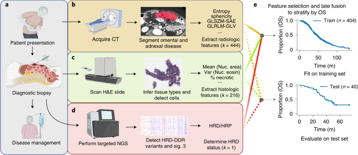
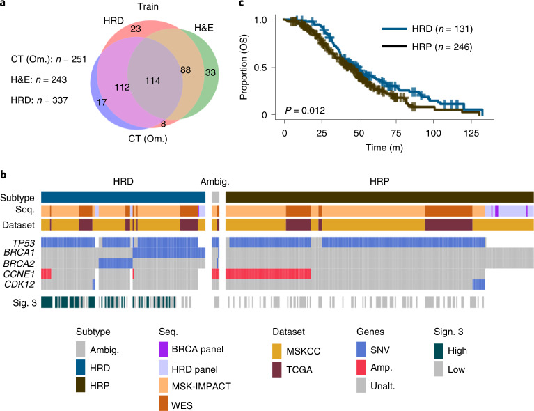
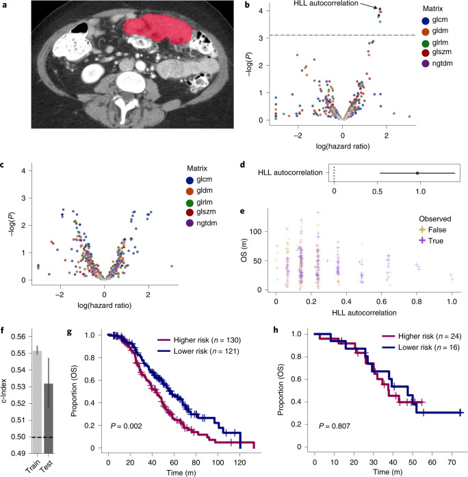
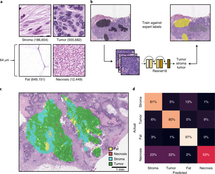
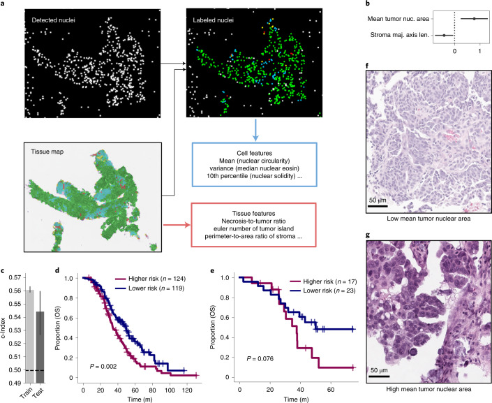
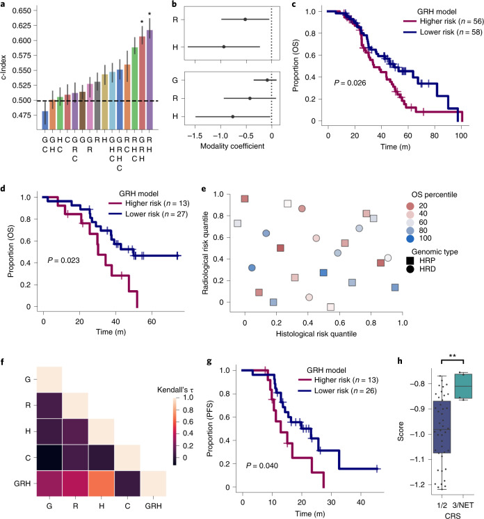
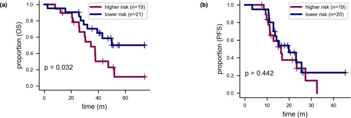
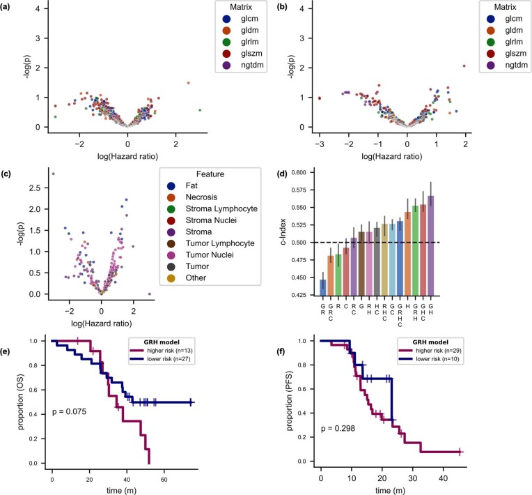
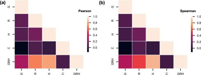
Patients with high-grade serous ovarian cancer suffer poor prognosis and variable response to treatment. Known prognostic factors for this disease include homologous recombination deficiency status, age, pathological stage and residual disease status after debulking surgery. Recent work has highlighted important prognostic information captured in computed tomography and histopathological specimens, which can be exploited through machine learning. However, little is known about the capacity of combining features from these disparate sources to improve prediction of treatment response. Here, we assembled a multimodal dataset of 444 patients with primarily late-stage high-grade serous ovarian cancer and discovered quantitative features, such as tumor nuclear size on staining with hematoxylin and eosin and omental texture on contrast-enhanced computed tomography, associated with prognosis. We found that these features contributed complementary prognostic information relative to one another and clinicogenomic features. By fusing histopathological, radiologic and clinicogenomic machine-learning models, we demonstrate a promising path toward improved risk stratification of patients with cancer through multimodal data integration.
Overview
The claims and statistics of this paper haven’t been checked yet.
Run the check — free, no account needed. It reads the open-access text and checks the sources it cites.
Integrity signals
Registry
Cites
Statistics
Figures
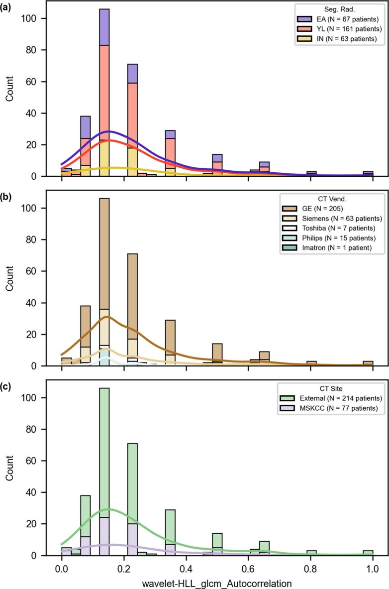
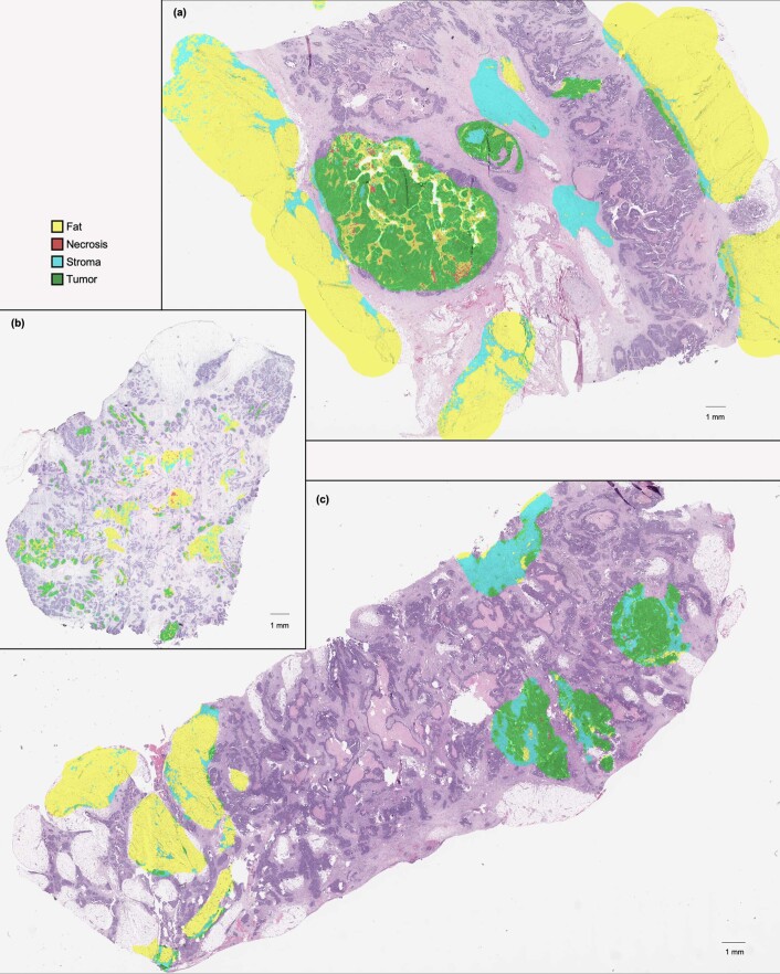
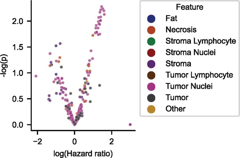
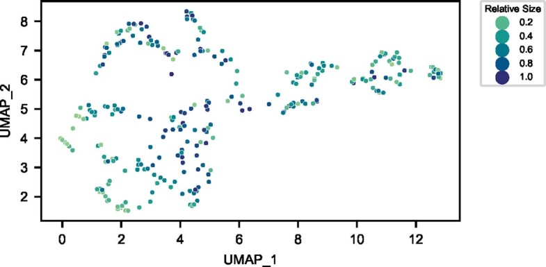
Verification
Claims the paper makes
The claims and statistics of this paper haven’t been checked yet.
Run the check — free, no account needed. It reads the open-access text and checks the sources it cites.
Claims other papers make about this one
Each is checked against this paper’s own text: does it say what the citing paper says it does?
The cited source says what the claim attributes to it.
In the citing paper
“Boehm et al. comprehensively characterized the multimodal datasets of high-grade serous ovarian cancer and proposed that a combination of histopathological, radiologic, and genomic features can better predict the prognosis of patients and aid in risk stratification [103].”
From Challenges in AI-driven Biomedical Multimodal Data Fusion and Analysis (2025) · cites this paper as [103]
In the citing paper
“Boehm and colleagues (170) used TCGA and an internal cohort to fuse genomic data, histopathology imaging, and radiology imaging to predict survival in high-grade serous ovarian cancer.”
From Artificial Intelligence in Oncology: Current Landscape, Challenges, and Future Directions (2024) · cites this paper as [170]
Self-check
Percentages, sums and reported differences re-computed from the paper’s own numbers.
The self-check needs the paper’s full text.
Citations
Cites
Controlling the False Discovery Rate: A Practical and Powerful Approach to Multiple Testing
Benjamini, Y., & Hochberg, Y. (1995) · Journal of the Royal Statistical Society Series B: Statistical Methodology · doi:10.1111/j.2517-6161.1995.tb02031.x · Cited by 109,482
Textural Features for Image Classification
Haralick, R. M., Shanmugam, K., & Dinstein, I. (1973) · IEEE Transactions on Systems, Man, and Cybernetics · doi:10.1109/tsmc.1973.4309314 · Cited by 22,886
Rudin, C. (2019) · Nature Machine Intelligence · doi:10.1038/s42256-019-0048-x · Cited by 10,561
QuPath: Open source software for digital pathology image analysis
Bankhead, P., Loughrey, M. B., Fernández, J. A., Dombrowski, Y., McArt, D. G., Dunne, P. D., McQuaid, S., Gray, R. T., Murray, L. J., Coleman, H. G., et al. (2017) · Scientific Reports · doi:10.1038/s41598-017-17204-5 · Cited by 8,908
Integrated genomic analyses of ovarian carcinoma
Perou, C. M. (2019) · Carolina Digital Repository (University of North Carolina at Chapel Hill) · doi:10.17615/hvp3-wg08 · Cited by 7,927
scikit-image: image processing in Python
(2014) · doi:10.7717/peerj.453 · Cited by 6,794
Computational Radiomics System to Decode the Radiographic Phenotype
(2017) · doi:10.1158/0008-5472.can-17-0339 · Cited by 6,593
Zwanenburg, A., Vallières, M., Abdalah, M. A., Aerts, H. J. W. L., Andrearczyk, V., Apte, A., Ashrafinia, S., Bakas, S., Beukinga, R. J., Boellaard, R., et al. (2020) · Radiology · doi:10.1148/radiol.2020191145 · Cited by 4,177
Liu, J., Lichtenberg, T., Hoadley, K. A., Poisson, L. M., Lazar, A. J., Cherniack, A. D., Kovatich, A. J., Benz, C. C., Levine, D. A., Lee, A. V., et al. (2018) · Cell · doi:10.1016/j.cell.2018.02.052 · Cited by 3,725
Self-citationThe repertoire of mutational signatures in human cancer
Alexandrov, L. B., Kim, J., Haradhvala, N. J., Huang, M. N., Tian Ng, A. W., Wu, Y., Boot, A., Covington, K. R., Gordenin, D. A., Bergstrom, E. N., et al. (2020) · Nature · doi:10.1038/s41586-020-1943-3 · Cited by 3,496
Cited by
Sorted by citations
Multistain deep learning for prediction of prognosis and therapy response in colorectal cancer
Foersch, S., Glasner, C., Woerl, A.-C., Eckstein, M., Wagner, D.-C., Schulz, S., Kellers, F., Fernandez, A., Tserea, K., Kloth, M., et al. (2023) · Nature Medicine · doi:10.1038/s41591-022-02134-1 · Cited by 251
A guide to artificial intelligence for cancer researchers
Pérez-López, R., Laleh, N. G., Mahmood, F., & Kather, J. N. (2024) · Nature reviews. Cancer · doi:10.1038/s41568-024-00694-7 · Cited by 227
Shaik, T., Tao, X., Li, L., Xie, H., & Velasquez, J. D. (2023) · Information Fusion · doi:10.1016/j.inffus.2023.102040 · Cited by 228
Artificial Intelligence in Oncology: Current Landscape, Challenges, and Future Directions
Lotter, W., Hassett, M. J., Schultz, N., Kehl, K. L., Van Allen, E. M., & Cerami, E. (2024) · Cancer Discovery · doi:10.1158/2159-8290.cd-23-1199 · Cited by 196
Metadata
- Authors
- Boehm, K. M., Aherne, E. A., Ellenson, L., Nikolovski, I., Alghamdi, M., Vázquez-García, I., Zamarin, D., Long Roche, K., Liu, Y., Patel, D., Aukerman, A., Pasha, A., Rose, D., Selenica, P., Causa Andrieu, P. I., Fong, C., Capanu, M., Reis-Filho, J. S., Vanguri, R., Veeraraghavan, H., Gangai, N., Sosa, R., Leung, S., McPherson, A., Gao, J., MSK MIND Consortium, Lakhman, Y., & Shah, S. P.
- Affiliations
- Memorial Sloan Kettering Cancer Center (US)
- Corresponding author
- Lakhman, Yulia
- Shah, Sohrab P.
- ORCID
- Boehm, Kevin M.
- Aherne, Emily A.
- Ellenson, Lora
- Nikolovski, Ines
- Alghamdi, Mohammed
- Vázquez-García, Ignacio
- Zamarin, Dmitriy
- Long Roche, Kara
- Liu, Ying
- Patel, Druv
- Aukerman, Andrew
- Rose, Doori
- Selenica, Pier
- Fong, Chris
- Capanu, Marinela
- Reis-Filho, Jorge S.
- Vanguri, Rami
- Veeraraghavan, Harini
- Gangai, Natalie
- McPherson, Andrew
- Gao, JianJiong
- Lakhman, Yulia
- Shah, Sohrab P.
- Venue
- Nature Cancer
- Published
- 2022-06-28
- Volume
- 3
- Issue
- 6
- Pages
- 723-733
- ISSN
- 2662-1347 (ISSN-L)
- PMC
- PMC9239907
- OpenAlex
- W4283645969
- Cited by
- 357
- References
- 54
- Citations by year
- 2026: 76 · 2025: 118 · 2024: 94 · 2023: 59 · 2022: 9
- FWCI
- 59.41
- Type
- journal article
- Language
- en
- Open access
- hybrid
- doi.org
- Locations
- Nature Cancer (publishedVersion, cc-by)
- PubMed Central (publishedVersion, cc-by)
- PubMed (publishedVersion)
- Funders
- U.S. Department of Health & Human Services | NIH | National Institute of General Medical Sciences (T32GM007739)
- U.S. Department of Health & Human Services | NIH | NCI | Division of Cancer Epidemiology and Genetics, National Cancer Institute (P30CA008748)
- Grayer Fellowship (MSKCC) Cycle for Survival
- Susan G. Komen Scholars Program Cycle for Survival
- Topics
- Health Sciences › Medicine › Oncology › Ovarian cancer diagnosis and treatment
- Health Sciences › Medicine › Radiology, Nuclear Medicine and Imaging › Radiomics and Machine Learning in Medical Imaging
- Physical Sciences › Computer Science › Artificial Intelligence › AI in cancer detection
- Keywords
- high-grade serous ovarian cancer, multimodal data integration, risk stratification, prognostic factors, treatment response prediction, homologous recombination deficiency, H&E staining, contrast-enhanced CT, histopathology
- MeSH terms
- Machine Learning, Female, Humans, Ovarian Neoplasms, Cystadenocarcinoma, Serous, Risk Assessment
About these checks
Registry, retraction, Cites and statistics results are computed, not judged, with one exception: a printed reference that no index lists is read once by a model before it counts as a citation error. Claims are extracted by a model and kept only when their quote appears verbatim in the text; each cited source is then checked, with a model’s help, for whether it says what the claim relies on. Those verdicts can be wrong — read the claim’s quote and the cited source before relying on one.
Statistics and arithmetic are recomputed, not judged.
Confidence bands are shown only for fields calibrated on our annotated test set. None is calibrated yet, so no band is shown.
What counts as a claim ›How we measure confidence ›How we check this ›
Are you an author, or do you see a problem with this page? Request a review
