

The DOI matches its Crossref record; 36 of the 37 references it cites were found in a registry or an index.
Elevated c-Src is linked to altered cell–matrix adhesion rather than proliferation in KM12C human colorectal cancer cells
Open accessAbstract
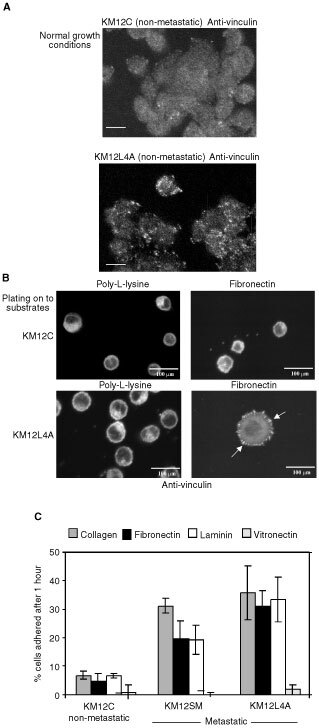
Elevated expression and/or activity of c-Src, the prototype of the Src family of protein tyrosine kinases, is associated with the development of human colon cancer. However, despite the known pleiotropic effects of these kinases in promoting (a) cell growth downstream of growth factor receptors, and (b) the dynamic regulation of integrin adhesions in fibroblast model systems, their precise role in epithelial cancer cells is unknown. Here we addressed whether elevated expression and activity of cellular Src alters cell proliferation and/or cell-matrix adhesion in cancer cells from the Fidler model of colorectal metastasis. Although elevated Src correlates with ability to metastasise to the liver after intrasplenic injection, we found that this was not linked to enhanced growth, either in vitro or in vivo as sub-cutaneous tumours. However, elevated Src was associated with enhanced attachment to extracellular matrix. In addition, adhesion to fibronectin, was suppressed by agents that inhibited Src activity, while enforced elevation of Src in non-metastatic cells was sufficient to stimulate adhesion to fibronectin and enhanced assembly of adhesion complexes, without influencing cell growth. Thus, we conclude that one role of elevated Src in human colon cancer cells is to modulate integrin-dependent cell-matrix attachment and formation of adhesion structures, which may, in turn, influence cell motility and integrin-dependent cellular responses.
Overview
The claims and statistics of this paper haven’t been checked yet.
Free: it reads the open-access text and checks the first 5 claims that cite a source we can check. Create a free account to check every claim.
Integrity signals
Registry
Cites
Statistics
Figures




Verification
Claims the paper makes
The claims and statistics of this paper haven’t been checked yet.
Free: it reads the open-access text and checks the first 5 claims that cite a source we can check. Create a free account to check every claim.
Claims other papers make about this one
Each is checked against this paper’s own text: does it say what the citing paper says it does?
The source backs part of the claim, not all of it — or backs it for a different population, condition or comparison.
In the citing paper
“Contradictory results show that elevated c-src does not directly influence proliferation either in vitro or in vivo in CRC.47”
From The molecular rationale of Src inhibition in colorectal carcinomas (2014) · cites this paper as [47]
Experimental — accuracy not yet measured
Self-check
Percentages, sums and reported differences re-computed from the paper’s own numbers.
The self-check needs the paper’s full text.
Citations
Cites
Src family kinases are required for integrin but not PDGFR signal transduction
Klinghoffer, R. A., Sachsenmaier, C., Cooper, J. A., & Soriano, P. (1999) · The EMBO Journal · doi:10.1093/emboj/18.9.2459 · Cited by 731 · cited 1×
“Recent work also showed that cells derived from triple Src/Fyn/Yes null embryos display impaired motility (Klinghoffer et al, 1999), while the dominant negative effect of kinase-deficient v-Src suppresses adhesion turnover and M o lecu lar an d C ellu lar P ath o lo gy Received 5 April 2002;…”
BackgroundSU6656, a Selective Src Family Kinase Inhibitor, Used To Probe Growth Factor Signaling
Blake, R. A., Broome, M. A., Liu, X., Wu, J., Gishizky, M., Sun, L., & Courtneidge, S. A. (2000) · Molecular and Cellular Biology · doi:10.1128/mcb.20.23.9018-9027.2000 · Cited by 584 · cited 8×
“The ability of SU6656 to suppress cell adhesion to fibronectin in 60 min was visualised microscopically and was quantitated as percentage inhibition of adhesion (lower panels).”
BackgroundMethodInfluentialActivating SRC mutation in a subset of advanced human colon cancers
Irby, R. B., Mao, W., Coppola, D., Kang, J., Loubeau, J. M., Trudeau, W., Karl, R., Fujita, D. J., Jove, R., & Yeatman, T. J. (1999) · Nature Genetics · doi:10.1038/5971 · Cited by 489 · cited 1×
“It was also recently reported that an activating mutation of c-Src, at tyrosine 527 in the carboxy-terminal regulatory region, occurred in a sub-set of advanced, metastatic colon cancers (Irby et al, 1999).”
BackgroundStat3-mediated Myc expression is required for Src transformation and PDGF-induced mitogenesis
Bowman, T., Broome, M. A., Sinibaldi, D., Wharton, W., Pledger, W. J., Sedivy, J. M., Irby, R., Yeatman, T., Courtneidge, S. A., & Jove, R. (2001) · Proceedings of the National Academy of Sciences · doi:10.1073/pnas.131568898 · Cited by 488 · cited 1×
“Specifically, c-Src is required for platelet-derived growth factor (PDGF)-induced mitogenesis (Roche et al, 1995; Twamley et al, 1993), most likely via its induction of a Stat3/c-Myc pathway (Barone and Courtneidge, 1995; Broome and Hunter, 1996; Wang et al, 2000b; Bowman et al, 2001).”
BackgroundIncrease in activity and level of pp60c-src in progressive stages of human colorectal cancer.
Talamonti, M. S., Roh, M. S., Curley, S. A., & Gallick, G. E. (1993) · Journal of Clinical Investigation · doi:10.1172/jci116200 · Cited by 399 · cited 1×
“…the prototypic member of the Src family of protein tyrosine kinases, during colorectal tumour progression, with an increasing trend from adenomas through to malignant carcinomas and metastases (Bolen et al, 1987; Cartwright et al, 1989, 1990, 1994; Talamonti et al, 1993; Termuhlen et al, 1993).”
BackgroundSrc Family Tyrosine Kinases and Growth Factor Signaling
Abram, C. L., & Courtneidge, S. A. (2000) · Experimental Cell Research · doi:10.1006/excr.1999.4732 · Cited by 391 · cited 2×
“Cells were visualised using a confocal microscope (model MRC600; BioRad Labs, Hercules, CA, USA).”
BackgroundMethodMorikawa, K., Walker, S. M., Jessup, J. M., & Fidler, I. J. (1988) · PubMed · Cited by 364
Src-induced de-regulation of E-cadherin in colon cancer cells requires integrin signalling
Avizienyte, E., Wyke, A. W., Jones, R. J., McLean, G. W., Westhoff, M. A., Brunton, V. G., & Frame, M. C. (2002) · Nature Cell Biology · doi:10.1038/ncb829 · Cited by 362 · cited 1×
“Our recent results also suggest that elevated c-Src de-regulates Ecadherin-mediated adhesion between colon cancer cells, and that the integrin adhesion formation we describe here is required for this effect (Avizienyte et al, 2002).”
BackgroundSelf-citationc-Src enhances the spreading of src-/- fibroblasts on fibronectin by a kinase-independent mechanism.
Kaplan, K. B., Swedlow, J. R., Morgan, D. O., & Varmus, H. E. (1995) · Genes & Development · doi:10.1101/gad.9.12.1505 · Cited by 317 · cited 1×
“…expression of c-Src can stimulate cell – matrix adhesion of c-Src 7/7 fibroblasts by a mechanism that is independent of its catalytic activity (Kaplan et al, 1995), while kinase-independent functions of v-Src are required for its incorporation into newly assembling integrin adhesions…”
BackgroundMyc but not Fos rescue of PDGF signalling block caused by kinase-inactive Src
Barone, M. V., & Courtneidge, S. A. (1995) · Nature · doi:10.1038/378509a0 · Cited by 309 · cited 1×
“Specifically, c-Src is required for platelet-derived growth factor (PDGF)-induced mitogenesis (Roche et al, 1995; Twamley et al, 1993), most likely via its induction of a Stat3/c-Myc pathway (Barone and Courtneidge, 1995; Broome and Hunter, 1996; Wang et al, 2000b; Bowman et al, 2001).”
Background
Cited by
Yeatman, T. J. (2004) · Nature Reviews Cancer · doi:10.1038/nrc1366 · Cited by 1,113
The interplay between Src and integrins in normal and tumor biology
Playford, M. P., & Schaller, M. D. (2004) · Oncogene · doi:10.1038/sj.onc.1208080 · Cited by 523
“Src-transformed cells are particularly characterized by a loss of actin reorganization and reduced cell–ECM adhesion leading to cell rounding and occasional cell detachment (Fincham et al., 1995, 1999; Jones et al., 2002).”
BackgroundThe ins and outs of fibronectin matrix assembly
Wierzbicka-Patynowski, I., & Schwarzbauer, J. E. (2003) · Journal of Cell Science · doi:10.1242/jcs.00670 · Cited by 473
“For example, increased Src expression and activity are associated with a decrease in the amount of FN matrix (Hynes, 1990; Olden and Yamada, 1977) and with changes in cell–FN interactions in human colon cancer (Jones et al., 2002).”
BackgroundNewest findings on the oldest oncogene; how activated src does it
Frame, M. C. (2004) · Journal of Cell Science · doi:10.1242/jcs.01111 · Cited by 367
“Since Src is often upregulated during the metastatic transition in colon cancers, for example in the Fidler model of colorectal metastasis (Jones et al., 2002; Mao et al., 1997), evaluating its mode of de-regulating the E-cadherin tumour suppressor protein might lead to new therapeutic targets.”
BackgroundSelf-citationSrc and FAK signalling controls adhesion fate and the epithelial-to-mesenchymal transition
Avizienyte, E., & Frame, M. C. (2005) · Current Opinion in Cell Biology · doi:10.1016/j.ceb.2005.08.007 · Cited by 309
Self-citation
Metadata
- Authors
- Jones, R. J., Avizienyte, E., Wyke, A. W., Owens, D. W., Brunton, V. G., & Frame, M. C.
- Affiliations
- Cancer Research UK (GB)
- University of Dundee (GB)
- Corresponding author
- Frame, M C
- Venue
- British Journal of Cancer
- Published
- 2002-10-29
- Volume
- 87
- Issue
- 10
- Pages
- 1128-1135
- ISSN
- 0007-0920 (ISSN-L)
- 1532-1827
- PMC
- PMC2376185
- OpenAlex
- W2164193456
- Cited by
- 80
- References
- 37
- Citations by year
- 2025: 1 · 2023: 2 · 2022: 1 · 2021: 3 · 2020: 2 · 2019: 1 · 2018: 1 · 2017: 3 · 2016: 4 · 2014: 3 · 2013: 4 · 2012: 3
- FWCI
- 3.50
- Type
- journal article
- Language
- en
- Open access
- hybrid
- doi.org
- Licence
- cc-by-nc-sa
- www.springer.com/tdm (unspecified)
- Locations
- British Journal of Cancer (publishedVersion, cc-by-nc-sa)
- Europe PMC (PubMed Central) (publishedVersion, cc-by)
- PubMed Central (publishedVersion, cc-by)
- PubMed (publishedVersion)
- ENLIGHTEN (Jurnal Bimbingan dan Konseling Islam) (submittedVersion)
- Research Explorer (The University of Manchester) (submittedVersion)
- Funders
- Cancer Research UK
- Topics
- Health Sciences › Medicine › Immunology and Allergy › Cell Adhesion Molecules Research
- Life Sciences › Biochemistry, Genetics and Molecular Biology › Molecular Biology › Protein Kinase Regulation and GTPase Signaling
- Life Sciences › Biochemistry, Genetics and Molecular Biology › Molecular Biology › Mechanisms of cancer metastasis
- Keywords
- Src kinase, Src family kinases, colorectal cancer, cell-matrix adhesion, integrin signaling, fibronectin, cell proliferation, metastasis, liver metastasis, extracellular matrix, adhesion complexes, cell motility
- MeSH terms
- Cell Adhesion, Humans, Neoplasm Metastasis, Protein-Tyrosine Kinases, Tumor Cells, Cultured, Colorectal Neoplasms, Oncogene Protein pp60(v-src), Cell-Matrix Junctions
About these checks
Registry, retraction, Cites and statistics results are computed, not judged, with one exception: a printed reference that no index lists is read once by a model before it counts as a citation error. Claims are extracted by a model and kept only when their quote appears verbatim in the text; each cited source is then checked, with a model’s help, for whether it says what the claim relies on. Those verdicts can be wrong — read the claim’s quote and the cited source before relying on one.
Statistics and arithmetic are recomputed, not judged.
Confidence bands are shown only for fields calibrated on our annotated test set. None is calibrated yet, so no band is shown.
What counts as a claim ›How we measure confidence ›How we check this ›
Are you an author, or do you see a problem with this page? Request a review
How to cite
Cite CoChat for the checks on this page, and the paper for its own findings.
- This page
- CoChat. (2026). Citation check: Elevated c-Src is linked to altered cell–matrix adhesion rather than proliferation in KM12C human colorectal cancer cells. https://app.cochat.ai/papers/10.1038/sj.bjc.6600594
- The paper
- Jones, R. J., Avizienyte, E., Wyke, A. W., Owens, D. W., Brunton, V. G., & Frame, M. C. (2002). Elevated c-Src is linked to altered cell–matrix adhesion rather than proliferation in KM12C human colorectal cancer cells. British Journal of Cancer. https://doi.org/10.1038/sj.bjc.6600594
Data sources
Bibliographic data: Semantic Scholar, OpenAlex, Crossref.
Contains information from the Semantic Scholar Academic Graph Datasets, made available under the ODC Attribution License (ODC-BY 1.0).
Semantic Scholar’s data is described in: Kinney, R., et al. (2023). The Semantic Scholar Open Data Platform. arXiv:2301.10140.
