

The DOI matches its Crossref record; 48 of the 48 references it cites were found in a registry or an index.
Computer-Aided Diagnosis with Deep Learning Architecture: Applications to Breast Lesions in US Images and Pulmonary Nodules in CT Scans
Open accessAbstract
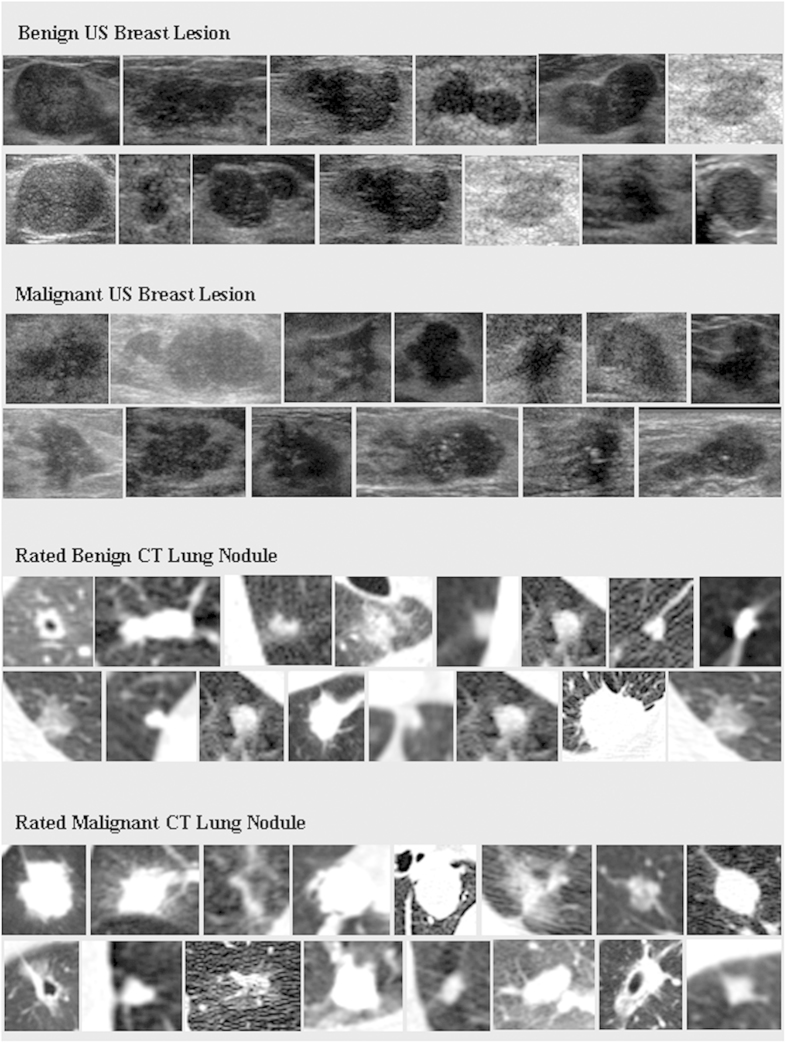
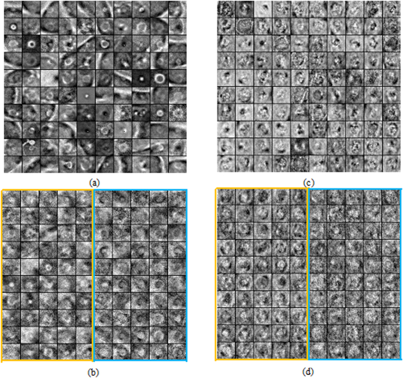
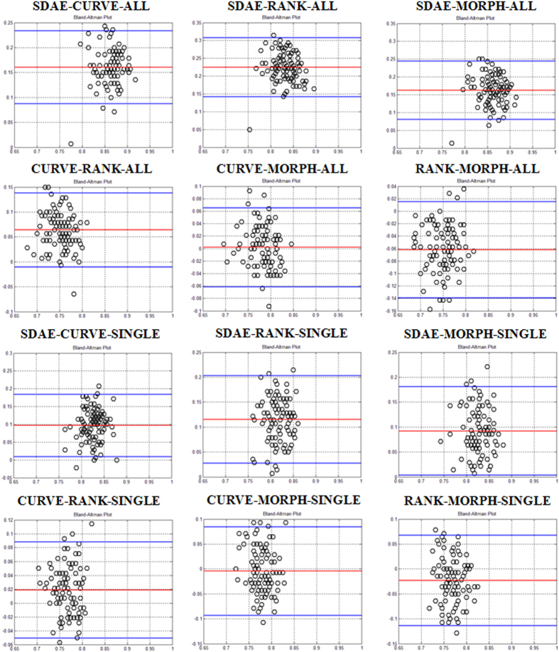
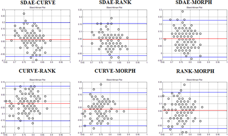
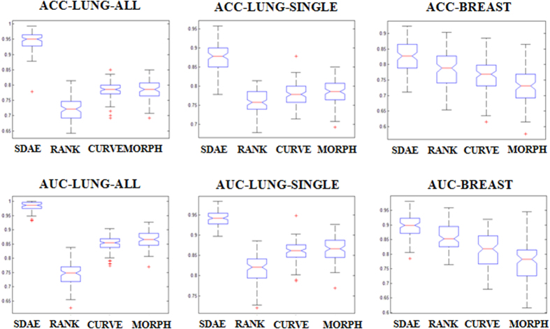
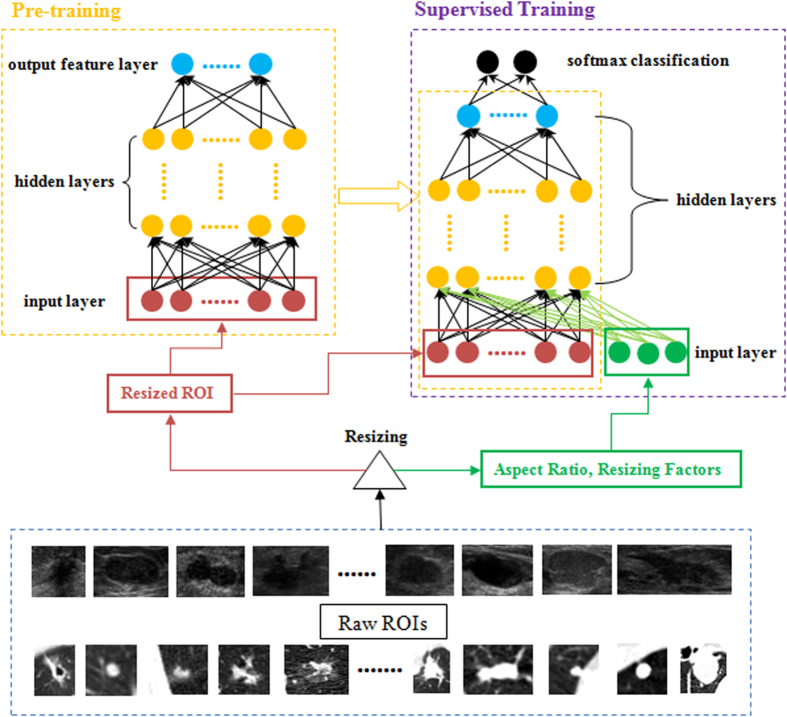
This paper performs a comprehensive study on the deep-learning-based computer-aided diagnosis (CADx) for the differential diagnosis of benign and malignant nodules/lesions by avoiding the potential errors caused by inaccurate image processing results (e.g., boundary segmentation), as well as the classification bias resulting from a less robust feature set, as involved in most conventional CADx algorithms. Specifically, the stacked denoising auto-encoder (SDAE) is exploited on the two CADx applications for the differentiation of breast ultrasound lesions and lung CT nodules. The SDAE architecture is well equipped with the automatic feature exploration mechanism and noise tolerance advantage, and hence may be suitable to deal with the intrinsically noisy property of medical image data from various imaging modalities. To show the outperformance of SDAE-based CADx over the conventional scheme, two latest conventional CADx algorithms are implemented for comparison. 10 times of 10-fold cross-validations are conducted to illustrate the efficacy of the SDAE-based CADx algorithm. The experimental results show the significant performance boost by the SDAE-based CADx algorithm over the two conventional methods, suggesting that deep learning techniques can potentially change the design paradigm of the CADx systems without the need of explicit design and selection of problem-oriented features.
Overview
The claims and statistics of this paper haven’t been checked yet.
Run the check — free, no account needed. It reads the open-access text and checks the sources it cites.
Integrity signals
Registry
Cites
Statistics
Figures
Verification
Claims the paper makes
The claims and statistics of this paper haven’t been checked yet.
Run the check — free, no account needed. It reads the open-access text and checks the sources it cites.
Claims other papers make about this one
Each is checked against this paper’s own text: does it say what the citing paper says it does?
In the citing paper
“In addition, the stacked denoising auto-encoder (SDAE) [88], a combination of the point-wise gated Boltzmann machine (PGBM) and RBM [89], and the GoogLeNet CNN [90] have been applied to breast US or shear-wave elastography images for breast cancer diagnosis; all of these obtained a superior performance when compared with human experts.”
From Deep Learning in Medical Ultrasound Analysis: A Review (2019) · cites this paper as [88]
Self-check
Percentages, sums and reported differences re-computed from the paper’s own numbers.
The self-check needs the paper’s full text.
Citations
Cites
STATISTICAL METHODS FOR ASSESSING AGREEMENT BETWEEN TWO METHODS OF CLINICAL MEASUREMENT
Martin Bland, J., & Altman, D. (1986) · The Lancet · doi:10.1016/s0140-6736(86)90837-8 · Cited by 47,965
Hinton, G., Deng, L., Yu, D., Dahl, G., Mohamed, A.-R., Jaitly, N., Senior, A., Vanhoucke, V., Nguyen, P., Sainath, T., et al. (2012) · IEEE Signal Processing Magazine · doi:10.1109/msp.2012.2205597 · Cited by 10,260 · cited 1×
“Although deep learning has been successfully applied to many image-and acoustic-related problems 46 , it is less explored in the context of CADx.”
Contour Detection and Hierarchical Image Segmentation
Arbeláez, P., Maire, M., Fowlkes, C. C., & Malik, J. (2010) · IEEE Transactions on Pattern Analysis and Machine Intelligence · doi:10.1109/tpami.2010.161 · Cited by 5,581 · cited 1×
“…image segmentation 4,6,14,15,22–24 for the morphological feature computing 4,6,7,14,15,22 , which unfortunately remains quite difficult to address 25 , and 2) image decomposition 12,19 followed with statistical summarizations and presentations 12,19,20,26 for the textural feature calculation 27 .”
Background(2010) · Journal of Machine Learning Research · doi:10.5555/1756006.1953039 · Cited by 4,051 · cited 1×
“In this study, we further exploit the deep learning model of the stacked denoising autoencoder (SDAE) 41 for the differentiation of distinctive types of lesions and nodules depicted with different imaging modalities.”
MethodArmato, S. G., McLennan, G., Bidaut, L., McNitt‐Gray, M. F., Meyer, C. R., Reeves, A. P., Zhao, B., Aberle, D. R., Henschke, C. I., Hoffman, E. A., et al. (2011) · Medical Physics · doi:10.1118/1.3528204 · Cited by 2,887
OverFeat: Integrated Recognition, Localization and Detection using Convolutional Networks
Sermanet, P., Eigen, D., Zhang, X., Mathieu, M., Fergus, R., & LeCun, Y. (2013) · arXiv · doi:10.48550/arxiv.1312.6229 · Cited by 2,680 · cited 1×
“For the problem of CADx, a specific convolutional neural network model, OverFeat 39 , was employed in the work 40 to classify the specific type of peri-fissural nodules with the ensemble fashion in AUC performance around 0.86.”
MethodInfluentialDistance Regularized Level Set Evolution and Its Application to Image Segmentation
Li, C., Xu, C., Gui, C., & Fox, M. D. (2010) · IEEE Transactions on Image Processing · doi:10.1109/tip.2010.2069690 · Cited by 2,106 · cited 2×
“The MORPH features are computed from either experts’ drawings or the computer-generated boundaries by level set 43 (DRLSE) and grow-cut 44 (GC) methods.”
MethodComputer-aided diagnosis in medical imaging: historical review, current status and future potential
Doi, K. (2007) · Computerized Medical Imaging and Graphics · doi:10.1016/j.compmedimag.2007.02.002 · Cited by 1,855
Exploring Strategies for Training Deep Neural Networks
Larochelle, H., Bengio, Y., Louradour, J., & Lamblin, P. (2009) · doi:10.1145/1577069.1577070 · Cited by 1,111
Deep convolutional neural networks for multi-modality isointense infant brain image segmentation
Zhang, W., Li, R., Deng, H., Wang, L., Lin, W., Ji, S., & Shen, D. (2015) · NeuroImage · doi:10.1016/j.neuroimage.2014.12.061 · Cited by 817 · cited 1×
“…to the medical image analysis domain with promising results on various applications, like the computerized prognosis for Alzheimer’s disease and mild cognitive impairment 32 , organ segmentations 33 and detection 34 , ultrasound standard plane selection 35 , etc., on 3D or 4D image data, etc.”
MethodImproving Computer-Aided Detection UsingConvolutional Neural Networks and Random View Aggregation
Roth, H. R., Lu, L., Liu, J., Yao, J., Seff, A., Cherry, K., Kim, L., & Summers, R. M. (2016) · IEEE Transactions on Medical Imaging · doi:10.1109/tmi.2015.2482920 · Cited by 608 · cited 1×
“In the context of CAD, most works focused on the problem of abnormality detection (CADe) 36–38 .”
BackgroundLatent feature representation with stacked auto-encoder for AD/MCI diagnosis
Initiative, T. A. D. N., Suk, H., Lee, S., & Shen, D. G. (2013) · Brain Structure and Function · doi:10.1007/s00429-013-0687-3 · Cited by 538 · cited 1×
“…to the medical image analysis domain with promising results on various applications, like the computerized prognosis for Alzheimer’s disease and mild cognitive impairment 32 , organ segmentations 33 and detection 34 , ultrasound standard plane selection 35 , etc., on 3D or 4D image data, etc.”
MethodShin, H.-C., Orton, M. R., Collins, D. J., Doran, S. J., & Leach, M. O. (2013) · IEEE Transactions on Pattern Analysis and Machine Intelligence · doi:10.1109/tpami.2012.277 · Cited by 488 · cited 1×
“…to the medical image analysis domain with promising results on various applications, like the computerized prognosis for Alzheimer’s disease and mild cognitive impairment 32 , organ segmentations 33 and detection 34 , ultrasound standard plane selection 35 , etc., on 3D or 4D image data, etc.”
MethodLung Image Database Consortium: Developing a Resource for the Medical Imaging Research Community
Armato, S. G., McLennan, G., McNitt‐Gray, M. F., Meyer, C. R., Yankelevitz, D. F., Aberle, D. R., Henschke, C. I., Hoffman, E. A., Kazerooni, E. A., MacMahon, H., et al. (2004) · Radiology · doi:10.1148/radiol.2323032035 · Cited by 381
Quantitative Analysis of Pulmonary Emphysema Using Local Binary Patterns
Srensen, L., Shaker, S. B., & de Bruijne, M. (2010) · IEEE Transactions on Medical Imaging · doi:10.1109/tmi.2009.2038575 · Cited by 374
Giger, M. L., Chan, H., & Boone, J. (2008) · Medical Physics · doi:10.1118/1.3013555 · Cited by 311
Computer-aided Diagnosis: How to Move from the Laboratory to the Clinic
van Ginneken, B., Schaefer-Prokop, C. M., & Prokop, M. (2011) · Radiology · doi:10.1148/radiol.11091710 · Cited by 306
Ciompi, F., de Hoop, B., van Riel, S. J., Chung, K., Scholten, E. T., Oudkerk, M., de Jong, P. A., Prokop, M., & Ginneken, B. V. (2015) · Medical Image Analysis · doi:10.1016/j.media.2015.08.001 · Cited by 289 · cited 1×
“For the problem of CADx, a specific convolutional neural network model, OverFeat 39 , was employed in the work 40 to classify the specific type of peri-fissural nodules with the ensemble fashion in AUC performance around 0.86.”
MethodInfluentialAutomated lung segmentation for thoracic CT
Armato, S. G., & Sensakovic, W. F. (2004) · Academic Radiology · doi:10.1016/j.acra.2004.06.005 · Cited by 259
Chen, C., Chou, Y., Han, K.-C., Hung, G.-S., Tiu, C., Chiou, H., & Chiou, S. (2003) · Radiology · doi:10.1148/radiol.2262011843 · Cited by 257
Joo, S., Yang, Y., Moon, W. K., & Kim, H. C. (2004) · IEEE Transactions on Medical Imaging · doi:10.1109/tmi.2004.834617 · Cited by 255 · cited 4×
“Accordingly, the extraction of useful features highly depends on the quality of each intermediate result in the image processing steps 4,6,14,22,27 , which often needs many passes of trial-and-error design to find satisfactory intermediate results.”
BackgroundMethodInfluentialGómez‐Flores, W., Pereira, W. C. D. A., & Infantosi, A. F. C. (2012) · IEEE Transactions on Medical Imaging · doi:10.1109/tmi.2012.2206398 · Cited by 248 · cited 3×
“…image segmentation 4,6,14,15,22–24 for the morphological feature computing 4,6,7,14,15,22 , which unfortunately remains quite difficult to address 25 , and 2) image decomposition 12,19 followed with statistical summarizations and presentations 12,19,20,26 for the textural feature calculation 27 .”
BackgroundMethodJourney toward Computer-aided Diagnosis: Role of Image Texture Analysis
Tourassi, G. D. (1999) · Radiology · doi:10.1148/radiology.213.2.r99nv49317 · Cited by 233
Way, T. W., Hadjiiski, L. M., Sahiner, B., Chan, H., Cascade, P. N., Kazerooni, E. A., Bogot, N., & Zhou, C. (2006) · Medical Physics · doi:10.1118/1.2207129 · Cited by 225 · cited 3×
“For a better quality of morphological feature computing, the computed lesion/nodule boundaries usually require manual intervention, including model initialization 13,15 , parameter adjustment 6,13,15 , boundary refinement 6 and selection 4,23 , to improve the segmentation results.”
MethodApplication of the mutual information criterion for feature selection in computer‐aided diagnosis
Tourassi, G. D., Frederick, E. D., Markey, M. K., & Floyd, C. E. (2001) · Medical Physics · doi:10.1118/1.1418724 · Cited by 222
Breast Image Analysis for Risk Assessment, Detection, Diagnosis, and Treatment of Cancer
Giger, M. L., Karssemeijer, N., & Schnabel, J. A. (2013) · Annual Review of Biomedical Engineering · doi:10.1146/annurev-bioeng-071812-152416 · Cited by 211
Automatic ultrasound segmentation and morphology based diagnosis of solid breast tumors
Chang, R., Wu, W.-J., Moon, W. K., & Chen, D. (2005) · Breast Cancer Research and Treatment · doi:10.1007/s10549-004-2043-z · Cited by 198 · cited 1×
“Meanwhile, the computerized image segmentation results often require various case-by-case user interventions, e.g., parameter adjustment 6,28 , manual refinement 6,9,28–31 , solution selection 4,23 , etc., to improve the contour correctness 4,6,13,15,23 .”
Chen, H., Dou, Q., Ni, D., Cheng, J.-Z., Qin, J., Li, S., & Heng, P.-A. (2015) · Lecture Notes in Computer Science · doi:10.1007/978-3-319-24553-9_62 · Cited by 180 · cited 1×
“…to the medical image analysis domain with promising results on various applications, like the computerized prognosis for Alzheimer’s disease and mild cognitive impairment 32 , organ segmentations 33 and detection 34 , ultrasound standard plane selection 35 , etc., on 3D or 4D image data, etc.”
MethodSelf-citationKubota, T., Jerebko, A. K., Dewan, M., Salganicoff, M., & Krishnan, A. V. (2010) · Medical Image Analysis · doi:10.1016/j.media.2010.08.005 · Cited by 178 · cited 1×
“Meanwhile, the computerized image segmentation results often require various case-by-case user interventions, e.g., parameter adjustment 6,28 , manual refinement 6,9,28–31 , solution selection 4,23 , etc., to improve the contour correctness 4,6,13,15,23 .”
Sahiner, B., Petrick, N., Chan, H., Hadjiiski, L. M., Paramagul, C., Helvie, M. A., & Gürcan, M. N. (2001) · IEEE Transactions on Medical Imaging · doi:10.1109/42.974922 · Cited by 174
Newell, D., Nie, K., Chen, J., Hsu, C., Yu, H. J., Nalcioğlu, O., & Su, M. (2009) · European Radiology · doi:10.1007/s00330-009-1616-y · Cited by 150
Way, T. W., Sahiner, B., Chan, H., Hadjiiski, L. M., Cascade, P. N., Chughtai, A. R., Bogot, N., & Kazerooni, E. A. (2009) · Medical Physics · doi:10.1118/1.3140589 · Cited by 147 · cited 2×
“For a better quality of morphological feature computing, the computed lesion/nodule boundaries usually require manual intervention, including model initialization 13,15 , parameter adjustment 6,13,15 , boundary refinement 6 and selection 4,23 , to improve the segmentation results.”
MethodTexture Classification Using Refined Histogram
Li, L., Tong, C. S., & Choy, S. K. (2010) · IEEE Transactions on Image Processing · doi:10.1109/tip.2010.2041414 · Cited by 141
McCarville, M. B., Lederman, H. M., Santana, V. M., Daw, N. C., Shochat, S. J., Li, C., & Kaufman, R. A. (2006) · Radiology · doi:10.1148/radiol.2392050631 · Cited by 139
Sahiner, B., Chan, H., Roubidoux, M. A., Hadjiiski, L. M., Helvie, M. A., Paramagul, C., Bailey, J. E., Nees, A. V., & Blane, C. E. (2007) · Radiology · doi:10.1148/radiol.2423051464 · Cited by 135
Computer‐aided diagnosis in high resolution CT of the lungs
Sluimer, I. C., van Waes, P. F. G. M., Viergever, M. A., & van Ginneken, B. (2003) · Medical Physics · doi:10.1118/1.1624771 · Cited by 134
Tajbakhsh, N., Gotway, M., & Liang, J. (2015) · International Conference on Medical Image Computing and Computer-Assisted Intervention · doi:10.1007/978-3-319-24571-3_8 · Cited by 129
Alvarenga, A. V., Pereira, W. C. D. A., Infantosi, A. F. C., & de Azevedo, C. M. (2007) · Medical Physics · doi:10.1118/1.2401039 · Cited by 118
Computer‐aided diagnosis using morphological features for classifying breast lesions on ultrasound
Huang, Y. ‐., Chen, D. ‐., Jiang, Y., Kuo, S. ‐., Wu, H., & Moon, W. K. (2008) · Ultrasound in Obstetrics and Gynecology · doi:10.1002/uog.5205 · Cited by 106
Yang, M.-C., Moon, W. K., Wang, Y.-C. F., Bae, M. S., Huang, C., Chen, J., & Chang, R. (2013) · IEEE Transactions on Medical Imaging · doi:10.1109/tmi.2013.2279938 · Cited by 97 · cited 9×
“In this case, the feature selection step has to be applied 19,20 to find a subset of the most useful features.”
BackgroundMethodInfluentialAwai, K., Murao, K., Ozawa, A., Nakayama, Y., Nakaura, T., Liu, D., Kawanaka, K., Funama, Y., Morishita, S., & Yamashita, Y. (2006) · Radiology · doi:10.1148/radiol.2383050167 · Cited by 85
Computer-Aided Diagnosis for Early-Stage Lung Cancer Based on Longitudinal and Balanced Data
Sun, T., Zhang, R., Wang, J., Li, X., & Guo, X. (2013) · PLoS ONE · doi:10.1371/journal.pone.0063559 · Cited by 49 · cited 5×
“For lung nodule differentiation, the latest lung CT CADx algorithm 12 , denoted as CURVE, is implemented.”
BackgroundMethodInfluentialSingh, S., Maxwell, J., Baker, J. A., Nicholas, J. L., & Lo, J. Y.-C. (2010) · Radiology · doi:10.1148/radiol.10081308 · Cited by 49
Automated Method for Improving System Performance of Computer-Aided Diagnosis in Breast Ultrasound
Drukker, K., Sennett, C. A., & Giger, M. L. (2009) · IEEE Transactions on Medical Imaging · doi:10.1109/tmi.2008.928178 · Cited by 48
Computer-aided US Diagnosis of Breast Lesions by Using Cell-based Contour Grouping
Cheng, J., Chou, Y., Huang, C., Chang, Y., Tiu, C., Chen, K., & Chen, C. (2010) · Radiology · doi:10.1148/radiol.09090001 · Cited by 47
Chen, C., Chou, Y., Chen, C. S. K., Cheng, J.-Z., Ou, Y.-F., Yeh, F.-C. F., & Chen, K. (2005) · Ultrasound in Medicine & Biology · doi:10.1016/j.ultrasmedbio.2005.09.011 · Cited by 46
Leveraging Mid-Level Semantic Boundary Cues for Automated Lymph Node Detection
Seff, A., Lu, L., Barbu, A., Roth, H., Shin, H.-C., & Summers, R. M. (2015) · Lecture Notes in Computer Science · doi:10.1007/978-3-319-24571-3_7 · Cited by 36
ACCOMP: Augmented cell competition algorithm for breast lesion demarcation in sonography
Cheng, J., Chou, Y., Huang, C., Chang, Y., Tiu, C., Yeh, F.-C. F., Chen, K., Tsou, C., & Chen, C. (2010) · Medical Physics · doi:10.1118/1.3512799 · Cited by 26 · cited 2×
“For a better quality of morphological feature computing, the computed lesion/nodule boundaries usually require manual intervention, including model initialization 13,15 , parameter adjustment 6,13,15 , boundary refinement 6 and selection 4,23 , to improve the segmentation results.”
BackgroundMethod
Research works printed but not linked: 1
- Vezhnevets, V. & Konouchine, V. GrowCut: Interactive multi-label ND image segmentation by cellular automata. Proc. of Graphicon. 1, 150–156 (2005).
Cited by
Sorted by citations
A survey on deep learning in medical image analysis
Litjens, G., Kooi, T., Bejnordi, B. E., Setio, A. A. A., Ciompi, F., Ghafoorian, M., van der Laak, J. A. W. M., van Ginneken, B., & Sánchez, C. I. (2017) · Medical Image Analysis · doi:10.1016/j.media.2017.07.005 · Cited by 15,332
“Janowczyk et al. (2016a) used SAEs to normalize H&E-stained histopathology images whereas Benou et al. (2016) used CNNs to perform de-noising in DCE-MRI time-series.”
BackgroundMethodInfluentialA guide to deep learning in healthcare
Esteva, A., Robicquet, A., Ramsundar, B., Kuleshov, V., DePristo, M., Chou, K., Cui, C., Corrado, G., Thrun, S., & Dean, J. (2019) · Nature Medicine · doi:10.1038/s41591-018-0316-z · Cited by 5,312
“Many studies have demonstrated promising results in complex diagnostics spanning dermatology 9,10 , radiology 11–14 , ophthalmology 15–17 , and pathology 18–21 (Fig.”
BackgroundDeep learning in medical image analysis
Shen, D., Wu, G., & Suk, H.-I. (2017) · Annual Review of Biomedical Engineering · doi:10.1146/annurev-bioeng-071516-044442 · Cited by 4,968
“To this end, research groups have devised various strategies, such as (i) to take image patches either 2D or 3D as input (25, 35, 36, 37, 38, 39, 40, 41), rather than the full-sized images, to reduce the input dimensionality, thus the number of model parameters; (ii) to expand their dataset by…”
MethodArtificial intelligence in radiology
Hosny, A., Parmar, C., Quackenbush, J., Schwartz, L. H., & Aerts, H. J. W. L. (2018) · Nature Reviews Cancer · doi:10.1038/s41568-018-0016-5 · Cited by 3,803
“Such performance gain is assessed using multiple metrics, including the area under receiver operating characteristic curve (AUC), accuracy, sensitivity and specificity, to name a few 35 .”
Deep learning for healthcare: review, opportunities and challenges
Miotto, R., Wang, F., Wang, S., Jiang, X., & Dudley, J. T. (2018) · Briefings in Bioinformatics · doi:10.1093/bib/bbx044 · Cited by 3,076
Metadata
- Authors
- Cheng, J.-Z., Ni, D., Chou, Y.-H., Qin, J., Tiu, C.-M., Chang, Y.-C., Huang, C.-S., Shen, D., & Chen, C.-M.
- Affiliations
- Shenzhen University (CN)
- Taipei Veterans General Hospital (TW)
- National Taiwan University (TW)
- University of North Carolina at Chapel Hill (US)
- Corresponding author
- Chou, Yi-Hong
- Shen, Dinggang
- Chen, Chung-Ming
- ORCID
- Venue
- Scientific Reports
- Published
- 2016-04-15
- Volume
- 6
- Issue
- 1
- Pages
- 24454-24454
- ISSN
- 2045-2322 (ISSN-L)
- PMC
- PMC4832199
- OpenAlex
- W2341106171
- Cited by
- 755
- References
- 48
- Citations by year
- 2026: 9 · 2025: 32 · 2024: 78 · 2023: 59 · 2022: 100 · 2021: 120 · 2020: 103 · 2019: 110 · 2018: 87 · 2017: 45 · 2016: 11
- FWCI
- 68.59
- Type
- journal article
- Language
- en
- Open access
- gold
- doi.org
- Locations
- Scientific Reports (publishedVersion, cc-by)
- PubMed Central (publishedVersion, cc-by)
- PubMed (publishedVersion)
- Carolina Digital Repository (University of North Carolina at Chapel Hill) (submittedVersion)
- UNC Libraries
- Funders
- National Science Council (NSC 100)
- Shenzhen Fundamental Research Program (JCYJ20130329105033277, JCYJ20150525092940982, 81571758, JCYJ20140509172609164, JSE201109150013A)
- National Cancer Institute (R01CA206100)
- Topics
- Physical Sciences › Computer Science › Artificial Intelligence › AI in cancer detection
- Health Sciences › Medicine › Radiology, Nuclear Medicine and Imaging › Radiomics and Machine Learning in Medical Imaging
- Physical Sciences › Computer Science › Computer Vision and Pattern Recognition › Medical Image Segmentation Techniques
- Keywords
- computer-aided diagnosis, stacked denoising autoencoder, pulmonary nodules, benign versus malignant differentiation, medical image denoising, feature extraction, noise immunity, differential diagnosis
- MeSH terms
- Machine Learning, Algorithms, Breast, Solitary Pulmonary Nodule, Diagnosis, Computer-Assisted, Female, Humans, Tomography, X-Ray Computed, Ultrasonography, Reproducibility of Results, Datasets as Topic
About these checks
Registry, retraction, Cites and statistics results are computed, not judged, with one exception: a printed reference that no index lists is read once by a model before it counts as a citation error. Claims are extracted by a model and kept only when their quote appears verbatim in the text; each cited source is then checked, with a model’s help, for whether it says what the claim relies on. Those verdicts can be wrong — read the claim’s quote and the cited source before relying on one.
Statistics and arithmetic are recomputed, not judged.
Confidence bands are shown only for fields calibrated on our annotated test set. None is calibrated yet, so no band is shown.
What counts as a claim ›How we measure confidence ›How we check this ›
Are you an author, or do you see a problem with this page? Request a review
