

The DOI matches its Crossref record; 48 of the 48 references it cites were found in a registry or an index.
Computer-Aided Diagnosis with Deep Learning Architecture: Applications to Breast Lesions in US Images and Pulmonary Nodules in CT Scans
Open accessAbstract
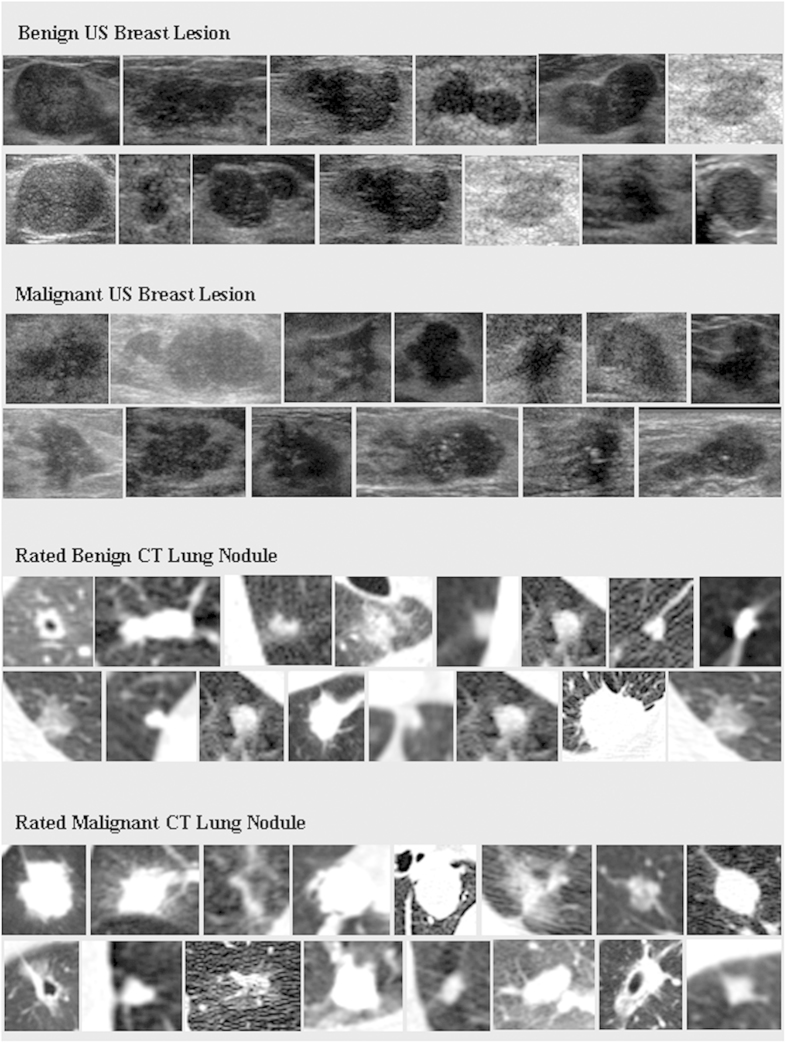
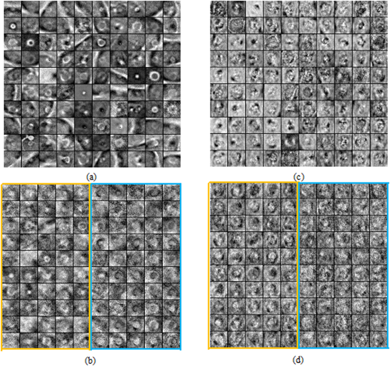
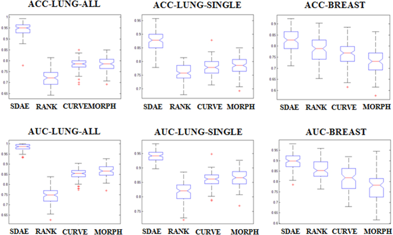
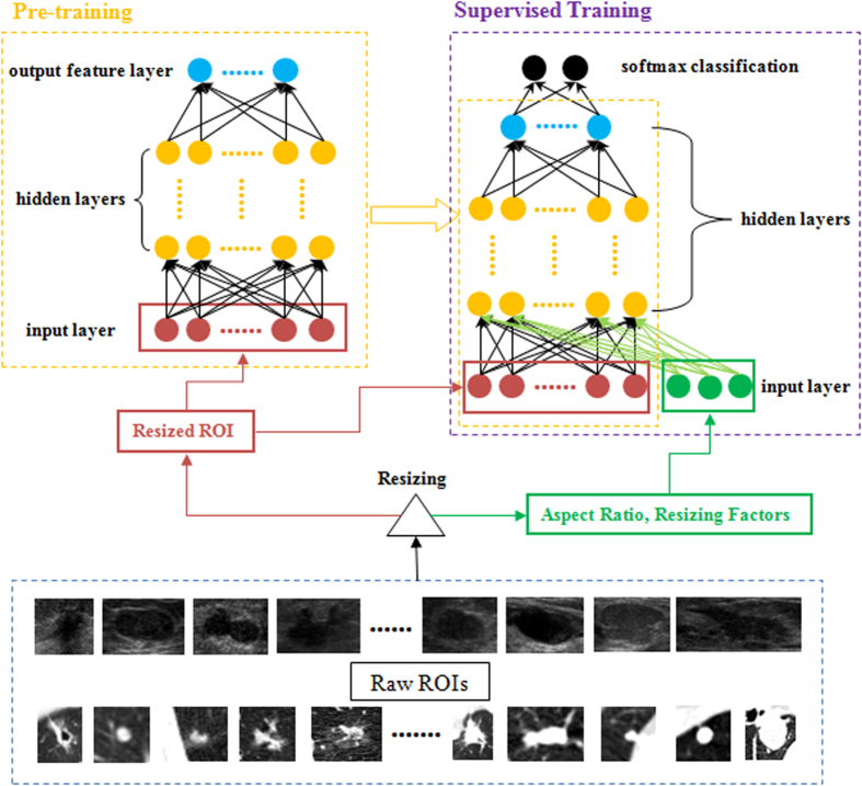
This paper performs a comprehensive study on the deep-learning-based computer-aided diagnosis (CADx) for the differential diagnosis of benign and malignant nodules/lesions by avoiding the potential errors caused by inaccurate image processing results (e.g., boundary segmentation), as well as the classification bias resulting from a less robust feature set, as involved in most conventional CADx algorithms. Specifically, the stacked denoising auto-encoder (SDAE) is exploited on the two CADx applications for the differentiation of breast ultrasound lesions and lung CT nodules. The SDAE architecture is well equipped with the automatic feature exploration mechanism and noise tolerance advantage, and hence may be suitable to deal with the intrinsically noisy property of medical image data from various imaging modalities. To show the outperformance of SDAE-based CADx over the conventional scheme, two latest conventional CADx algorithms are implemented for comparison. 10 times of 10-fold cross-validations are conducted to illustrate the efficacy of the SDAE-based CADx algorithm. The experimental results show the significant performance boost by the SDAE-based CADx algorithm over the two conventional methods, suggesting that deep learning techniques can potentially change the design paradigm of the CADx systems without the need of explicit design and selection of problem-oriented features.
Overview
The claims and statistics of this paper haven’t been checked yet.
Run the check — free, no account needed. It reads the open-access text and checks the sources it cites.
Integrity signals
Registry
Cites
Statistics
Figures
Verification
Claims the paper makes
The claims and statistics of this paper haven’t been checked yet.
Run the check — free, no account needed. It reads the open-access text and checks the sources it cites.
Claims other papers make about this one
Each is checked against this paper’s own text: does it say what the citing paper says it does?
In the citing paper
“In addition, the stacked denoising auto-encoder (SDAE) [88], a combination of the point-wise gated Boltzmann machine (PGBM) and RBM [89], and the GoogLeNet CNN [90] have been applied to breast US or shear-wave elastography images for breast cancer diagnosis; all of these obtained a superior performance when compared with human experts.”
From Deep Learning in Medical Ultrasound Analysis: A Review (2019) · cites this paper as [88]
Self-check
Percentages, sums and reported differences re-computed from the paper’s own numbers.
The self-check needs the paper’s full text.
Citations
Cites
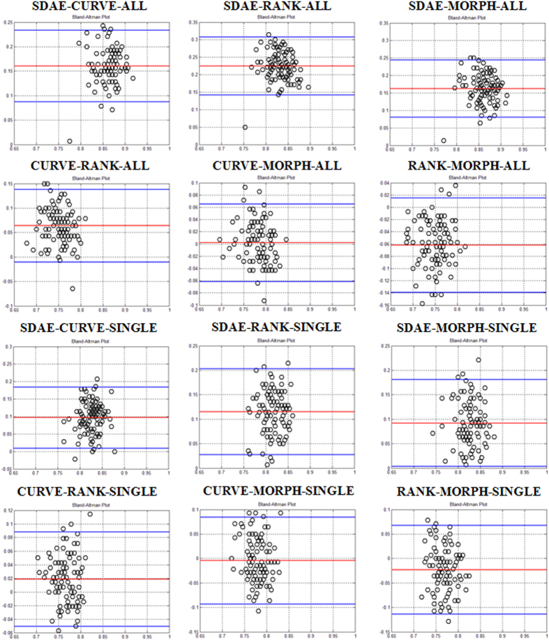
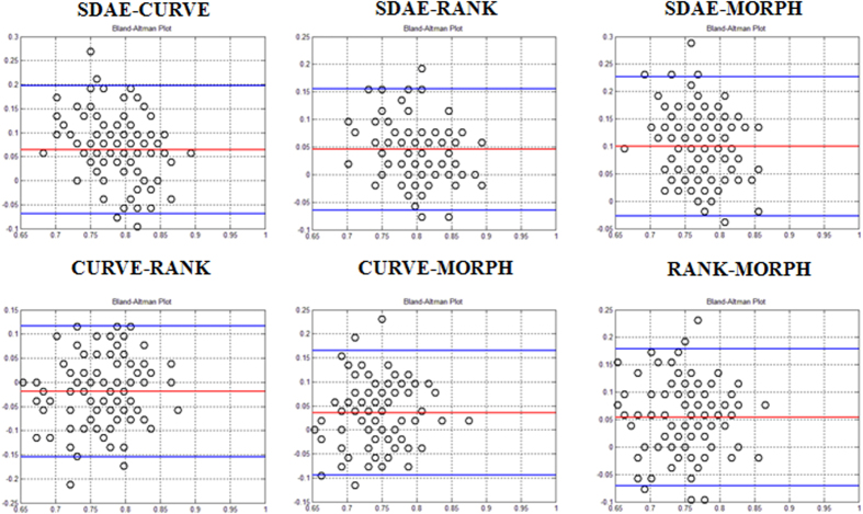
STATISTICAL METHODS FOR ASSESSING AGREEMENT BETWEEN TWO METHODS OF CLINICAL MEASUREMENT
Martin Bland, J., & Altman, D. (1986) · The Lancet · doi:10.1016/s0140-6736(86)90837-8 · Cited by 47,965
Hinton, G., Deng, L., Yu, D., Dahl, G., Mohamed, A.-R., Jaitly, N., Senior, A., Vanhoucke, V., Nguyen, P., Sainath, T., et al. (2012) · IEEE Signal Processing Magazine · doi:10.1109/msp.2012.2205597 · Cited by 10,260
Contour Detection and Hierarchical Image Segmentation
Arbeláez, P., Maire, M., Fowlkes, C. C., & Malik, J. (2010) · IEEE Transactions on Pattern Analysis and Machine Intelligence · doi:10.1109/tpami.2010.161 · Cited by 5,581
(2010) · Journal of Machine Learning Research · doi:10.5555/1756006.1953039 · Cited by 4,051
Armato, S. G., McLennan, G., Bidaut, L., McNitt‐Gray, M. F., Meyer, C. R., Reeves, A. P., Zhao, B., Aberle, D. R., Henschke, C. I., Hoffman, E. A., et al. (2011) · Medical Physics · doi:10.1118/1.3528204 · Cited by 2,887
OverFeat: Integrated Recognition, Localization and Detection using Convolutional Networks
Sermanet, P., Eigen, D., Zhang, X., Mathieu, M., Fergus, R., & LeCun, Y. (2013) · arXiv · doi:10.48550/arxiv.1312.6229 · Cited by 2,680
Distance Regularized Level Set Evolution and Its Application to Image Segmentation
Li, C., Xu, C., Gui, C., & Fox, M. D. (2010) · IEEE Transactions on Image Processing · doi:10.1109/tip.2010.2069690 · Cited by 2,106
Computer-aided diagnosis in medical imaging: historical review, current status and future potential
Doi, K. (2007) · Computerized Medical Imaging and Graphics · doi:10.1016/j.compmedimag.2007.02.002 · Cited by 1,855
Exploring Strategies for Training Deep Neural Networks
Larochelle, H., Bengio, Y., Louradour, J., & Lamblin, P. (2009) · doi:10.1145/1577069.1577070 · Cited by 1,111
Deep convolutional neural networks for multi-modality isointense infant brain image segmentation
Zhang, W., Li, R., Deng, H., Wang, L., Lin, W., Ji, S., & Shen, D. (2015) · NeuroImage · doi:10.1016/j.neuroimage.2014.12.061 · Cited by 817
Research works printed but not linked: 1
- Vezhnevets, V. & Konouchine, V. GrowCut: Interactive multi-label ND image segmentation by cellular automata. Proc. of Graphicon. 1, 150–156 (2005).
Cited by
Sorted by citations
A survey on deep learning in medical image analysis
Litjens, G., Kooi, T., Bejnordi, B. E., Setio, A. A. A., Ciompi, F., Ghafoorian, M., van der Laak, J. A. W. M., van Ginneken, B., & Sánchez, C. I. (2017) · Medical Image Analysis · doi:10.1016/j.media.2017.07.005 · Cited by 15,332
A guide to deep learning in healthcare
Esteva, A., Robicquet, A., Ramsundar, B., Kuleshov, V., DePristo, M., Chou, K., Cui, C., Corrado, G., Thrun, S., & Dean, J. (2019) · Nature Medicine · doi:10.1038/s41591-018-0316-z · Cited by 5,312
Deep learning in medical image analysis
Shen, D., Wu, G., & Suk, H.-I. (2017) · Annual Review of Biomedical Engineering · doi:10.1146/annurev-bioeng-071516-044442 · Cited by 4,968
Artificial intelligence in radiology
Hosny, A., Parmar, C., Quackenbush, J., Schwartz, L. H., & Aerts, H. J. W. L. (2018) · Nature Reviews Cancer · doi:10.1038/s41568-018-0016-5 · Cited by 3,803
Deep learning for healthcare: review, opportunities and challenges
Miotto, R., Wang, F., Wang, S., Jiang, X., & Dudley, J. T. (2018) · Briefings in Bioinformatics · doi:10.1093/bib/bbx044 · Cited by 3,076
Coudray, N., Ocampo, P. S., Sakellaropoulos, T., Narula, N., Snuderl, M., Fenyö, D., Moreira, A. L., Razavian, N., & Tsirigos, A. (2018) · Nature Medicine · doi:10.1038/s41591-018-0177-5 · Cited by 2,930
The practical implementation of artificial intelligence technologies in medicine
He, J., Baxter, S. L., Xu, J., Xu, J., Zhou, X., & Zhang, K. (2019) · Nature Medicine · doi:10.1038/s41591-018-0307-0 · Cited by 2,246
Deep Learning for Health Informatics
Ravì, D., Wong, C., Deligianni, F., Berthelot, M., Andreu-Perez, J., Lo, B., & Yang, G.-Z. (2017) · IEEE Journal of Biomedical and Health Informatics · doi:10.1109/jbhi.2016.2636665 · Cited by 2,000
The rise of deep learning in drug discovery
Chen, H., Engkvist, O., Wang, Y., Olivecrona, M., & Blaschke, T. (2018) · Drug Discovery Today · doi:10.1016/j.drudis.2018.01.039 · Cited by 1,770
Deep Learning Techniques for Medical Image Segmentation: Achievements and Challenges
Hesamian, M. H., Jia, W., He, X., & Kennedy, P. J. (2019) · Journal of Imaging Informatics in Medicine · doi:10.1007/s10278-019-00227-x · Cited by 1,710
Deep Learning in Medical Imaging: General Overview
Lee, J., Jun, S., Cho, Y.-W., Lee, H., Kim, G. B., Seo, J. B., & Kim, N. (2017) · Korean Journal of Radiology · doi:10.3348/kjr.2017.18.4.570 · Cited by 1,328
Deep learning for brain MRI segmentation: state of the art and future directions
Akkus, Z., Galimzianova, A., Hoogi, A., Rubin, D. L., & Erickson, B. J. (2017) · Journal of Digital Imaging · doi:10.1007/s10278-017-9983-4 · Cited by 1,129
Deep learning algorithms for detection of critical findings in head CT scans: a retrospective study
Chilamkurthy, S., Ghosh, R., Tanamala, S., Biviji, M., Campeau, N. G., Venugopal, V. K., Mahajan, V., Rao, P., & Warier, P. (2018) · The Lancet · doi:10.1016/s0140-6736(18)31645-3 · Cited by 1,020
Applications of deep learning and reinforcement learning to biological data
Mahmud, M., Kaiser, M. S., Hussain, A., & Vassanelli, S. (2018) · IEEE Transactions on Neural Networks and Learning Systems · doi:10.1109/tnnls.2018.2790388 · Cited by 765
Pesapane, F., Codari, M., & Sardanelli, F. (2018) · European Radiology Experimental · doi:10.1186/s41747-018-0061-6 · Cited by 902
Deep Learning in Medical Ultrasound Analysis: A Review
Liu, S., Wang, Y., Yang, X., Lei, B., Liu, L., Li, S. X., Ni, D., & Wang, T. (2019) · Engineering · doi:10.1016/j.eng.2018.11.020 · Cited by 798
Limkin, E. J., Sun, R., Dercle, L., Zacharaki, E. I., Robert, C., Reuzé, S., Schernberg, A., Nikos Paragios, A., Deutsch, É., & Ferté, C. (2017) · Annals of Oncology · doi:10.1093/annonc/mdx034 · Cited by 756
Rahimzadeh, M., & Attar, A. (2020) · Informatics in Medicine Unlocked · doi:10.1016/j.imu.2020.100360 · Cited by 695
A critical review on computer vision and artificial intelligence in food industry
Kakani, V., Nguyen, V. H., Kumar, B. P., Kim, H., & Pasupuleti, V. R. (2020) · Journal of Agriculture and Food Research · doi:10.1016/j.jafr.2020.100033 · Cited by 565
Aung, Y. Y. M., Wong, D. C. S., & Ting, D. S. W. (2021) · British Medical Bulletin · doi:10.1093/bmb/ldab016 · Cited by 511
Deep Learning and Medical Diagnosis: A Review of Literature
Bakator, M., & Radosav, D. (2018) · Multimodal Technologies and Interaction · doi:10.3390/mti2030047 · Cited by 520
McBee, M. P., Awan, O. A. R., Colucci, A. T., Ghobadi, C. W., Kadom, N., Kansagra, A. P., Tridandapani, S., & Auffermann, W. F. (2018) · Academic Radiology · doi:10.1016/j.acra.2018.02.018 · Cited by 512
Artificial intelligence and machine learning for medical imaging: A technology review
Montero, A. M. B., Javaid, U., Valdés, G., Nguyen, D. T. D., Desbordes, P., Macq, B. M. M., Willems, S., Vandewinckele, L., Holmström, M., Löfman, F., et al. (2021) · Physica Medica · doi:10.1016/j.ejmp.2021.04.016 · Cited by 510
Convolutional neural network improvement for breast cancer classification
Ting, F. F., Tan, Y. J., & Sim, K. S. (2018) · Expert Systems with Applications · doi:10.1016/j.eswa.2018.11.008 · Cited by 451
Bhandary, A., Prabhu, G. A., Rajinikanth, V., Thanaraj, K. P., Satapathy, S. C., Robbins, D. E., Shasky, C., Zhang, Y., Tavares, J. M. R. S., & Raja, N. S. M. (2019) · Pattern Recognition Letters · doi:10.1016/j.patrec.2019.11.013 · Cited by 462
Neural network analysis of sleep stages enables efficient diagnosis of narcolepsy
Stephansen, J. B., Olesen, A. N., Olsen, M., Ambati, A., Leary, E. B., Moore, H. E., Carrillo, O., Lin, L., Han, F., Yan, H., et al. (2018) · Nature Communications · doi:10.1038/s41467-018-07229-3 · Cited by 455
Deep and machine learning techniques for medical imaging-based breast cancer: A comprehensive review
Houssein, E. H., Emam, M. M., Ali, A. A., & Suganthan, P. N. (2020) · Expert Systems with Applications · doi:10.1016/j.eswa.2020.114161 · Cited by 429
Breast cancer detection using deep learning: Datasets, methods, and challenges ahead
Din, N. M. U., Dar, R. A., Rasool, M., & Assad, A. (2022) · Computers in Biology and Medicine · doi:10.1016/j.compbiomed.2022.106073 · Cited by 385
A deep learning framework for supporting the classification of breast lesions in ultrasound images
Han, S., Kang, H.-K., Jeong, J.-Y., Park, M.-H., Kim, W., Bang, W.-C., & Seong, Y.-K. (2017) · Physics in Medicine & Biology · doi:10.1088/1361-6560/aa82ec · Cited by 358
A survey on deep learning in medicine: Why, how and when?
Piccialli, F., Somma, V. D., Giampaolo, F., Cuomo, S., & Fortino, G. (2020) · Information Fusion · doi:10.1016/j.inffus.2020.09.006 · Cited by 369
From Handcrafted to Deep-Learning-Based Cancer Radiomics: Challenges and Opportunities
Afshar, P., Mohammadi, A., Plataniotis, K. N., Oikonomou, A., & Benali, H. (2019) · IEEE Signal Processing Magazine · doi:10.1109/msp.2019.2900993 · Cited by 334
Triple-Negative Breast Cancer: A Review of Conventional and Advanced Therapeutic Strategies
Salcedo, J. M. M., Oza, G., Sharma, A. K., Arriaga, L., Hernández, J. M. H., Rotello, V. M., & Tapia-Ramı́rez, J. (2020) · International Journal of Environmental Research and Public Health · doi:10.3390/ijerph17062078 · Cited by 348
Çelik, Y., Talo, M., Yıldırım, Ö., Karabatak, M., & Acharya, U. R. (2020) · Pattern Recognition Letters · doi:10.1016/j.patrec.2020.03.011 · Cited by 328
Rahimzadeh, M., Attar, A., & Sakhaei, S. M. (2021) · Biomedical Signal Processing and Control · doi:10.1016/j.bspc.2021.102588 · Cited by 321
Akkus, Z., Cai, J. C., Boonrod, A., Zeinoddini, A., Weston, A. D., Philbrick, K. A., & Erickson, B. J. (2019) · Journal of the American College of Radiology · doi:10.1016/j.jacr.2019.06.004 · Cited by 342
Murtaza, G., Shuib, L., Abdul Wahab, A. W., Mujtaba, G., Mujtaba, G., Nweke, H. F., Al-garadi, M. A., Zulfiqar, F., Raza, G., & Azmi, N. A. (2020) · Artificial Intelligence Review · doi:10.1007/s10462-019-09716-5 · Cited by 342
Rahaman, M. M., Li, C., Yao, Y., Kulwa, F., Rahman, M. A., Wang, Q., Qi, S., Kong, F., Zhu, X., & Zhao, X. (2020) · Journal of X-Ray Science and Technology · doi:10.3233/xst-200715 · Cited by 311
Implementing Machine Learning in Radiology Practice and Research
Kohli, M. D., Prevedello, L. M. S., Filice, R. W., & Geis, J. R. (2017) · American Journal of Roentgenology · doi:10.2214/ajr.16.17224 · Cited by 305
Qureshi, I., Yan, J., Abbas, Q., Shaheed, K., Riaz, A. B., Wahid, A., Khan, M. W. J., & Szczuko, P. (2022) · Information Fusion · doi:10.1016/j.inffus.2022.09.031 · Cited by 317
An overview of deep learning in the field of dentistry
Hwang, J. J., Jung, Y., Cho, B., & Heo, M.-S. (2019) · Imaging Science in Dentistry · doi:10.5624/isd.2019.49.1.1 · Cited by 297
Deep Learning Algorithms for Detection of Lymph Node Metastases From Breast Cancer
Golden, J. A. (2017) · JAMA · doi:10.1001/jama.2017.14580 · Cited by 313
Byra, M., Styczyński, G., Szmigielski, C. A., Kalinowski, P., Michałowski, Ł., Paluszkiewicz, R., Ziarkiewicz‐Wróblewska, B., Zieniewicz, K., Sobieraj, P., & Nowicki, A. (2018) · International Journal of Computer Assisted Radiology and Surgery · doi:10.1007/s11548-018-1843-2 · Cited by 314
Miglani, A., & Kumar, N. (2019) · Vehicular Communications · doi:10.1016/j.vehcom.2019.100184 · Cited by 295
Going Deep in Medical Image Analysis: Concepts, Methods, Challenges, and Future Directions
Altaf, F., Islam, S. M. S., Akhtar, N., & Janjua, N. K. (2019) · IEEE Access · doi:10.1109/access.2019.2929365 · Cited by 301
Automatic breast ultrasound image segmentation: a survey
Xian, M., Zhang, Y., Cheng, H. D., Xu, F., Zhang, B., & Ding, J. (2018) · Pattern Recognition · doi:10.1016/j.patcog.2018.02.012 · Cited by 284
“Deep learning: in the last several years, deep learning has demonstrated impressive performance for many tasks such as object recognition [158], image classification [159], semantic segmentation [160], facial expression recognition [163], speech recognition [164], medical applications [161, 170], etc.”
BackgroundBreast Cancer Classification From Histopathological Images Using Patch-Based Deep Learning Modeling
Hirra, I., Ahmad, M., Hussain, A., Ashraf, M. U., Saeed, I. A., Qadri, S. F., Alghamdi, A. M., & Alfakeeh, A. S. (2021) · IEEE Access · doi:10.1109/access.2021.3056516 · Cited by 277
Shen, Y., Shamout, F. E., Oliver, J. R., Witowski, J. S., Kannan, K., Park, J., Wu, N., Huddleston, C., Wolfson, S., Millet, A., et al. (2021) · Nature Communications · doi:10.1038/s41467-021-26023-2 · Cited by 278
Breast Cancer Detection and Diagnosis Using Mammographic Data: Systematic Review
Gardezi, S. J. S., Elazab, A., Lei, B., & Wang, T. (2019) · Journal of Medical Internet Research · doi:10.2196/14464 · Cited by 283
A deep learning method for classifying mammographic breast density categories
Mohamed, A. A., Berg, W. A., Peng, H., Luo, Y., Jankowitz, R. C., & Wu, S. (2017) · Medical Physics · doi:10.1002/mp.12683 · Cited by 269
Li, Z., Keel, S., Liu, C., He, Y., Meng, W., Scheetz, J., Lee, P. Y., Shaw, J., Ting, D., Wong, T. Y., et al. (2018) · Diabetes Care · doi:10.2337/dc18-0147 · Cited by 255
Physician perspectives on integration of artificial intelligence into diagnostic pathology
Sarwar, S., Dent, A., Faust, K., Richer, M., Djuric, U., Van Ommeren, R., & Diamandis, P. (2019) · npj Digital Medicine · doi:10.1038/s41746-019-0106-0 · Cited by 297
Byra, M., Galperin, M., Ojeda‐Fournier, H., Olson, L., O'Boyle, M., Comstock, C., & Andre, M. (2019) · Medical Physics · doi:10.1002/mp.13361 · Cited by 253
Wang, H., Shen, Y., Wang, S., Xiao, T., Deng, L., Wang, X., & Zhao, X. (2018) · Neurocomputing · doi:10.1016/j.neucom.2018.12.018 · Cited by 261
Qian, X., Pei, J., Zheng, H., Xie, X., Lin, Y., Zhang, H., Han, C., Gao, X., Zhang, H., Zheng, W., et al. (2021) · Nature Biomedical Engineering · doi:10.1038/s41551-021-00711-2 · Cited by 257
FUIQA: Fetal Ultrasound Image Quality Assessment with Deep Convolutional Networks
Wu, L., Cheng, J.-Z., Li, S., Lei, B., Wang, T., & Ni, D. (2017) · IEEE Transactions on Cybernetics · doi:10.1109/tcyb.2017.2671898 · Cited by 242
Exploring the computational methods for protein-ligand binding site prediction
Zhao, J., Cao, Y., & Zhang, L. (2020) · Computational and Structural Biotechnology Journal · doi:10.1016/j.csbj.2020.02.008 · Cited by 245
Automated diagnosis of breast ultrasonography images using deep neural networks
Qi, X., Zhang, L., Yao, C., Pi, Y., Chen, Y., Lv, Q., & Zhang, Y. (2018) · Medical Image Analysis · doi:10.1016/j.media.2018.12.006 · Cited by 238
From CNN to Transformer: A Review of Medical Image Segmentation Models
Yao, W., Bai, J., Liao, W., Chen, Y., Liu, M., & Xie, Y. (2024) · Journal of Imaging Informatics in Medicine · doi:10.1007/s10278-024-00981-7 · Cited by 277
Deep Learning Based Methods for Breast Cancer Diagnosis: A Systematic Review and Future Direction
Nasser, M., & Yusof, U. K. (2023) · Diagnostics · doi:10.3390/diagnostics13010161 · Cited by 249
The state of the art of deep learning models in medical science and their challenges
Bhatt, C., Kumar, I., Vijayakumar, V., Singh, K. U., & Kumar, A. (2021) · Multimedia Systems · doi:10.1007/s00530-020-00694-1 · Cited by 222
Yamanakkanavar, N., Choi, J. Y., & Lee, B. (2020) · Sensors · doi:10.3390/s20113243 · Cited by 235
Artificial intelligence in ultrasound
Shen, Y., Chen, L., Yue, W., & Xu, H. (2021) · European Journal of Radiology · doi:10.1016/j.ejrad.2021.109717 · Cited by 226
Understanding artificial intelligence based radiology studies: What is overfitting?
Mutasa, S., Sun, S., & Ha, R. (2020) · Clinical Imaging · doi:10.1016/j.clinimag.2020.04.025 · Cited by 216
Radiological images and machine learning: Trends, perspectives, and prospects
Zhang, Z., & Sejdić, E. (2019) · Computers in Biology and Medicine · doi:10.1016/j.compbiomed.2019.02.017 · Cited by 249
Deep Learning Techniques to Diagnose Lung Cancer
Wang, L. (2022) · Cancers · doi:10.3390/cancers14225569 · Cited by 224
Highly accurate model for prediction of lung nodule malignancy with CT scans
Causey, J., Zhang, J., Ma, S., Jiang, B., Qualls, J., Politte, D. G., Prior, F., Zhang, S., & Huang, X. (2018) · Scientific Reports · doi:10.1038/s41598-018-27569-w · Cited by 214
Benefits and Risks of AI in Health Care: Narrative Review
Chustecki, M. (2024) · Interactive Journal of Medical Research · doi:10.2196/53616 · Cited by 208
“In addition, AI algorithms, such as deep learning, aid in disease classification and enable more personalized care [21].”
Vial, A., Stirling, D. A., Field, M., Ros, M., Ritz, C. H., Carolan, M., Holloway, L. C., & Miller, A. (2018) · Translational Cancer Research · doi:10.21037/tcr.2018.05.02 · Cited by 207
Machine Learning in Ultrasound Computer-Aided Diagnostic Systems: A Survey
Huang, Q., Zhang, F., & Li, X. (2018) · BioMed Research International · doi:10.1155/2018/5137904 · Cited by 206
A survey of computer-aided diagnosis of lung nodules from CT scans using deep learning
Gu, Y., Chi, J., Liu, J., Yang, L., Zhang, B., Yu, D., Zhao, Y., & Lu, X. (2021) · Computers in Biology and Medicine · doi:10.1016/j.compbiomed.2021.104806 · Cited by 212
Wang, X., Yang, W. H., Weinreb, J. C., Han, J., Li, Q., Kong, X., Yan, Y., Ke, Z., Luo, B., Liu, T., et al. (2017) · Scientific Reports · doi:10.1038/s41598-017-15720-y · Cited by 200
Deep learning in generating radiology reports: A survey
Monshi, M. M. A., Poon, J., & Chung, Y. Y. (2020) · Artificial Intelligence in Medicine · doi:10.1016/j.artmed.2020.101878 · Cited by 201
Precision medicine for human cancers with Notch signaling dysregulation (Review)
Katoh, M., & Katoh, M. (2019) · International Journal of Molecular Medicine · doi:10.3892/ijmm.2019.4418 · Cited by 201
VM-UNET-V2: Rethinking Vision Mamba UNet for Medical Image Segmentation
Zhang, M., Yu, Y., Sun, J., Gu, L., Ling, T., & Tao, X. (2024) · Lecture notes in computer science · doi:10.1007/978-981-97-5128-0_27 · Cited by 186
An introduction to deep learning in medical physics: advantages, potential, and challenges
Shen, C., Nguyen, D. T. D., Zhou, Z., Jiang, S. B., Dong, B., & Jia, X. (2020) · Physics in Medicine and Biology · doi:10.1088/1361-6560/ab6f51 · Cited by 194
Agile convolutional neural network for pulmonary nodule classification using CT images
Zhao, X., Liu, L., Qi, S., Teng, Y., Li, J., & Qian, W. (2018) · International Journal of Computer Assisted Radiology and Surgery · doi:10.1007/s11548-017-1696-0 · Cited by 190
Machine learning meets omics: applications and perspectives
Li, R., Li, L., Xu, Y., & Yang, J. (2021) · Briefings in Bioinformatics · doi:10.1093/bib/bbab460 · Cited by 186
Convolutional neural network-based PSO for lung nodule false positive reduction on CT images
da Silva, G. L. F., Valente, T. L. A., Silva, A. C., de Paiva, A. C., & Gattass, M. (2018) · Computer Methods and Programs in Biomedicine · doi:10.1016/j.cmpb.2018.05.006 · Cited by 182
Lee, J.-S., Adhikari, S. P., Liu, L., Jeong, H.-G., Kim, H., & Yoon, S.-J. (2018) · Dentomaxillofacial Radiology · doi:10.1259/dmfr.20170344 · Cited by 178
Using deep learning techniques in medical imaging: a systematic review of applications on CT and PET
Domingues, I., Pereira, G. C., Martins, P., Duarte, H., Santos, J. A. M., & Abreu, P. H. (2019) · Artificial Intelligence Review · doi:10.1007/s10462-019-09788-3 · Cited by 176
Burt, J. R., Torosdagli, N., Khosravan, N., RaviPrakash, H., Mortazi, A., Tissavirasingham, F., Hussein, S., & Bağcı, U. (2018) · British Journal of Radiology · doi:10.1259/bjr.20170545 · Cited by 172
Gao, J. A., Jiang, Q., Zhou, B., & Chen, D. (2019) · Mathematical Biosciences & Engineering · doi:10.3934/mbe.2019326 · Cited by 180
Intelligent Health Care: Applications of Deep Learning in Computational Medicine
Yang, S., Zhu, F., Ling, X., Liu, Q., & Zhao, P. (2021) · Frontiers in Genetics · doi:10.3389/fgene.2021.607471 · Cited by 186
Cellular Senescence: From Mechanisms to Current Biomarkers and Senotherapies
Lucas, V., Cavadas, C., & Aveleira, C. A. (2023) · Pharmacological Reviews · doi:10.1124/pharmrev.122.000622 · Cited by 172
A Survey on Multimodal Data-Driven Smart Healthcare Systems: Approaches and Applications
Cai, Q., Wang, H. H., Li, Z., & Liu, X. (2019) · IEEE Access · doi:10.1109/access.2019.2941419 · Cited by 172
Shin, S. Y., Lee, S., Yun, I. D., Kim, S. M., & Lee, K. M. (2018) · IEEE Transactions on Medical Imaging · doi:10.1109/tmi.2018.2872031 · Cited by 160
Deep Learning in Breast Cancer Imaging: A Decade of Progress and Future Directions
Luo, L., Wang, X., Lin, Y., Ma, X., Tan, A., Chan, R., Vardhanabhuti, V., Chu, C. W., Cheng, K.-T. T., & Chen, H. (2024) · IEEE Reviews in Biomedical Engineering · doi:10.1109/rbme.2024.3357877 · Cited by 192
Artificial intelligence, machine learning and the evolution of healthcare
Jones, L. D., Golan, D., Hanna, S. A., & Ramachandran, M. (2018) · Bone and Joint Research · doi:10.1302/2046-3758.73.bjr-2017-0147.r1 · Cited by 160
A survey on brain tumor detection techniques for MR images
Chahal, P. K., Pandey, S., & Goel, S. (2020) · Multimedia Tools and Applications · doi:10.1007/s11042-020-08898-3 · Cited by 165
Deep Learning in Medical Ultrasound Image Analysis: A Review
Wang, Y., Ge, X., Ma, H., Qi, S., Zhang, G., & Yao, Y. (2021) · IEEE Access · doi:10.1109/access.2021.3071301 · Cited by 173
Yan, K., Wang, X., Lu, L., Zhang, L., Harrison, A. P., Bagheri, M., & Summers, R. M. (2018) · 2018 IEEE/CVF Conference on Computer Vision and Pattern Recognition · doi:10.1109/cvpr.2018.00965 · Cited by 150
Thrift, W. J., Ronaghi, S., Samad, M., Wei, H. L., Nguyen, D. G., Cabuslay, A. S., Groome, C. E., Santiago, P. J., Baldi, P., Hochbaum, A. I., et al. (2020) · ACS Nano · doi:10.1021/acsnano.0c05693 · Cited by 151
Interactive computer-aided diagnosis on medical image using large language models
Wang, S., Zhao, Z., Ouyang, X., Liu, T., Wang, Q., & Shen, D. G. (2024) · Communications Engineering · doi:10.1038/s44172-024-00271-8 · Cited by 157
Rapid Intraoperative Diagnosis of Pediatric Brain Tumors Using Stimulated Raman Histology
Hollon, T. C., Lewis, S., Pandian, B., Niknafs, Y. S., Garrard, M. R., Garton, H. J. L., Maher, C. O., McFadden, K. A., Snuderl, M., Lieberman, A. P., et al. (2017) · Cancer Research · doi:10.1158/0008-5472.can-17-1974 · Cited by 155
Deep learning for detection of focal epileptiform discharges from scalp EEG recordings
Tjepkema‐Cloostermans, M. C., de Carvalho, R. C. V., & van Putten, M. J. A. M. (2018) · Clinical Neurophysiology · doi:10.1016/j.clinph.2018.06.024 · Cited by 146
Artificial intelligence for breast cancer analysis: Trends & directions
Shah, S. M., Khan, R. A., Arif, S., & Sajid, U. (2022) · Computers in Biology and Medicine · doi:10.1016/j.compbiomed.2022.105221 · Cited by 160
Chen, S., Qin, J., Ji, X., Lei, B., Wang, T., Ni, D., & Cheng, J. (2016) · IEEE Transactions on Medical Imaging · doi:10.1109/tmi.2016.2629462 · Cited by 144
Polat, H., & Mehr, H. D. (2019) · Applied Sciences · doi:10.3390/app9050940 · Cited by 142
Convolution- and Attention-Based Neural Network for Automated Sleep Stage Classification
Zhu, T., Luo, W., & Yu, F. N. (2020) · International Journal of Environmental Research and Public Health · doi:10.3390/ijerph17114152 · Cited by 142
Chen, C., Wang, Y., Niu, J., Liu, X., Li, Q., & Gong, X. (2021) · IEEE Transactions on Medical Imaging · doi:10.1109/tmi.2021.3078370 · Cited by 142
Metadata
- Authors
- Cheng, J.-Z., Ni, D., Chou, Y.-H., Qin, J., Tiu, C.-M., Chang, Y.-C., Huang, C.-S., Shen, D., & Chen, C.-M.
- Affiliations
- Shenzhen University (CN)
- Taipei Veterans General Hospital (TW)
- National Taiwan University (TW)
- University of North Carolina at Chapel Hill (US)
- Corresponding author
- Chou, Yi-Hong
- Shen, Dinggang
- Chen, Chung-Ming
- ORCID
- Venue
- Scientific Reports
- Published
- 2016-04-15
- Volume
- 6
- Issue
- 1
- Pages
- 24454-24454
- ISSN
- 2045-2322 (ISSN-L)
- PMC
- PMC4832199
- OpenAlex
- W2341106171
- Cited by
- 755
- References
- 48
- Citations by year
- 2026: 9 · 2025: 32 · 2024: 78 · 2023: 59 · 2022: 100 · 2021: 120 · 2020: 103 · 2019: 110 · 2018: 87 · 2017: 45 · 2016: 11
- FWCI
- 68.59
- Type
- journal article
- Language
- en
- Open access
- gold
- doi.org
- Locations
- Scientific Reports (publishedVersion, cc-by)
- PubMed Central (publishedVersion, cc-by)
- PubMed (publishedVersion)
- Carolina Digital Repository (University of North Carolina at Chapel Hill) (submittedVersion)
- UNC Libraries
- Funders
- National Science Council (NSC 100)
- Shenzhen Fundamental Research Program (JCYJ20130329105033277, JCYJ20150525092940982, 81571758, JCYJ20140509172609164, JSE201109150013A)
- National Cancer Institute (R01CA206100)
- Topics
- Physical Sciences › Computer Science › Artificial Intelligence › AI in cancer detection
- Health Sciences › Medicine › Radiology, Nuclear Medicine and Imaging › Radiomics and Machine Learning in Medical Imaging
- Physical Sciences › Computer Science › Computer Vision and Pattern Recognition › Medical Image Segmentation Techniques
- Keywords
- computer-aided diagnosis, stacked denoising autoencoder, pulmonary nodules, benign versus malignant differentiation, medical image denoising, feature extraction, noise immunity, differential diagnosis
- MeSH terms
- Machine Learning, Algorithms, Breast, Solitary Pulmonary Nodule, Diagnosis, Computer-Assisted, Female, Humans, Tomography, X-Ray Computed, Ultrasonography, Reproducibility of Results, Datasets as Topic
About these checks
Registry, retraction, Cites and statistics results are computed, not judged, with one exception: a printed reference that no index lists is read once by a model before it counts as a citation error. Claims are extracted by a model and kept only when their quote appears verbatim in the text; each cited source is then checked, with a model’s help, for whether it says what the claim relies on. Those verdicts can be wrong — read the claim’s quote and the cited source before relying on one.
Statistics and arithmetic are recomputed, not judged.
Confidence bands are shown only for fields calibrated on our annotated test set. None is calibrated yet, so no band is shown.
What counts as a claim ›How we measure confidence ›How we check this ›
Are you an author, or do you see a problem with this page? Request a review
