

The DOI matches its Crossref record; 3 of the 3 references it cites were found in a registry or an index.
Chymase‐producing cells of the innate immune system are required for decidual vascular remodeling and fetal growth
Abstract
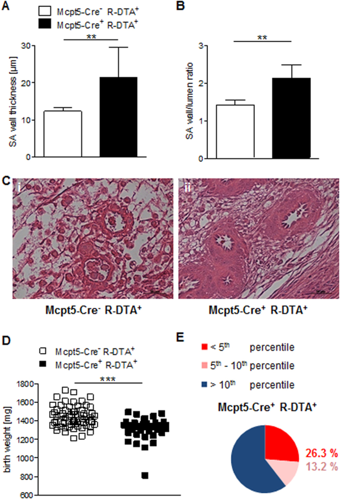
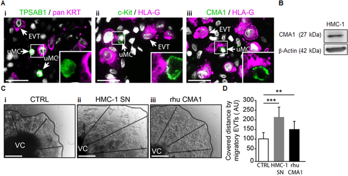
Intrauterine growth restriction (IUGR) is caused by insufficient remodeling of spiral arteries (SAs). The mechanism underlying the relevance of natural killer cells (NKs) and mast cells (MCs) for SA remodeling and its effects on pregnancy outcome are not well understood. We show that NK depletion arrested SA remodeling without affecting pregnancy. MC depletion resulted in abnormally remodeled SAs and IUGR. Combined absence of NKs and MCs substantially affected SA remodeling and impaired fetal growth. We found that α-chymase mast cell protease (Mcpt) 5 mediates apoptosis of uterine smooth muscle cells, a key feature of SA remodeling. Additionally, we report a previously unknown source for Mcpt5: uterine (u) NKs. Mice with selective deletion of Mcpt5+cells had un-remodeled SAs and growth-restricted progeny. The human α-chymase CMA1, phylogenetic homolog of Mcpt5, stimulated the ex vivo migration of human trophoblasts, a pre-requisite for SA remodeling. Our results show that chymases secreted by uMCs and uNKs are pivotal to the vascular changes required to support pregnancy. Understanding the mechanisms underlying pregnancy-induced vascular changes is essential for developing therapeutic options against pregnancy complications associated with poor vascular remodeling.
Overview
The claims and statistics of this paper haven’t been checked yet.
Run the check — free, no account needed. It reads the open-access text and checks the sources it cites.
Integrity signals
Registry
Cites
Statistics
Figures
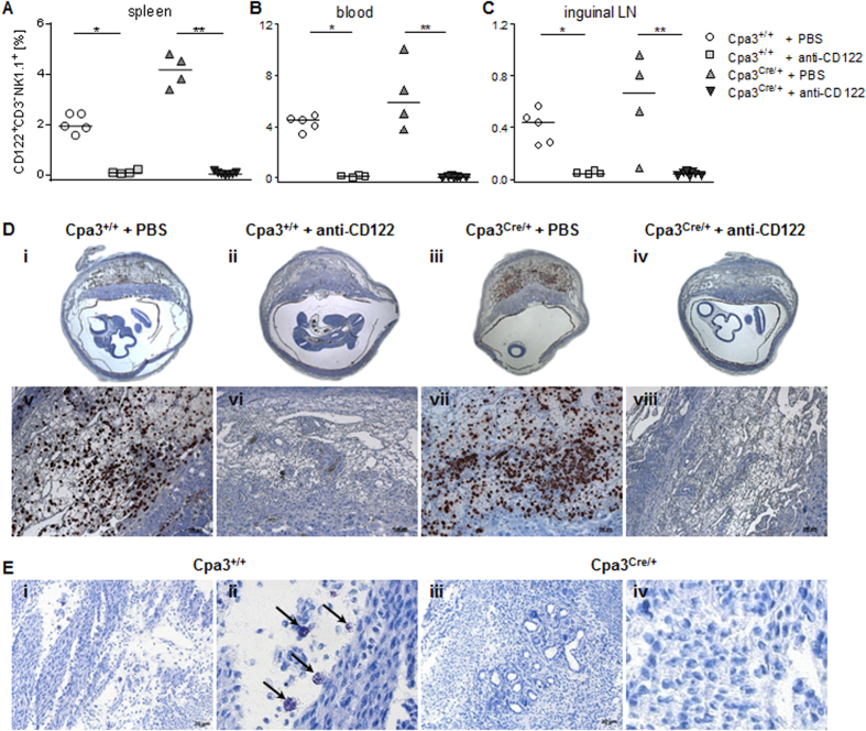
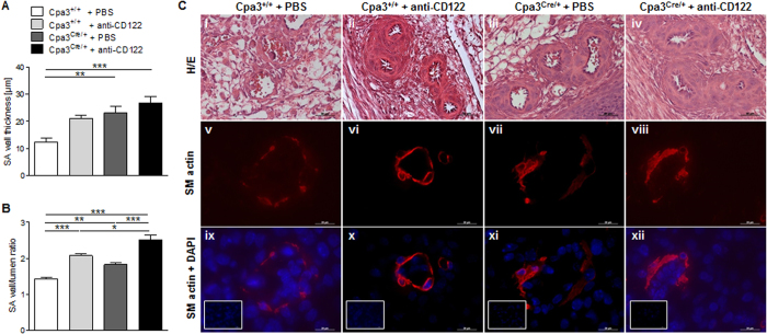
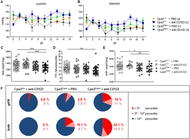
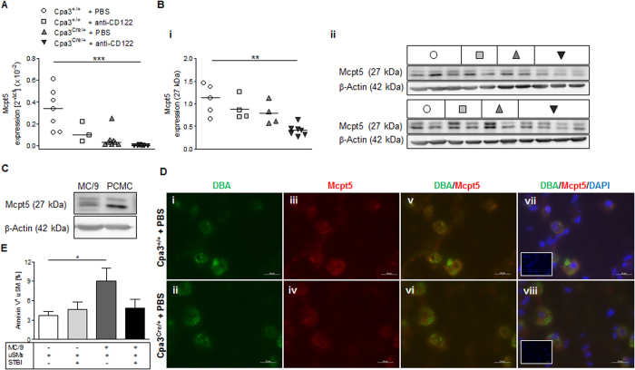
Verification
Claims the paper makes
The claims and statistics of this paper haven’t been checked yet.
Run the check — free, no account needed. It reads the open-access text and checks the sources it cites.
Claims other papers make about this one
Each is checked against this paper’s own text: does it say what the citing paper says it does?
In the citing paper
“Mast cell‐deficient female mice display defective spiral artery remodeling and impaired implantation, collectively resulting in fetal growth restriction. 122 , 124 , 125 Interestingly, mast cells appear to mediate these processes cooperatively with uterine NK cells, since both can (partially) compensate in the absence of the other. 125”
From Mast cell ontogeny: From fetal development to life‐long health and disease (2023) · cites this paper as [124]
Self-check
Percentages, sums and reported differences re-computed from the paper’s own numbers.
The self-check needs the paper’s full text.
Citations
Cites
Jensen, F., Woudwyk, M., Teles, A., Woidacki, K., Taran, F., Costa, S., Malfertheiner, S. F., & Zenclussen, A. C. (2010) · PLoS ONE · doi:10.1371/journal.pone.0014409 · Cited by 145
Self-citationDog mastocytoma cells secrete a 92-kD gelatinase activated extracellularly by mast cell chymase.
Fang, K. C., Raymond, W. W., Lazarus, S. C., & Caughey, G. H. (1996) · Journal of Clinical Investigation · doi:10.1172/jci118583 · Cited by 125
Fetal-derived adrenomedullin mediates the innate immune milieu of the placenta
Li, M., Schwerbrock, N. M. J., Lenhart, P. M., Fritz-Six, K. L., Kadmiel, M., Christine, K. S., Kraus, D. M., Espenschied, S. T., Willcockson, H. H., Mack, C. P., et al. (2013) · Journal of Clinical Investigation · doi:10.1172/jci67039 · Cited by 65
Research works printed but not linked: 58
- Moll, W., Künzel, W. & Herberger, J. Hemodynamic implications of hemochorial placentation. European journal of obstetrics, gynecology, and reproductive biology 5, 67–74 (1975). · DOI
- Whitley, G. S. J. & Cartwright, J. E. Trophoblast-mediated spiral artery remodelling: a role for apoptosis. Journal of anatomy 215, 21–26 (2009). · DOI
- Khong, T. Y., Wolf, F., Robertson, W. B. & Brosens, I. Inadequate maternal vascular response to placentation in pregnancies complicated by pre-eclampsia and by small-for-gestational age infants. British journal of obstetrics and gynaecology 93, 1049–1059 (1986). · DOI · Paper page
- Brosens, I. A., Robertson, W. B. & Dixon, H. G. The role of the spiral arteries in the pathogenesis of preeclampsia. Obstetrics and gynecology annual 1, 177–191 (1972).
- Barker, D. J., Martyn, C. N., Osmond, C., Hales, C. N. & Fall, C. H. Growth in utero and serum cholesterol concentrations in adult life. BMJ (Clinical research ed.) 307, 1524–1527 (1993). · DOI
- Ashkar, A. A., Di Santo, J. P. & Croy, B. A. Interferon gamma contributes to initiation of uterine vascular modification, decidual integrity, and uterine natural killer cell maturation during normal murine pregnancy. The Journal of experimental medicine 192, 259–270 (2000). · DOI
- Croy, B. A., van den Heuvel, Marianne J., Borzychowski, A. M. & Tayade, C. Uterine natural killer cells: a specialized differentiation regulated by ovarian hormones. Immunological reviews 214, 161–185 (2006). · DOI
- Bulmer, J. N. & Lash, G. E. Human uterine natural killer cells: a reappraisal. Molecular immunology 42, 511–521 (2005). · DOI
- Chakraborty, D., Rumi, M. A. K., Konno, T. & Soares, M. J. Natural killer cells direct hemochorial placentation by regulating hypoxia-inducible factor dependent trophoblast lineage decisions. Proceedings of the National Academy of Sciences of the United States of America 108, 16295–16300 (2011). · DOI
- Lash, G. E., Robson, S. C. & Bulmer, J. N. Review: Functional role of uterine natural killer (uNK) cells in human early pregnancy decidua. Placenta 31 Suppl, S87–92 (2010). · DOI
- Hanna, J. et al. Decidual NK cells regulate key developmental processes at the human fetal-maternal interface. Nature medicine 12, 1065–1074 (2006). · DOI
- Woidacki, K. et al. Mast cells rescue implantation defects caused by c-kit deficiency. Cell death & disease 4, e462 (2013). · DOI · Paper page
- Woidacki, K. et al. Transfer of regulatory T cells into abortion-prone mice promotes the expansion of uterine mast cells and normalizes early pregnancy angiogenesis. Scientific reports 5, 13938 (2015). · DOI
- Galli, S. J. et al. Mast cells as “tunable” effector and immunoregulatory cells: recent advances. Annual review of immunology 23, 749–786 (2005). · DOI · Paper page
- Moon, T. C. et al. Advances in mast cell biology: new understanding of heterogeneity and function. Mucosal immunology 3, 111–128 (2010). · DOI
- Scandiuzzi, L. et al. Mouse mast cell protease-4 deteriorates renal function by contributing to inflammation and fibrosis in immune complex-mediated glomerulonephritis. Journal of immunology (Baltimore, Md. 1950) 185, 624–633 (2010). · DOI
- Caughey, G. H. Mast cell tryptases and chymases in inflammation and host defense. Immunological reviews 217, 141–154 (2007). · DOI · Paper page
- Urata, H., Kinoshita, A., Misono, K. S., Bumpus, F. M. & Husain, A. Identification of a highly specific chymase as the major angiotensin II-forming enzyme in the human heart. The Journal of biological chemistry 265, 22348–22357 (1990). · DOI
- Tchougounova, E. et al. A key role for mast cell chymase in the activation of pro-matrix metalloprotease-9 and pro-matrix metalloprotease-2. The Journal of biological chemistry 280, 9291–9296 (2005). · DOI
- Fang, K. C., Raymond, W. W., Blount, J. L. & Caughey, G. H. Dog mast cell alpha-chymase activates progelatinase B by cleaving the Phe88-Gln89 and Phe91-Glu92 bonds of the catalytic domain. The Journal of biological chemistry 272, 25628–25635 (1997). · DOI
- Leskinen, M. J., Lindstedt, K. A., Wang, Y. & Kovanen, P. T. Mast cell chymase induces smooth muscle cell apoptosis by a mechanism involving fibronectin degradation and disruption of focal adhesions. Arteriosclerosis, thrombosis, and vascular biology 23, 238–243 (2003). · DOI
- Banovac, K. & Forteza, R. The effect of mast cell chymase on extracellular matrix: studies in autoimmune thyroiditis and in cultured thyroid cells. International archives of allergy and immunology 99, 141–149 (1992). · DOI
- Wang, Y., Shiota, N., Leskinen, M. J., Lindstedt, K. A. & Kovanen, P. T. Mast cell chymase inhibits smooth muscle cell growth and collagen expression in vitro: transforming growth factor-beta1-dependent and -independent effects. Arteriosclerosis, thrombosis, and vascular biology 21, 1928–1933 (2001). · DOI
- den Dekker, W. K. et al. Mast cells induce vascular smooth muscle cell apoptosis via a toll-like receptor 4 activation pathway. Arteriosclerosis, thrombosis, and vascular biology 32, 1960–1969 (2012). · DOI
- Leskinen, M., Wang, Y., Leszczynski, D., Lindstedt, K. A. & Kovanen, P. T. Mast cell chymase induces apoptosis of vascular smooth muscle cells. Arteriosclerosis, thrombosis, and vascular biology 21, 516–522 (2001). · DOI
- Feyerabend, T. B. et al. Cre-mediated cell ablation contests mast cell contribution in models of antibody- and T cell-mediated autoimmunity. Immunity 35, 832–844 (2011). · DOI
- Dilworth, M. R. et al. Defining fetal growth restriction in mice: A standardized and clinically relevant approach. Placenta 32, 914–916 (2011). · DOI
- Reynolds, D. S. et al. Different mouse mast cell populations express various combinations of at least six distinct mast cell serine proteases. Proceedings of the National Academy of Sciences of the United States of America 87, 3230–3234 (1990). · DOI
- Paffaro, V. A., Bizinotto, M. C., Joazeiro, P. P. & Yamada, A. T. Subset classification of mouse uterine natural killer cells by DBA lectin reactivity. Placenta 24, 479–488 (2003). · DOI
- Whitley, G. S. J. & Cartwright, J. E. Cellular and molecular regulation of spiral artery remodelling: lessons from the cardiovascular field, Placenta 31, 465–474 (2010). · DOI
- Muramatsu, M., Katada, J., Hattori, M., Hayashi, I. & Majima, M. Chymase mediates mast cell-induced angiogenesis in hamster sponge granulomas. European journal of pharmacology 402, 181–191 (2000). · DOI
- He, S., Gaça, M. D., McEuen, A. R. & Walls, A. F. Inhibitors of chymase as mast cell-stabilizing agents: contribution of chymase in the activation of human mast cells. The Journal of pharmacology and experimental therapeutics 291, 517–523 (1999). · DOI
- Ware, J. H., Wan, X. S., Rubin, H., Schechter, N. M. & Kennedy, A. R. Soybean Bowman-Birk protease inhibitor is a highly effective inhibitor of human mast cell chymase. Archives of biochemistry and biophysics 344, 133–138 (1997). · DOI
- Sommerhoff, C. P., Ruoss, S. J. & Caughey, G. H. Mast cell proteoglycans modulate the secretagogue, proteoglycanase, and amidolytic activities of dog mast cell chymase. Journal of immunology (Baltimore, Md. 1950) 148, 2859–2866 (1992). · DOI
- Sommerhoff, C. P. et al. Mast cell chymase. A potent secretagogue for airway gland serous cells. Journal of immunology (Baltimore, Md. 1950) 142, 2450–2456 (1989). · DOI
- Scholten, J. et al. Mast cell-specific Cre/loxP-mediated recombination in vivo . Transgenic research 17, 307–315 (2008). · DOI
- Knoeller, S. et al. Distribution of immunocompetent cells in decidua of controlled and uncontrolled (choriocarcinoma/hydatidiform mole) trophoblast invasion. American journal of reproductive immunology (New York, N.Y. 1989) 50, 41–47 (2003).
- Bauer, S. et al. Tumor necrosis factor-alpha inhibits trophoblast migration through elevation of plasminogen activator inhibitor-1 in first-trimester villous explant cultures. The Journal of clinical endocrinology and metabolism 89, 812–822 (2004). · DOI
- Wallace, A. E., Fraser, R. & Cartwright, J. E. Extravillous trophoblast and decidual natural killer cells: a remodelling partnership. Human reproduction update 18, 458–471 (2012). · DOI
- Wallace, A. E., Host, A. J., Whitley, G. S. & Cartwright, J. E. Decidual natural killer cell interactions with trophoblasts are impaired in pregnancies at increased risk of preeclampsia. The American journal of pathology 183, 1853–1861 (2013). · DOI
- Zenclussen, A. C. & Hammerling, G. J. Cellular Regulation of the Uterine Microenvironment That Enables Embryo Implantation. Frontiers in immunology 6, 321 (2015). · DOI
- Barber, E. M. & Pollard, J. W. The uterine NK cell population requires IL-15 but these cells are not required for pregnancy nor the resolution of a Listeria monocytogenes infection. Journal of immunology (Baltimore, Md. 1950) 171, 37–46 (2003). · DOI
- Burke, S. D. et al. Cardiovascular adaptations of pregnancy in T and B cell-deficient mice. Biology of reproduction 85, 605–614 (2011). · DOI
- Khalife, N. et al. Placental size is associated with mental health in children and adolescents. PLoS One 7(7), e40534 (2012). · DOI
- He, S. & Walls, A. F. Human mast cell chymase induces the accumulation of neutrophils, eosinophils and other inflammatory cells in vivo . British journal of pharmacology 125, 1491–1500 (1998). · DOI
- Chen, Z. et al. DBA-lectin reactivity defines mouse uterine natural killer cell subsets with biased gene expression. Biology of reproduction 87, 81 (2012). · DOI
- Wada, T. et al. Selective ablation of basophils in mice reveals their nonredundant role in acquired immunity against ticks. The Journal of clinical investigation 120, 2867–2875 (2010). · DOI · Paper page
- Ugajin, T. et al. Basophils preferentially express mouse Mast Cell Protease 11 among the mast cell tryptase family in contrast to mast cells. Journal of leukocyte biology 86, 1417–1425 (2009). · DOI
- Ratsep, M. T. et al. Uterine natural killer cells: supervisors of vasculature construction in early decidua basalis. Reproduction (Cambridge, England) 149, R91–102 (2015). · DOI
- Croy, B. A. et al. Uterine natural killer cells: insights into their cellular and molecular biology from mouse modelling. Reproduction (Cambridge, England) 126, 149–160 (2003). · DOI
- Marx, L. et al. Decidual mast cells might be involved in the onset of human first-trimester abortion. Am J Reprod Immunol. 41(1), 34–40 (1999). · DOI
- Garfield, R. E. et al. Structural and functional comparison of mast cells in the pregnant versus nonpregnant human uterus. Am J Obstet Gynecol. 194(1), 261–7 (2006). · DOI
- Aplin, J. D., Haigh, T., Jones, C. J., Church, H. J. & Vićovac, L. Development of cytotrophoblast columns from explanted first-trimester human placental villi: role of fibronectin and integrin alpha5beta1. Biology of reproduction 60, 828–838 (1999). · DOI
- Pijnenborg, R., Vercruysse, L. & Hanssens, M. The uterine spiral arteries in human pregnancy: facts and controversies. Placenta 27, 939–958 (2006). · DOI
- Dudeck, A. et al. Mast cells are key promoters of contact allergy that mediate the adjuvant effects of haptens. Immunity 34, 973–984 (2011). · DOI
- Tournoy, K. G., Depraetere, S., Pauwels, R. A. & Leroux-Roels, G. G. Mouse strain and conditioning regimen determine survival and function of human leucocytes in immunodeficient mice. Clinical and experimental immunology 119, 231–239 (2000). · DOI
- Yokoyama, S. et al. Antibody-mediated blockade of IL-15 reverses the autoimmune intestinal damage in transgenic mice that overexpress IL-15 in enterocytes. Proceedings of the National Academy of Sciences of the United States of America 106, 15849–15854 (2009). · DOI
- Zenclussen, A. C. et al. Abnormal T-cell reactivity against paternal antigens in spontaneous abortion: adoptive transfer of pregnancy-induced CD4+CD25+ T regulatory cells prevents fetal rejection in a murine abortion model. The American journal of pathology 166, 811–822 (2005). · DOI
Cited by
Sorted by citations
Mast cell ontogeny: From fetal development to life‐long health and disease
Chia, S. L., Kapoor, S., Carvalho, C., Bajénoff, M., & Gentek, R. (2023) · Immunological Reviews · doi:10.1111/imr.13191 · Cited by 60
Metadata
- Authors
- Meyer, N., Woidacki, K., Knöfler, M., Meinhardt, G., Nowak, D., Velicky, P., Pollheimer, J., & Zenclussen, A. C.
- Venue
- Scientific Reports
- Published
- 2017-03-22
- Volume
- 7
- Issue
- 1
- ISSN
- 2045-2322
- PMC
- PMC5361184
- Cited by
- 62
- References
- 3
- Language
- en
About these checks
Registry, retraction, Cites and statistics results are computed, not judged, with one exception: a printed reference that no index lists is read once by a model before it counts as a citation error. Claims are extracted by a model and kept only when their quote appears verbatim in the text; each cited source is then checked, with a model’s help, for whether it says what the claim relies on. Those verdicts can be wrong — read the claim’s quote and the cited source before relying on one.
Statistics and arithmetic are recomputed, not judged.
Confidence bands are shown only for fields calibrated on our annotated test set. None is calibrated yet, so no band is shown.
What counts as a claim ›How we measure confidence ›How we check this ›
Are you an author, or do you see a problem with this page? Request a review
