

The DOI matches its Crossref record; 2 of the 2 references it cites were found in a registry or an index.
Nanoparticle mediated alteration of EMT dynamics: an approach to modulate cancer therapeutics
Abstract
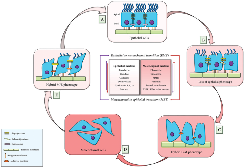
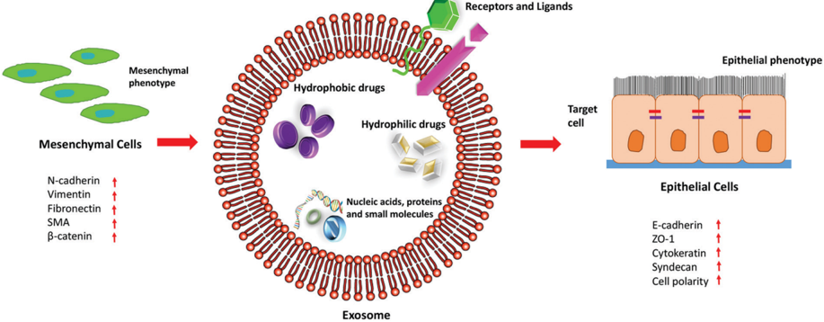
Metastasis is the cause of approximately 90% of cancer-related morbidities and mortalities, which is ascribed to the phenomenon of EMT (epithelial to mesenchymal transition). The nanoparticle mediated suppression of EMT exhibit strong therapeutic modalities.
Overview
The claims and statistics of this paper haven’t been checked yet.
Run the check — free, no account needed. It reads the open-access text and checks the sources it cites.
Integrity signals
Registry
Cites
Statistics
Figures
Verification
Claims the paper makes
The claims and statistics of this paper haven’t been checked yet.
Run the check — free, no account needed. It reads the open-access text and checks the sources it cites.
Claims other papers make about this one
Each is checked against this paper’s own text: does it say what the citing paper says it does?
In the citing paper
“Several types of ligand have been utilized including protein, monoclonal antibodies, folic acid, aptamers, small molecules, nucleic acid and polysaccharides.154”
From Current Advances in Chitosan Nanoparticles Based Oral Drug Delivery for Colorectal Cancer Treatment (2022) · cites this paper as [154]
Self-check
Percentages, sums and reported differences re-computed from the paper’s own numbers.
The self-check needs the paper’s full text.
Citations
Cites
Bunney, P. E., Zink, A. N., Holm, A. A., Billington, C. J., & Kotz, C. M. (2017) · Physiology & Behavior · doi:10.1016/j.physbeh.2017.03.040 · Cited by 2,104
Polymeric micelles: nanocarriers for cancer-targeted drug delivery
Zhang, Y., Huang, Y., & Li, S. (2014) · AAPS PharmSciTech · doi:10.1208/s12249-014-0113-z · Cited by 367
Research works printed but not linked: 144
- Boareto, 2016, J. R. Soc. Interface, 13, 20151106, 10.1098/rsif.2015.1106 · DOI
- Portillo, 2008, Oncogene, 6958, 10.1038/onc.2008.346 · DOI
- Min, 2008, J. Cell Biochem., 744, 733, 10.1002/jcb.21695 · DOI
- Terry, 2017, Mol. Oncol., 11, 824, 10.1002/1878-0261.12093 · DOI · Paper page
- Cao, 2016, Crit. Rev. Oncog., 21, 155, 10.1615/CritRevOncog.2016016955 · DOI
- Lamouille, 2014, Nat. Rev. Mol. Cell Biol., 15, 178, 10.1038/nrm3758 · DOI · Paper page
- Haynes, 2011, Mol. Biol. Cell, 22, 4750, 10.1091/mbc.e11-02-0119 · DOI
- Shankar, 2010, Cancer Res., 70, 3780, 10.1158/0008-5472.CAN-09-4439 · DOI
- Fife, 2014, Br. J. Pharmacol., 171, 5507, 10.1111/bph.12704 · DOI
- Sun, 2015, Biomed. Rep., 3, 603, 10.3892/br.2015.494 · DOI
- Golubovskaya, 2014, Front. Biosci., 19, 687, 10.2741/4236 · DOI
- Ackland, 2003, Lab. Investig., 83, 435, 10.1097/01.LAB.0000059927.97515.FD · DOI
- Strutz, 2002, Kidney Int., 61, 1714, 10.1046/j.1523-1755.2002.00333.x · DOI
- Farrell, 2014, J. Proteome Res., 13, 2874, 10.1021/pr5000285 · DOI
- Wu, 2013, Cancer Treat. Rev., 39, 640, 10.1016/j.ctrv.2012.11.006 · DOI
- Natsuizaka, 2017, Nat. Commun., 8, 1758, 10.1038/s41467-017-01500-9 · DOI
- Chen, 2010, Br. J. Cancer, 351, 10.1038/sj.bjc.6605486 · DOI · Paper page
- Jiang, 2007, Int. J. Urol., 14, 1034, 10.1111/j.1442-2042.2007.01866.x · DOI
- Cao, 2015, Kaohsiung J. Med. Sci., 31, 70, 10.1016/j.kjms.2014.11.008 · DOI
- Gui, 2012, J. Signal Transduct., 2012, 1, 10.1155/2012/289243 · DOI
- Hsu, 2016, Physiol. Behav., 176, 139
- Yang, 2008, Cell Cycle, 7, 2090, 10.4161/cc.7.14.6324 · DOI
- Suarez-Carmona, 2017, Mol. Oncol., 11, 805, 10.1002/1878-0261.12095 · DOI
- Sophie Mokas, 2009, Mol. Biol. Cell, 20, 2673, 10.1091/mbc.e08-10-1061 · DOI
- Shao, 2015, Mol. Cancer, 1, 10.1186/s12943-015-0295-3 · DOI
- Wang, 2014, Curr. Cancer Drug Targets, 13, 963, 10.2174/15680096113136660102 · DOI
- Jordà, 2005, J. Cell Sci., 118, 3371, 10.1242/jcs.02465 · DOI
- Albanell, 2013, Nat. Cell Biol., 11, 943
- Yang, 2010, Nat. Cell Biol., 12, 982, 10.1038/ncb2099 · DOI
- Castillon, 2003, Curr. Biol., 13, 654, 10.1016/S0960-9822(03)00247-1 · DOI
- Postigo, 2003, EMBO J., 22, 2453, 10.1093/emboj/cdg226 · DOI
- Dave, 2011, J. Biol. Chem., 286, 12024, 10.1074/jbc.M110.168625 · DOI
- Olea-Flores, 2019, Int. J. Mol. Sci., 20, 1, 10.3390/ijms20122885 · DOI
- Wang, 2010, Curr. Drug Targets, 11, 745, 10.2174/138945010791170860 · DOI
- Islam, 2016, Mol. Carcinog., 55, 537, 10.1002/mc.22300 · DOI
- Larue, 2005, Oncogene, 24, 7443, 10.1038/sj.onc.1209091 · DOI
- Manuscript, A. NIH Public Access, 2010, 19 , 128–139
- Hao, 2019, Int. J. Mol. Sci., 20, 2767, 10.3390/ijms20112767 · DOI
- Fe, 2004, Curr. Opin. Cell Biol., 16, 415, 10.1016/j.ceb.2004.06.003 · DOI · Paper page
- Vella, 2014, Front. Oncol., 4, 1, 10.3389/fonc.2014.00361 · DOI
- Conigliaro, 2019, J. Clin Med., 8, 26, 10.3390/jcm8010026 · DOI
- Chen, 2018, Cell Death Dis., 9, 513, 10.1038/s41419-018-0534-9 · DOI
- Xiao, 2017, Cancer Lett., 376, 318, 10.1016/j.canlet.2016.03.050 · DOI
- He, 2019, Exp. Cell Res., 379, 203, 10.1016/j.yexcr.2019.03.035 · DOI
- W. Zhou , et al. , NIH Public Access , 2015, 25 , 501515
- Li, 2017, Oncotarget., 8, 96035, 10.18632/oncotarget.21635 · DOI
- Pastushenko, 2018, Nature, 556, 463, 10.1038/s41586-018-0040-3 · DOI
- Gunasinghe, 2012, Cancer Metastasis Rev., 31, 469, 10.1007/s10555-012-9377-5 · DOI
- Lu, 2013, Proc. Natl. Acad. Sci. U. S. A., 110, 18144, 10.1073/pnas.1318192110 · DOI
- Jolly, 2015, Front. Oncol., 5, 155, 10.3389/fonc.2015.00155 · DOI
- Arvizo, 2013, Proc. Natl. Acad. Sci. U. S. A., 110, 6700, 10.1073/pnas.1214547110 · DOI
- Yang, 2017, Int. J. Nanomed., 3509
- Xiong, 2014, Oncotarget., 5, 6453, 10.18632/oncotarget.2203 · DOI
- Huai, 2019, Cell Stress, 3, 267, 10.15698/cst2019.08.195 · DOI
- Therapy, 2018, HHS Public Access., 12, 9279
- Kumar, 2016, Biomaterials, 87, 118, 10.1016/j.biomaterials.2016.02.014 · DOI
- Bhat, 2016, Cell Prolif., 678, 10.1111/cpr.12296 · DOI
- Loutfy, 2015, Asian Pacific J. Cancer Prev., 16, 6039, 10.7314/APJCP.2015.16.14.6039 · DOI
- Sunil Gowda, 2018, Toxicol. Vitr., 52, 170, 10.1016/j.tiv.2018.06.015 · DOI
- Vila, 2017, Nanotoxicology, 11, 771
- Khurana, 2019, Biomed. Pharmacother., 111, 802, 10.1016/j.biopha.2018.12.146 · DOI
- Kumari, 2017, Eur. J. Pharm. Biopharm., 117, 346, 10.1016/j.ejpb.2017.05.003 · DOI
- Kumari, 2018, Eur. J. Pharm. Biopharm., 130, 185, 10.1016/j.ejpb.2018.06.030 · DOI
- Li, 2018, ACS Appl. Mater. Interfaces, 10, 3449, 10.1021/acsami.7b18986 · DOI
- Peng, 2018, ACS Appl. Mater. Interfaces, 10, 31938, 10.1021/acsami.8b10527 · DOI
- Wahab, 2016, Nat. Publ. Gr., 1, 10.1038/srep19950 · DOI
- Wang, 2013, Cell Death Dis., 4, e783, 10.1038/cddis.2013.314 · DOI
- Thoidingjam, 2017, Adv. Nat. Sci., 8, 023002
- Malekigorji, 2014, J. Nanomed. Res., 1, 1
- Saveleva, 2019, Front. Chem., 7, 1, 10.3389/fchem.2019.00179 · DOI
- Ananikov, 2019, Nanomaterials, 9, 1197, 10.3390/nano9091197 · DOI
- Xu, 2016, Acta Biomater., 36, 152, 10.1016/j.actbio.2016.02.041 · DOI
- Galkina, 2015, J. Mater. Chem. B, 3, 1688, 10.1039/C4TB01823K · DOI
- Hao, 2020, Mater. Chem. Front., 4, 2571, 10.1039/D0QM00323A · DOI
- Pang, 2019, Nanomedicine, 14, 1291, 10.2217/nnm-2018-0390 · DOI
- Shukla, 2020, Biomater. Sci., 8, 1298, 10.1039/C9BM01246J · DOI
- Saleem, 2018, Adv. Healthcare Mater., 1800525, 1
- Su, 2020, J. Phys. Chem. Lett., 11, 1357, 10.1021/acs.jpclett.9b03891 · DOI
- Manuscript, A. rsc.li/njc, 2018, DOI: 10.1039/C8NJ02092B · DOI
- Xie, 2019, HHS Public Access., 38, 2967
- Mehra, 2014, Nanostruct. Drug Delivery, 4, 327
- Singh, 2013, J. Drug Target., 21, 581, 10.3109/1061186X.2013.778264 · DOI
- Datir, 2012, Bioconjugate Chem., 23, 2201, 10.1021/bc300248t · DOI
- Yang, 2011, Eur. J. Cancer, 7, 0
- Yang, 2009, Chem. Commun., 4447, 10.1039/b908012k · DOI
- Yao, 2014, Biomaterials, 35, 9208, 10.1016/j.biomaterials.2014.07.033 · DOI
- A. Manuscript NIH Public Access, 2012, 8, 136–146
- Liu, 2015, Nat. Commun., 6, 1
- Mohammed, 2017, Pharmaceutics, 9, 53, 10.3390/pharmaceutics9040053 · DOI
- Potdar, 2016, J. Cancer Metastasis Treat., 2, 259, 10.20517/2394-4722.2016.25 · DOI
- Manuscript, A. Nanoscale, 2020, DOI: 10.1039/C9NR10673A · DOI
- Yu, 2017, Biomed. Pharmacother., 96, 650, 10.1016/j.biopha.2017.10.026 · DOI
- Zhuo, 2015, Am. J. Cancer Res., 5, 1571
- Sadreddini, 2017, Immunol. Lett., 181, 79, 10.1016/j.imlet.2016.11.013 · DOI · Paper page
- Kay, 2005, Cell. Mol. Biol., 51, 117
- Houde, 2004, Blood, 104, 3697, 10.1182/blood-2003-12-4114 · DOI
- Fang, 2014, Nanoscale, 6, 10084, 10.1039/C4NR01518E · DOI
- Sousa, 2019, Pharm. Res., 36, 83, 10.1007/s11095-019-2615-6 · DOI
- Rezvantalab, 2018, Front. Pharmacol., 9, 1, 10.3389/fphar.2018.01260 · DOI
- Daman, 2018, Drug Dev. Ind. Pharm., 44, 1434, 10.1080/03639045.2018.1459674 · DOI
- Sureban, 2011, J. Nanobiotechnol., 9, 1, 10.1186/1477-3155-9-40 · DOI
- Wang, 2018, Biomaterials, 175, 110, 10.1016/j.biomaterials.2018.05.034 · DOI
- Verma, 2016, Sci. Rep., 6, 1, 10.1038/s41598-016-0001-8 · DOI · Paper page
- Verma, 2015, Nanomedicine, 11, 2061, 10.1016/j.nano.2015.07.001 · DOI
- Das, 2019, Mol. Cancer Ther., 18, 680, 10.1158/1535-7163.MCT-18-0409 · DOI
- Fan, 2017, Biomaterials, 139, 116, 10.1016/j.biomaterials.2017.06.007 · DOI
- Manuscript, A. Nanoscale, 2018, DOI: 10.1039/C8NR02708K · DOI
- Huang, 2019, Theranostics, 9, 4391, 10.7150/thno.32462 · DOI
- Lu, 2019, J. Exp. Clin. Cancer Res., 3, 1
- Shahin, 2019, HHS Public Access., 14, 1381
- Roberts, 2016, Nanomedicine, 13, 965, 10.1016/j.nano.2016.11.010 · DOI
- Finlay, 2016, HHS Public Access., 11, 1657
- Finlay, 2015, Biomed. Res. Int., 2015, 382745, 10.1155/2015/382745 · DOI
- Parvani, 2016, HHS Public Access., 75, 2316
- Lu, 2020, HHS Public Access., 30, 907
- Ma, 2017, Biomater. Sci., 5, 600, 10.1039/C7BM90004J · DOI
- Ma, 2015, PLos One, 1, 10.1371/journal.pone.0124924 · DOI
- Suresh, 2019, Nanomedicine, 20, 102007, 10.1016/j.nano.2019.04.010 · DOI
- Li, 2017, Cancer Sci., 108, 1493, 10.1111/cas.13267 · DOI
- Nanobiotechnol, 2019, J. Nanobiotechnol., 1, 10.1186/s12951-019-0519-6 · DOI
- Liu, 2013, Biomaterials, 34, 7191, 10.1016/j.biomaterials.2013.06.004 · DOI
- Tang, 2015, Biomaterials, 48, 1, 10.1016/j.biomaterials.2015.01.049 · DOI
- Shen, 2013, Biomaterials, 34, 1581, 10.1016/j.biomaterials.2012.10.057 · DOI
- Alavi, 2017, Adv. Pharm. Bull., 7, 3, 10.15171/apb.2017.002 · DOI
- Kullberg, 2019, Pharm. Front., 3, 1
- Yao, 2020, Drug Dev. Ind. Pharm., 9045, 1
- Song, 2019, Nanoscale Res. Lett., 14, 1, 10.1186/s11671-018-2843-4 · DOI
- Chang, 2018, Cell Death Discovery, 4, 100, 10.1038/s41420-018-0116-8 · DOI
- Shen, 2016, Radiother. Oncol., 119, 282, 10.1016/j.radonc.2016.02.007 · DOI
- Guo, 2018, Drug Delivery, 25, 112, 10.1080/10717544.2017.1417511 · DOI
- Zheng, 2014, Asian Pacific J. Cancer Prev., 15, 8679, 10.7314/APJCP.2014.15.20.8679 · DOI
- Jin, 2019, Theranostics, 9, 265, 10.7150/thno.27246 · DOI
- Kim, 2016, Nanomedicine, 12, 655, 10.1016/j.nano.2015.10.012 · DOI · Paper page
- Alvarez-erviti, 2011, Nat. Biotechnol., 29, 341, 10.1038/nbt.1807 · DOI · Paper page
- Srivastava, 2016, Nat. Publ. Gr., 1, 10.1038/srep38541 · DOI
- Wang, 2019, Biomed. Pharmacother., 117, 109109, 10.1016/j.biopha.2019.109109 · DOI
- Xie, 2019, Mol. Cell. Biochem., 458, 11, 10.1007/s11010-019-03526-7 · DOI
- Guo, 2014, Oncol. Rep., 2021, 10.3892/or.2014.3106 · DOI
- Li, 2018, J. Cell Physiol., 1, 10.1002/jcp.27111 · DOI
- Zhang, 2017, Biomark. Res., 1, 10.1186/s40364-017-0081-z · DOI
- Ota, 2018, Oncotarget., 9, 16400, 10.18632/oncotarget.24711 · DOI
- Tan, 2018, PLoS Bio., 16, e3000051, 10.1371/journal.pbio.3000051 · DOI
- Yuan, 2019, Front. Oncol., 9, 1, 10.3389/fonc.2019.00001 · DOI
- Bunggulawa, 2018, J. Nanobiotechnol., 16, 1, 10.1186/s12951-018-0403-9 · DOI
Cited by
Sorted by citations
Current Advances in Chitosan Nanoparticles Based Oral Drug Delivery for Colorectal Cancer Treatment
Choukaife, H., Seyam, S., Alallam, B., Doolaanea, A. A., & Alfatama, M. (2022) · International Journal of Nanomedicine · doi:10.2147/ijn.s375229 · Cited by 127
Metadata
- Authors
- Sen, P., Saha, M., & Ghosh, S. S.
- Venue
- Materials Advances
- Publisher
- Royal Society of Chemistry (RSC)
- Published
- 2020
- Volume
- 1
- Issue
- 8
- Pages
- 2614-2630
- ISSN
- 2633-5409
- Cited by
- 21
- References
- 2
- Language
- en
- Funders
- Department of Biotechnology, Ministry of Science and Technology (BT/PR13560/COE/34/44/2015, BT/PR25095/NER/95/1011/2017)
About these checks
Registry, retraction, Cites and statistics results are computed, not judged, with one exception: a printed reference that no index lists is read once by a model before it counts as a citation error. Claims are extracted by a model and kept only when their quote appears verbatim in the text; each cited source is then checked, with a model’s help, for whether it says what the claim relies on. Those verdicts can be wrong — read the claim’s quote and the cited source before relying on one.
Statistics and arithmetic are recomputed, not judged.
Confidence bands are shown only for fields calibrated on our annotated test set. None is calibrated yet, so no band is shown.
What counts as a claim ›How we measure confidence ›How we check this ›
Are you an author, or do you see a problem with this page? Request a review
