

The DOI matches its Crossref record; 208 of the 208 references it cites were found in a registry or an index.
Regulation of cellular iron metabolism
Open accessAbstract
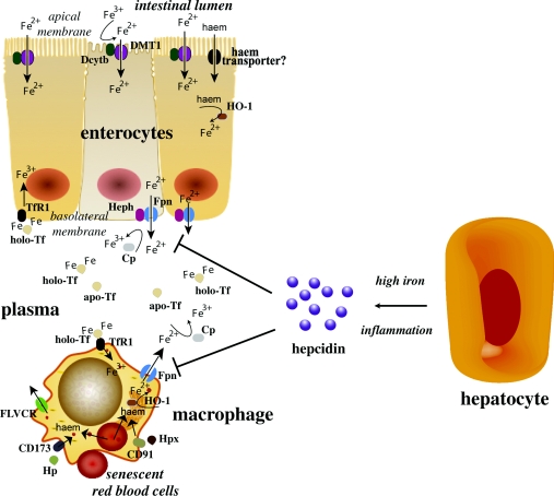
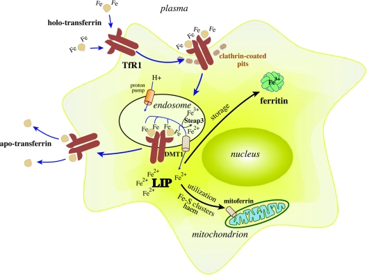
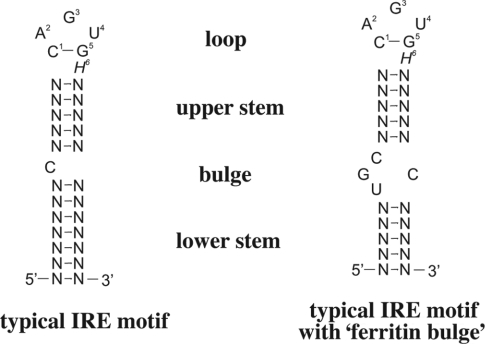
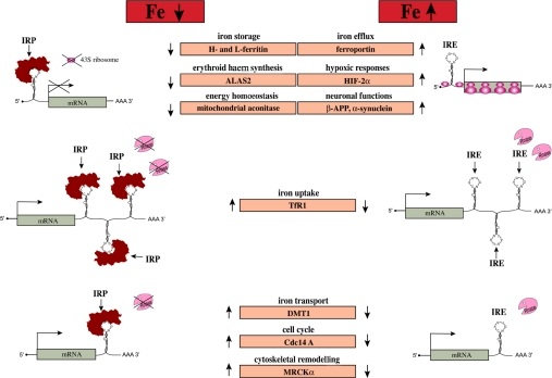
Iron is an essential but potentially hazardous biometal. Mammalian cells require sufficient amounts of iron to satisfy metabolic needs or to accomplish specialized functions. Iron is delivered to tissues by circulating transferrin, a transporter that captures iron released into the plasma mainly from intestinal enterocytes or reticuloendothelial macrophages. The binding of iron-laden transferrin to the cell-surface transferrin receptor 1 results in endocytosis and uptake of the metal cargo. Internalized iron is transported to mitochondria for the synthesis of haem or iron-sulfur clusters, which are integral parts of several metalloproteins, and excess iron is stored and detoxified in cytosolic ferritin. Iron metabolism is controlled at different levels and by diverse mechanisms. The present review summarizes basic concepts of iron transport, use and storage and focuses on the IRE (iron-responsive element)/IRP (iron-regulatory protein) system, a well known post-transcriptional regulatory circuit that not only maintains iron homoeostasis in various cell types, but also contributes to systemic iron balance.
Overview
The claims and statistics of this paper haven’t been checked yet.
Run the check — free, no account needed. It reads the open-access text and checks the sources it cites.
Integrity signals
Registry
Cites
Statistics
Figures
Verification
Claims the paper makes
The claims and statistics of this paper haven’t been checked yet.
Run the check — free, no account needed. It reads the open-access text and checks the sources it cites.
Claims other papers make about this one
Each is checked against this paper’s own text: does it say what the citing paper says it does?
The source backs part of the claim, not all of it — or backs it for a different population, condition or comparison.
In the citing paper
“IRPs, in addition to modulating ferritin and TfR1 levels can regulate the mRNAs or other proteins required for iron utilization (erythroid 5-aminolevulanata synthase), iron uptake (DMT1) and export (FPN1).DMT1 mRNA has IRE in its 3 ' UTR and is upregulated by iron deficiency while the other two proteins have IREs in their 5 ' UTR and are therefore upregulated under high iron conditions”
From Iron, Oxidative Stress and Health (2012) · cites this paper as Wang and Pantopoulos, 2011
Experimental — accuracy not yet measuredThe cited source says what the claim attributes to it.
In the citing paper
“Conversely, when iron concentration is high, IRPs are inactivated resulting in degradation of TfR1 mRNA while translation of ferritin mRNA occurs.Thus when iron levels exceed the cellular needs, the IRE/IRP switch minimizes iron uptake and promotes iron storage”
From Iron, Oxidative Stress and Health (2012) · cites this paper as Wang and Pantopoulos, 2011
The cited source says what the claim attributes to it.
In the citing paper
“When iron is depleted, IRE-IRP interaction at the 3 ' UTR of TfR1 stabilizes mRNA, while imposing a steric blockade to ferritin mRNA translation.As a result, TfR1 is upregulated and stimulates acquisition of iron from plasma Tf to counteract low iron levels.Simultaneously ferritin synthesis is inhibited as storage of iron is not needed.”
From Iron, Oxidative Stress and Health (2012) · cites this paper as Wang and Pantopoulos, 2011
The cited source says what the claim attributes to it.
In the citing paper
“However, in erythroid progenitor cells which require high amounts of iron for heme synthesis, TfR1 stability is uncoupled from iron supply and TfR1 level is transcriptionally regulated by erythroid active element in its promoter region (Wang and Pantopoulos, 2011).”
From Iron, Oxidative Stress and Health (2012) · cites this paper as Wang and Pantopoulos, 2011
In the citing paper
“Iron homoeostasis is needed to maintain normal physiological brain function, whereas misregulation of iron homoeostasis can cause neurotoxicity through different mechanisms.”
From The role of iron in brain ageing and neurodegenerative disorders (2014) · cites this paper as [3]
In the citing paper
“Iron homoeostasis is needed to maintain normal physiological brain function, whereas misregulation of iron homoeostasis can cause neurotoxicity through different mechanisms.”
From The role of iron in brain ageing and neurodegenerative disorders (2014) · cites this paper as [3]
In the citing paper
“Circulating hepcidin (panel) regulates systemic iron homoeostasis by interacting with ferroportin when iron is abundant, which results in ferroportin internalisation and degradation, thereby blocking iron exportation from cells.3”
From The role of iron in brain ageing and neurodegenerative disorders (2014) · cites this paper as [3]
Self-check
Percentages, sums and reported differences re-computed from the paper’s own numbers.
The self-check needs the paper’s full text.
Citations
Cites
Weiss, G., & Goodnough, L. T. (2005) · New England Journal of Medicine · doi:10.1056/nejmra041809 · Cited by 3,800
Cloning and characterization of a mammalian proton-coupled metal-ion transporter
Gunshin, H., Mackenzie, B., Berger, U. V., Gunshin, Y., Romero, M. F., Boron, W. F., Nussberger, S., Gollan, J. L., & Hediger, M. A. (1997) · Nature · doi:10.1038/41343 · Cited by 3,166
Andrews, N. C. (1999) · New England Journal of Medicine · doi:10.1056/nejm199912233412607 · Cited by 2,130
Two to tango: Regulation of Mammalian iron metabolism
Hentze, M. W., Muckenthaler, M. U., Galy, B., & Camaschella, C. (2010) · Cell · doi:10.1016/j.cell.2010.06.028 · Cited by 2,120
Lipocalin 2 mediates an innate immune response to bacterial infection by sequestrating iron
Flo, T. H., Smith, K. D., Sato, S., Rodriguez, D. J., Holmes, M. A., Strong, R. K., Akira, S., & Aderem, A. (2004) · Nature · doi:10.1038/nature03104 · Cited by 1,877
Iron, brain ageing and neurodegenerative disorders
Zecca, L., Youdim, M. B. H., Riederer, P., Connor, J. R., & Crichton, R. R. (2004) · Nature Reviews Neuroscience · doi:10.1038/nrn1537 · Cited by 1,801
Identification of the haemoglobin scavenger receptor
Kristiansen, M., Graversen, J. H., Jacobsen, C., Sonne, O., Hoffman, H.-J., Law, S. K. A., & Moestrup, S. K. (2001) · Nature · doi:10.1038/35051594 · Cited by 1,712
Structure, function, and formation of biological iron-sulfur clusters
Johnson, D. C., Dean, D. R., Smith, A. D., & Johnson, M. K. (2005) · Annual Review of Biochemistry · doi:10.1146/annurev.biochem.74.082803.133518 · Cited by 1,438
McKie, A. T., Marciani, P., Rolfs, A., Brennan, K., Wehr, K., Barrow, D., Miret, S., Bomford, A., Peters, T. J., Farzaneh, F., et al. (2000) · Molecular Cell · doi:10.1016/s1097-2765(00)80425-6 · Cited by 1,422
Goetz, D. H., Holmes, M. A., Borregaard, N., Bluhm, M. E., Raymond, K. N., & Strong, R. K. (2002) · Molecular Cell · doi:10.1016/s1097-2765(02)00708-6 · Cited by 1,355
Cited by
Sorted by citations
Items 501–600 of 1,001
Proteomic analysis of Hemoglobin H-Constant Spring (Hb H-CS) erythroblasts
Sriiam, S., Leecharoenkiat, A., Lithanatudom, P., Wannatung, T., Svasti, S., Fucharoen, S., Svasti, J., Chokchaichamnankit, D., Srisomsap, C., & Smith, D. R. (2011) · Blood Cells Molecules and Diseases · doi:10.1016/j.bcmd.2011.11.004 · Cited by 18
Iron metabolism in patients with Graves’ hyperthyroidism
Fischli, S., von Wyl, V., Trummler, M., Konrad, D., Wueest, S., Ruefer, A., Heering, K., Streuli, R., Steuer, C., Bernasconi, L., et al. (2017) · Clinical Endocrinology · doi:10.1111/cen.13450 · Cited by 18
Micronutrients that Affect Immunosenescence
Abiri, B., & Vafa, M. (2020) · Advances in experimental medicine and biology · doi:10.1007/978-3-030-42667-5_2 · Cited by 18
Alternative ferritin mRNA translation via internal initiation
Daba, A., Koromilas, A. E., & Pantopoulos, K. (2012) · RNA · doi:10.1261/rna.029322.111 · Cited by 17
Self-citationIron, Oxidative Stress and Health
Udipi, S., Ghugre, P., & Gokhale, C. (2012) · Oxidative Stress - Molecular Mechanisms and Biological Effects · doi:10.5772/34975 · Cited by 17
“Thus when iron levels exceed the cellular needs, the IRE/IRP switch minimizes iron uptake and promotes iron storage (Mackenzie, Iwasaki and Tsuji, 2008; Wang and Pantopoulos, 2011).”
BackgroundInfluentialImportance of Iron Absorption in Human Health: An Overview
Dixit, S. P., Logesh, R., Palaniswamy, D. S., & Mohankumar, S. K. (2020) · Current Nutrition & Food Science · doi:10.2174/1573401316999200801021752 · Cited by 18
Srivastava, N. K., Mukherjee, S., & Mishra, V. N. (2023) · Clinical Nutrition ESPEN · doi:10.1016/j.clnesp.2023.10.006 · Cited by 17
Chatzikalil, E., Arvanitakis, K., Kalopitas, G., Florentin, M., Germanidis, G. S., Koufakis, T., & Solomou, E. E. (2025) · Cancers · doi:10.3390/cancers17030392 · Cited by 17
Fibroblast growth factor 23 and acute kidney injury
Neyra, J. A., Moe, O. W., & Hu, M. C. (2014) · Pediatric Nephrology · doi:10.1007/s00467-014-3006-1 · Cited by 17
Ferrolho, J., Antunes, S., Sanches, G. S., Couto, J., Évora, P. M., Rosa, C., André, M. R., Machado, R. Z., Bechara, G. H., & Domingos, A. (2016) · Ticks and Tick-borne Diseases · doi:10.1016/j.ttbdis.2016.10.015 · Cited by 17
Yu, H., Zhang, S.-Z., Ma, Y., Fei, D.-Q., Li, B., Yang, L.-A., Wang, J., Li, Z., Muhammad, A., & Xu, J. (2017) · International Journal of Molecular Sciences · doi:10.3390/ijms18102126 · Cited by 17
Self-citationSeidel, U., Lüersen, K., Huebbe, P., & Rimbach, G. H. (2020) · Antioxidants · doi:10.3390/antiox9111071 · Cited by 17
Liang, Y., Zhang, Y., Li, M., Meng, Z., Gao, Y., Yin, J., Yang, Y., Wang, Z., & Wang, S. (2021) · Analytical and Bioanalytical Chemistry · doi:10.1007/s00216-021-03581-4 · Cited by 17
Nisar, M. F., Yousaf, M., Saleem, M. H., Khalid, H., Niaz, K., Yaqub, M., Waqas, M. Y., Ahmed, A., Abaid-Ullah, M., Chen, J., et al. (2021) · Oxidative Medicine and Cellular Longevity · doi:10.1155/2021/6072631 · Cited by 17
Sen, S., Bal, S. K., Yadav, S., Mishra, P., Vishnu Vivek, G., Rastogi, R., & Mukhopadhyay, C. K. (2022) · Journal of Biological Chemistry · doi:10.1016/j.jbc.2022.102646 · Cited by 17
Creanga, D. E., Oprica, L. A., Nădejde, C., Andries, M., Puscasu, E., & Balasoiu, M. (2015) · Environmental Engineering and Management Journal · doi:10.30638/eemj.2015.063 · Cited by 17
Baltacı, N. G., Güler, Ç., Ceylan, H., Kalın, Ş. N., Adem, Ş., Koçpınar, E. F., Erdoğan, O., & Budak, H. (2018) · Journal of Biochemical and Molecular Toxicology · doi:10.1002/jbt.22229 · Cited by 17
Iron regulatory protein 2 is a suppressor of mutant p53 in tumorigenesis
Zhang, Y., Feng, X., Zhang, J., Chen, M., Huang, E. C., & Chen, X. (2019) · Oncogene · doi:10.1038/s41388-019-0876-5 · Cited by 17
Nam, D., Matsumoto, Y., Uchida, T., O’Brian, M. R., & Ishimori, K. (2020) · Journal of Biological Chemistry · doi:10.1074/jbc.ra119.011855 · Cited by 17
Pauluhn, J., & Wiemann, M. (2011) · Inhalation Toxicology · doi:10.3109/08958378.2011.606431 · Cited by 16
Orchestrating a heist: uptake and storage of metals by apicomplexan parasites
Sloan, M. A., Aghabi, D., & Harding, C. R. (2021) · Microbiology · doi:10.1099/mic.0.001114 · Cited by 16
Fischer, C., de Souza, L. V., Komlódi, T., Garcia-Souza, L. F., Volani, C., Tymoszuk, P., Demetz, E., Seifert, M., Auer, K., Hilbe, R., et al. (2022) · Metabolites · doi:10.3390/metabo12030270 · Cited by 16
Liao, Z., Tong, B., Ou, Z., Wei, J., Lei, M., & Cao, Y. (2023) · Traffic · doi:10.1111/tra.12905 · Cited by 16
Sjögren, B., Bigert, C., & Gustavsson, P. (2014) · Elsevier eBooks · doi:10.1016/b978-0-444-59453-2.00016-0 · Cited by 16
Xie, F., Dai, S., Zhao, Y., Huang, P., Shen, Y., Ren, B., Wang, Q., Ji, Z., Alterovitz, G., Zhang, Q., et al. (2020) · Cell chemical biology · doi:10.1016/j.chembiol.2020.10.009 · Cited by 16
An indolino-spironaphthooxazine probe for colorimetric detection of ferric ions in drinking water
Pattaweepaiboon, S., Phiromphu, N., Kaewchangwat, N., Suttisintong, K., & Sirisaksoontorn, W. (2021) · New Journal of Chemistry · doi:10.1039/d1nj01166a · Cited by 16
Ceruloplasmin Deficiency Impaired Brain Iron Metabolism and Behavior in Mice
Niu, L., Zhou, Y., Lu, L., Su, A., & Guo, X. (2022) · Cell Biochemistry and Biophysics · doi:10.1007/s12013-022-01061-9 · Cited by 16
Heme‐related gene expression signatures of meat intakes in lung cancer tissues
Lam, T. K., Rotunno, M., Ryan, B. M., Pesatori, A. C., Bertazzi, P. A., Spitz, M. R., Caporaso, N. E., & Landi, M. T. (2013) · Molecular Carcinogenesis · doi:10.1002/mc.22006 · Cited by 16
Sakane, H., Horii, Y., Nogami, S., Kawano, Y., Kaneko‐Kawano, T., & Shirataki, H. (2014) · PLoS ONE · doi:10.1371/journal.pone.0093509 · Cited by 16
The regulation of iron metabolism in the mononuclear phagocyte system
Kong, W.-N., Lei, Y., & Chang, Y. (2013) · Expert Review of Hematology · doi:10.1586/17474086.2013.814840 · Cited by 16
Therapeutic potential of ATP-sensitive potassium channels in Parkinson’s disease
Zhao, S., Wang, M., & Ma, Z. (2021) · Brain Research Bulletin · doi:10.1016/j.brainresbull.2021.01.003 · Cited by 16
Redina, O. E., Smolenskaya, S. É., Klimov, L. O., & Markel, А. L. (2015) · BMC Genetics · doi:10.1186/1471-2156-16-s1-s1 · Cited by 16
HapX-mediated H2B deub1 and SreA-mediated H2A.Z deposition coordinate in fungal iron resistance
Sun, K., Li, Y., Gai, Y., Wang, J., Jian, Y., Liu, X., Wu, L., Shim, W., Lee, Y., Ma, Z., et al. (2023) · Nucleic Acids Research · doi:10.1093/nar/gkad708 · Cited by 15
Self-citationThe miR-182-5p/GPX4 Pathway Contributes to Sevoflurane-Induced Ototoxicity via Ferroptosis
Jin, L., Yu, X., Zhou, X., Li, G., Li, W., He, Y., Li, H., & Shen, X. (2024) · International Journal of Molecular Sciences · doi:10.3390/ijms25126774 · Cited by 15
Iron-mediated post-transcriptional regulation in Toxoplasma gondii
Sloan, M. A., Scott, A., Aghabi, D., Mrvova, L., & Harding, C. R. (2025) · PLoS Pathogens · doi:10.1371/journal.ppat.1012857 · Cited by 15
Lack of Plasma Protein Hemopexin Results in Increased Duodenal Iron Uptake
Fiorito, V., Crich, S. G., Silengo, L., Aime, S., Altruda, F., & Tolosano, E. (2013) · PLoS ONE · doi:10.1371/journal.pone.0068146 · Cited by 15
Oh, J. H., Moon, H., & Choe, E. S. (2012) · Environmental Toxicology and Pharmacology · doi:10.1016/j.etap.2012.01.007 · Cited by 15
Role of Pharmacogenetics in Hematopoietic Stem Cell Transplantation Outcome in Children
Franca, R., Stocco, G., Favretto, D., Giurici, N., Decorti, G., & Rabusin, M. (2015) · International Journal of Molecular Sciences · doi:10.3390/ijms160818601 · Cited by 15
de la Fuente-Ortega, E., Plaza-Briceño, W., Vargas-Robert, S., & Haeger, P. A. (2019) · Frontiers in Pharmacology · doi:10.3389/fphar.2019.01312 · Cited by 15
Martin, M., Sandhu, P., Kumar, R., & Buchkovich, N. J. (2022) · Journal of Virology · doi:10.1128/jvi.01806-21 · Cited by 15
Calla, J., Figueroa-Angulo, E. E., Ávila‐González, L., & Arroyo, R. (2014) · BioMed Research International · doi:10.1155/2014/424767 · Cited by 15
Yang, X.-Y., Zhang, L., Liu, J., Li, N., Yu, G., Cao, K., Han, J., Zeng, G., Pan, Y., Sun, X., et al. (2014) · Journal of Proteomics · doi:10.1016/j.jprot.2014.11.018 · Cited by 15
Pilat, A. K., Herrnreiter, A. M., Skumatz, C. M. B., Sarna, T., & Burke, J. M. (2012) · Investigative Ophthalmology & Visual Science · doi:10.1167/iovs.12-11153 · Cited by 15
Fibroblasts From Long-Lived Rodent Species Exclude Cadmium
Dostál, L., Kohler, W. M., Penner‐Hahn, J. E., Miller, R. A., & Fierke, C. A. (2014) · The Journals of Gerontology Series A · doi:10.1093/gerona/glu001 · Cited by 15
Genetically encoded iron‐associated proteins as MRI reporters for molecular and cellular imaging
Naumova, A. V., & Velde, G. V. (2017) · Wiley Interdisciplinary Reviews Nanomedicine and Nanobiotechnology · doi:10.1002/wnan.1482 · Cited by 15
Critical Role for Molecular Iron in Coxiella burnetii Replication and Viability
Sanchez, S. E., & Omsland, A. (2020) · mSphere · doi:10.1128/msphere.00458-20 · Cited by 15
Inflammation Mediated Hepcidin-Ferroportin Pathway and Its Therapeutic Window in Breast Cancer
Shibabaw, T., Teferi, B., Molla, M. D., & Ayelign, B. (2020) · Breast Cancer Targets and Therapy · doi:10.2147/bctt.s276404 · Cited by 15
Khatun, M., Ghorai, P., Mandal, J., Chowdhury, S. G., Karmakar, P., Blasco, S., García‐España, E., & Saha, A. (2023) · ACS Omega · doi:10.1021/acsomega.2c06549 · Cited by 14
Zhang, Y., Zhang, X., Zafar, M. H., Zhang, J., Wang, J., Xiang, Y., Liu, W., & Wang, M. (2023) · Frontiers in Plant Science · doi:10.3389/fpls.2023.1164363 · Cited by 14
Self-citationThe Role of Iron Metabolism in Sepsis-associated Encephalopathy: a Potential Target
Liu, Y., Hu, S., Shi, B., Yu, B., Luo, W., Peng, S., & Du, X. (2023) · Molecular Neurobiology · doi:10.1007/s12035-023-03870-2 · Cited by 14
Grubwieser, P., Brigo, N., Seifert, M., Grander, M., Theurl, I., Nairz, M., Weiß, G., & Pfeifhofer‐Obermair, C. (2024) · BIO-PROTOCOL · doi:10.21769/bioprotoc.4929 · Cited by 14
The metabolites from traditional Chinese medicine targeting ferroptosis for cancer therapy
Tang, Y., Zhuang, Y., Zhao, C., Gu, S., Zhang, J., Bi, S., Wang, M., Bao, L., Li, M., Zhang, W., et al. (2024) · Frontiers in Pharmacology · doi:10.3389/fphar.2024.1280779 · Cited by 14
Dysregulation of Labile Iron Predisposes Chemotherapy Resistant Cancer Cells to Ferroptosis
Loftus, L. V., Rolle, L. T. A., Cherry, C., Patatanian, M., Orzolek, L., Wang, B., Pienta, K. J., & Amend, S. R. (2025) · International Journal of Molecular Sciences · doi:10.3390/ijms26094193 · Cited by 14
Pelham, C. J., Jiménez, T., Rodova, M., Rudolph, A., Chipps, E., & Islam, M. R. (2013) · Biochimica et Biophysica Acta (BBA) - Gene Regulatory Mechanisms · doi:10.1016/j.bbagrm.2013.10.002 · Cited by 14
A disease-modifying treatment for Alzheimer’s disease: focus on the trans-sulfuration pathway
Berry, T. M., Hamza, E. A., & Moustafa, A. A. (2019) · Reviews in the Neurosciences · doi:10.1515/revneuro-2019-0076 · Cited by 14
Potential clinical implications of iron metabolism in ovarian endometriosis
Liu, M.-N., Chen, L., Xu, T., & Zhang, K. (2022) · Journal of Trace Elements in Medicine and Biology · doi:10.1016/j.jtemb.2022.127017 · Cited by 14
Fetal Alcohol Spectrum Disorder and Iron Homeostasis
Bradley, R., Lakpa, K. L., Burd, M., Mehta, S., Katusic, M. Z., & Greenmyer, J. R. (2022) · Nutrients · doi:10.3390/nu14204223 · Cited by 14
Interpreting Iron Homeostasis in Congenital and Acquired Disorders
Scaramellini, N., Fischer, D. P., Agarvas, A. R., Motta, I., Muckenthaler, M. U., & Mertens, C. (2023) · Pharmaceuticals · doi:10.3390/ph16030329 · Cited by 14
Stepanova, M. V., Sotnikova, L. F., & Zaitsev, S. Y. (2023) · Animals · doi:10.3390/ani13050852 · Cited by 14
Nsiri, K., & Krouma, A. (2023) · Agronomy · doi:10.3390/agronomy13082148 · Cited by 14
Al-Freah, M. A. B., Kriese, S., Foxton, M. R., Quaglia, A., Bomford, A. B., Heaton, N. D., O’Grady, J. G., Agarwal, K., Wendon, J. A., & Heneghan, M. A. (2013) · Transplant International · doi:10.1111/tri.12164 · Cited by 14
Tvrdá, E., Lukáč, N., Lukáčová, J., Jambor, T., & Massányi, P. (2015) · Biological Trace Element Research · doi:10.1007/s12011-015-0288-5 · Cited by 14
Shahramian, I., Razzaghian, M., Ramazani, A. A., Ahmadi, G. A., Noori, N. M., & Rezaee, A. R. (2013) · Europe PMC (PubMed Central) · Cited by 14
Kohjima, M., Yoshimoto, T., Enjoji, M., Fukushima, N., Fukuizumi, K., Nakamura, T., Kurokawa, M., Fujimori, N., Sasaki, Y., Shimonaka, Y., et al. (2015) · World Journal of Gastroenterology · doi:10.3748/wjg.v21.i11.3291 · Cited by 14
Jansová, H., Bureš, J., Macháček, M., Hašková, P., Jirkovská, A., Roh, J., Qin, W., Franz, K. J., Kovařı́ková, P., & Šimůnek, T. (2016) · Toxicology · doi:10.1016/j.tox.2016.03.004 · Cited by 14
Iron-Modulated Virulence Factors of Entamoeba Histolytica
Gastelum-Martínez, A. A., León-Sicairos, C., Plata-Guzmán, L. Y., Soto-Castro, L., León-Sicairos, N. M., & de la Garza, M. (2018) · Future Microbiology · doi:10.2217/fmb-2018-0066 · Cited by 14
Zhang, W., Wang, R., Li, P., Zhang, W., Pang, X., Wang, H., & Tang, B. (2019) · Sensors and Actuators B Chemical · doi:10.1016/j.snb.2019.01.106 · Cited by 14
Diagnostics and Treatments of Iron-Related CNS Diseases
Xiong, H., Tuo, Q., Guo, Y., & Lei, P. (2019) · Advances in experimental medicine and biology · doi:10.1007/978-981-13-9589-5_10 · Cited by 14
Lim, J., Lee, J., Boo, Y., & Kim, W. J. (2023) · Nanoscale · doi:10.1039/d3nr04733d · Cited by 13
The secreted micropeptide C4orf48 enhances renal fibrosis via an RNA-binding mechanism
Yang, J., Zhuang, H., Li, J., Nunez-Nescolarde, A. B., Luo, N., Chen, H., Li, A., Qu, X., Wang, Q., Fan, J., et al. (2024) · Journal of Clinical Investigation · doi:10.1172/jci178392 · Cited by 13
Fernández-Martín, K., Álvarez-Sánchez, M. E., Arana‐Argáez, V. E., Alvarez-Sánchez, L. C., Lara‐Riegos, J., & Torres‐Romero, J. C. (2017) · BioMetals · doi:10.1007/s10534-017-0034-x · Cited by 13
Jing, T., Wang, P., Liu, Y., Zhao, J., Niu, X., & Wang, X. (2017) · Andrologia · doi:10.1111/and.12878 · Cited by 13
Role of Gender in Regulation of Redox Homeostasis in Pulmonary Arterial Hypertension
Rafikov, R., James, J., McClain, N., Tofovic, S. P., & Rafikova, O. (2019) · Antioxidants · doi:10.3390/antiox8050135 · Cited by 13
Yuan, J., Huang, X., Gu, J., Yuan, Y., Liu, Z., Zou, H., & Bian, J. (2022) · The Science of The Total Environment · doi:10.1016/j.scitotenv.2022.159626 · Cited by 13
Feng, M., Gao, B., Garcia, L. R., & Sun, Q. H. (2023) · Frontiers in Microbiology · doi:10.3389/fmicb.2023.1035582 · Cited by 13
Recent Advances in Delivery of Peptide and Protein Therapeutics to the Brain
Arora, S., Bajaj, T., Kumar, J., Goyal, M., Singh, A., & Singh, C. (2023) · Journal of Pharmacology and Experimental Therapeutics · doi:10.1124/jpet.123.001690 · Cited by 13
Does Erythropoietin Have a Role in the Treatment of β-Hemoglobinopathies?
Fibach, E., & Rachmilewitz, E. A. (2014) · Hematology/Oncology Clinics of North America · doi:10.1016/j.hoc.2013.11.002 · Cited by 13
Dassi, E., & Quattrone, A. (2012) · RNA Biology · doi:10.4161/rna.22035 · Cited by 13
Characterization of Salmonella enterica serovar Typhimurium aconitase A
Baothman, O. A. S., Rolfe, M. D., & Green, J. D. (2013) · Microbiology · doi:10.1099/mic.0.067934-0 · Cited by 13
Effects of extracts from Gynura bicolor (Roxb. & Willd.) DC. on iron bioavailability in rats
Wu, C., Chang, W.-L., Lu, C.-H., Chang, Y.-P., Wang, J., & Hsieh, S. (2015) · Journal of Food and Drug Analysis · doi:10.1016/j.jfda.2014.11.001 · Cited by 13
Self-citationIn ovo toxico-teratological effects of aluminum on embryonic chick heart and vascularization
ElMazoudy, R. H., & Bekhet, G. M. (2016) · Environmental Science and Pollution Research · doi:10.1007/s11356-016-7461-z · Cited by 13
Iron regulatory protein 2 in ovarian endometrial cysts
Takenaka, M., Suzuki, N., Mori, M., Hirayama, T., Nagasawa, H., & Morishige, K. (2017) · Biochemical and Biophysical Research Communications · doi:10.1016/j.bbrc.2017.04.115 · Cited by 13
El-Maleky, N. F., & Ebrahim, R. H. (2018) · Electromagnetic Biology and Medicine · doi:10.1080/15368378.2018.1531423 · Cited by 13
Остроушко, А. А., Гетте, И. Ф., Бриллиант, С. А., & Danilova, I. G. (2019) · Nanotechnologies in Russia · doi:10.1134/s1995078019020101 · Cited by 13
Plett, C., Klauer, L. K., Amberger, D. C., Ugur, S., Rabe, A., Fischer, Z., Deen, D., Hirn-Lopez, A., Gunsilius, C., Werner, J.-O., et al. (2022) · Clinical Immunology · doi:10.1016/j.clim.2022.109083 · Cited by 13
The role of HFE genotype in macrophage phenotype
Nixon, A. M., Neely, E. B., Simpson, I. A., & Connor, J. R. (2018) · Journal of Neuroinflammation · doi:10.1186/s12974-018-1057-0 · Cited by 13
Waheed, M. M., Meligy, A. M. A., & Dhalam, S. A. (2018) · Reproduction in Domestic Animals · doi:10.1111/rda.13268 · Cited by 13
Iron metabolism, ferroptosis and cancer
Вартанян, А. А. (2017) · Russian Journal of Biotherapy · doi:10.17650/1726-9784-2017-16-3-14-20 · Cited by 13
Skrypnik, K., Bogdański, P., Sobieska, M., & Suliburska, J. (2020) · Molecules · doi:10.3390/molecules25071674 · Cited by 13
Dietary and Sentinel Factors Leading to Hemochromatosis
Oh, C.-K., & Moon, Y. (2019) · Nutrients · doi:10.3390/nu11051047 · Cited by 12
Evidence for alcohol-mediated hemolysis and erythrophagocytosis
Zheng, C., Li, S., Mueller, J., Chen, C., Lyu, H., Yuan, G., Zamalloa, A., Adofina, L., Srinivasan, P., Menon, K., et al. (2025) · Redox Biology · doi:10.1016/j.redox.2025.103742 · Cited by 12
Onofre, C., Tomé, F., Barbosa, C., Silva, A. L., & Romão, L. (2015) · Molecular and Cellular Biology · doi:10.1128/mcb.01462-14 · Cited by 12
Mobilia, N., Donzé, A., Moulis, J., & Fanchon, É. (2012) · Electronic Proceedings in Theoretical Computer Science · doi:10.4204/eptcs.92.4 · Cited by 12
Iron–Sulfur Cluster Metabolism Impacts Iron Homeostasis, Ferroptosis Sensitivity, and Human Disease
Sviderskiy, V. O., Terzi, E. M., & Possemato, R. (2019) · Springer eBooks · doi:10.1007/978-3-030-26780-3_12 · Cited by 12
Toxicological Aspects of Iron Oxide Nanoparticles
Fernández‐Bertólez, N., Costa, C., Brandão, F., Teixeira, J. P., Pásaro, E., Valdiglesias, V., & Laffón, B. (2022) · Advances in experimental medicine and biology · doi:10.1007/978-3-030-88071-2_13 · Cited by 12
A role for ceruloplasmin in the control of human glioblastoma cell responses to radiation
Roy, C., Avril, S., Legendre, C., Lelièvre, B., Vellenriter, H., Boni, S., Cayon, J., Guillet, C., Guilloux, Y., Chérel, M., et al. (2022) · BMC Cancer · doi:10.1186/s12885-022-09808-6 · Cited by 12
Cao, J., Zhu, J., Zhou, Q., Zhao, L., Zou, C., Guo, Y., Curtin, B., Ji, F., Liu, B., & Yu, D. (2022) · Animal Bioscience · doi:10.5713/ab.22.0086 · Cited by 12
Response of genes related to iron and porphyrin transport in Porphyromonas gingivalis to blue light
Yuan, L., Wang, Y., Zong, Y., Dong, F., Zhang, L., Wang, G., Dong, H., & Wang, Y. (2023) · Journal of Photochemistry and Photobiology B Biology · doi:10.1016/j.jphotobiol.2023.112670 · Cited by 12
The role of ion channels in the relationship between the immune system and cancer
Erdoğan, M. A., Ugo, D., & Fasolino, I. (2023) · Current topics in membranes · doi:10.1016/bs.ctm.2023.09.001 · Cited by 12
Ćurko‐Cofek, B., Kezele, T. G., & Barac-Latas, V. (2017) · Medical Hypotheses · doi:10.1016/j.mehy.2017.07.022 · Cited by 12
Metadata
- Authors
- Wang, J., & Pantopoulos, K.
- Affiliations
- Jewish General Hospital (CA)
- Corresponding author
- Pantopoulos, Kostas
- ORCID
- Pantopoulos, Kostas
- Venue
- Biochemical Journal
- Publisher
- Portland Press Ltd.
- Published
- 2011-02-24
- Volume
- 434
- Issue
- 3
- Pages
- 365-381
- ISSN
- 0264-6021 (ISSN-L)
- 1470-8728
- PMC
- PMC3048577
- OpenAlex
- W2158115535
- Cited by
- 986
- References
- 208
- Citations by year
- 2026: 38 · 2025: 41 · 2024: 54 · 2023: 72 · 2022: 69 · 2021: 70 · 2020: 71 · 2019: 72 · 2018: 67 · 2017: 64 · 2016: 56 · 2015: 67 · 2014: 77 · 2013: 76 · 2012: 60
- FWCI
- 10.28
- Type
- journal article
- Language
- en
- Open access
- hybrid
- doi.org
- Licence
- Locations
- Biochemical Journal (publishedVersion, cc-by-nc)
- PubMed Central (publishedVersion, cc-by-nc)
- PubMed (publishedVersion)
- Funders
- Canadian Institutes of Health Research (MOP-86514)
- Topics
- Health Sciences › Medicine › Hematology › Iron Metabolism and Disorders
- Life Sciences › Neuroscience › Neurology › Neurological diseases and metabolism
- Health Sciences › Nursing › Nutrition and Dietetics › Trace Elements in Health
- Keywords
- iron homeostasis, transferrin, transferrin receptor 1, iron absorption, endocytosis, mitochondrial iron transport, haem synthesis, iron-sulfur clusters, ferritin, iron storage, post-transcriptional regulation
- MeSH terms
- Animals, Biological Transport, Ferritins, Humans, Iron, Mitochondria, Neoplasms, Oxidation-Reduction, RNA, Messenger, Transferrin, Response Elements, Iron-Regulatory Proteins
About these checks
Registry, retraction, Cites and statistics results are computed, not judged, with one exception: a printed reference that no index lists is read once by a model before it counts as a citation error. Claims are extracted by a model and kept only when their quote appears verbatim in the text; each cited source is then checked, with a model’s help, for whether it says what the claim relies on. Those verdicts can be wrong — read the claim’s quote and the cited source before relying on one.
Statistics and arithmetic are recomputed, not judged.
Confidence bands are shown only for fields calibrated on our annotated test set. None is calibrated yet, so no band is shown.
What counts as a claim ›How we measure confidence ›How we check this ›
Are you an author, or do you see a problem with this page? Request a review
