Cited by 303

The DOI matches its Crossref record; 2 of the 2 references it cites were found in a registry or an index.

Ceramide Activates the Stress-activated Protein Kinases

Westwick, John K., Bielawska, Alicja E., Dbaibo, Ghassan, Hannun, Yusuf A., Brenner, David A.·1995·Journal of Biological Chemistry

Overview

The claims and statistics of this paper haven’t been checked yet.

Run the check — free, no account needed. It reads the open-access text and checks the sources it cites.

A few free checks a day · papers already verified open instantly · results are public

Integrity signals

Registry

Crossref · 10.1074/jbc.270.39.22689

Cites

2 of 2 references verified0 citation errors · 0 not checkable

Statistics

Figures

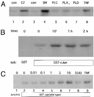
FIG. 1 Stimulation of JNK/SAPK activity by sphingomyelinase ( SM ) and D-e-C2-ceramide. A , HL-60 cells were cultured as described (18) and treated with D-e-C 2 -ceramide ( C2 , 5 m M, lane 2 ) or ethanol only ( con , lane 1 ) or the enzymes indicated (300 milliunits/ml, lanes 4-7 ) or vehicle ( con , lane 3 ) or TNF a (10 ng/ml, lane 8 ) for 20 min. The single prominent band visible following 10% SDS-polyacrylamide gel electrophoresis and autoradiography represents phosphorylated GST-c-Jun. Equal substrate loading was confirmed in all cases by Coomassie Blue staining of the gels prior to autoradiography. PL , phospholipase. B , time course of JNK/SAPK induction following D-e-C 2 -ceramide addition (5 m M) to HL-60 cells. The substrate ( sub ) for the kinase assays was GST-cJun or GST alone as control. C , response of SAPK activity to D-e-C 2 -ceramide treatment. All cells received the indicated concentration in an equal volume of ethanol or ethanol alone 5(di), 5 m M dihydroC2-ceramide. Extracts were prepared after 10 min of treatment. The substrate for the kinase assay was wild-type GST-cJun ( lanes 2-9 ) or GST-cJun(AA) ( lane 1 ) in which Ser-63 and Ser-73 are mutated to Ala (10).
FIG. 1 Stimulation of JNK/SAPK activity by sphingomyelinase ( SM ) and D-e-C2-ceramide. A , HL-60 cells were cultured as described (18) and treated with D-e-C 2 -ceramide ( C2 , 5 m M, lane 2 ) or ethanol only ( con , lane 1 ) or the enzymes indicated (300 milliunits/ml, lanes 4-7 ) or vehicle ( con , lane 3 ) or TNF a (10 ng/ml, lane 8 ) for 20 min. The single prominent band visible following 10% SDS-polyacrylamide gel electrophoresis and autoradiography represents phosphorylated GST-c-Jun. Equal substrate loading was confirmed in all cases by Coomassie Blue staining of the gels prior to autoradiography. PL , phospholipase. B , time course of JNK/SAPK induction following D-e-C 2 -ceramide addition (5 m M) to HL-60 cells. The substrate ( sub ) for the kinase assays was GST-cJun or GST alone as control. C , response of SAPK activity to D-e-C 2 -ceramide treatment. All cells received the indicated concentration in an equal volume of ethanol or ethanol alone 5(di), 5 m M dihydroC2-ceramide. Extracts were prepared after 10 min of treatment. The substrate for the kinase assay was wild-type GST-cJun ( lanes 2-9 ) or GST-cJun(AA) ( lane 1 ) in which Ser-63 and Ser-73 are mutated to Ala (10).CC-BY · From the PDF
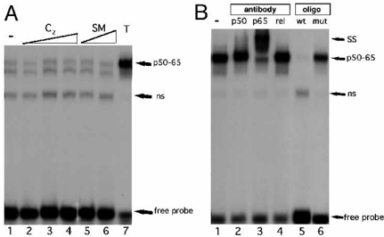
FIG. 2 TNF a , but not sphingomyelinase or D-e-C2-ceramide, activates NFk B binding activity. A , HL-60 cells were treated with vehicle, 0.1, 1, or 10 m M C2-ceramide ( lanes 1-4 ), or with 10 or 100 milliunits/ml sphingomyelinase ( SM ) ( B. cereus, Boehringer Mannheim) ( lanes 5 and 6 ) or recombinant TNF a ( T , 10 nM, lane 7 ) for 30 min prior to nuclear extract preparation and gel mobility shift analysis using a consensus NFk B binding site probe. ns , nonspecific shifted complex; p50-65 , heterodimeric NFk B. B , extracts from TNF a -treated HL-60 cells were used in binding assays, with antibodies to p50 ( lane 2 ), p65 ( lane 3 ), or c-Rel ( lane 4 ), or with a 50-fold excess of unlabeled wild-type ( wt , lane 5 ) or mutant NFk B binding site ( mut , lane 6 ) included in the reaction. After incubation on ice for 1 h, products were resolved on non-denaturing polyacrylamide gels. SS , supershifted NFk B; ns , nonspecific shifted complex (not competed by cold excess oligonucleotide).
FIG. 2 TNF a , but not sphingomyelinase or D-e-C2-ceramide, activates NFk B binding activity. A , HL-60 cells were treated with vehicle, 0.1, 1, or 10 m M C2-ceramide ( lanes 1-4 ), or with 10 or 100 milliunits/ml sphingomyelinase ( SM ) ( B. cereus, Boehringer Mannheim) ( lanes 5 and 6 ) or recombinant TNF a ( T , 10 nM, lane 7 ) for 30 min prior to nuclear extract preparation and gel mobility shift analysis using a consensus NFk B binding site probe. ns , nonspecific shifted complex; p50-65 , heterodimeric NFk B. B , extracts from TNF a -treated HL-60 cells were used in binding assays, with antibodies to p50 ( lane 2 ), p65 ( lane 3 ), or c-Rel ( lane 4 ), or with a 50-fold excess of unlabeled wild-type ( wt , lane 5 ) or mutant NFk B binding site ( mut , lane 6 ) included in the reaction. After incubation on ice for 1 h, products were resolved on non-denaturing polyacrylamide gels. SS , supershifted NFk B; ns , nonspecific shifted complex (not competed by cold excess oligonucleotide).CC-BY · From the PDF
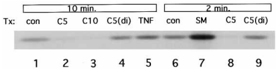
FIG. 3 MAPkinase activity is stimulated by sphingomyelinase but not ceramide. An immune complex MAP kinase assay was performed with HL-60 cell extracts prepared following cell treatment ( Tx ) with the indicated agonists for 10 min ( lanes 1-5 ) or 2 min ( lanes 6-9 ). C5 and C10 , 5 and 10 m M D-e-C 2 -ceramide; C5(di) , 5 m M DLerythro -dihydro-C 2 -ceramide; SM , 100 milliunits/ml Staphylococcus aureus sphingomyelinase; TNF , 50 ng/ml recombinant human TNF a ; con , control. GST-ElkC was used as the substrate. Equal substrate loading was confirmed in all cases by Coomassie Blue staining of the gels prior to autoradiography.
FIG. 3 MAPkinase activity is stimulated by sphingomyelinase but not ceramide. An immune complex MAP kinase assay was performed with HL-60 cell extracts prepared following cell treatment ( Tx ) with the indicated agonists for 10 min ( lanes 1-5 ) or 2 min ( lanes 6-9 ). C5 and C10 , 5 and 10 m M D-e-C 2 -ceramide; C5(di) , 5 m M DLerythro -dihydro-C 2 -ceramide; SM , 100 milliunits/ml Staphylococcus aureus sphingomyelinase; TNF , 50 ng/ml recombinant human TNF a ; con , control. GST-ElkC was used as the substrate. Equal substrate loading was confirmed in all cases by Coomassie Blue staining of the gels prior to autoradiography.CC-BY · From the PDF
Image 4
CC-BY · From the PDF
Image 5
CC-BY · From the PDF
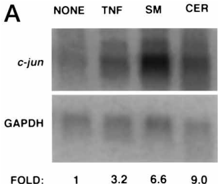
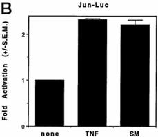
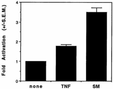
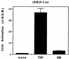
FIG. 4 Stimulation of cjun transcriptional activity. A , cjun mRNA levels are increased following TNF a , sphingomyelinase, and C2-ceramide treatment. HL-60 cells were incubated with TNF a ( TNF , 20 ng/ml), sphingomyelinase ( SM , 100 milliunits/ml), C2-ceramide ( CER , 10 m M), or untreated ( NONE ) for 1 h, and total RNA was prepared followed by electrophoresis and Northern blotting. The blot was hybridized first with a radioactively labeled human cjun probe ( upper panel ) and then with a labeled human glyceraldehyde-3-phosphate dehydrogenase ( GAPDH ) probe. Blots were quantitated by phosphor image analysis, and -fold induction of cjun mRNA normalized to GAPDH mRNA ( FOLD ) is indicated. B , activity of luciferase reporter genes in the L929 fibrosarcoma cell line. Jun-luciferase ( Luc ), or 5XGalluciferase plus Gal-ElkC, or (KB)3-luciferase (three copies of the consensus NFk B response element linked to luciferase) was transfected into L929 cells. Cells were then treated with TNF a (20 ng/ml) or sphingomyelinase (100 milliunits/ml) for 5 h, and extracts were prepared for determination of luciferase activity. -Fold activation represents reporter activity relative to cells treated with vehicle only. Results from one representative experiment performed in duplicate are shown ( 6 S.E.). Results were consistent over at least three experiments.
FIG. 4 Stimulation of cjun transcriptional activity. A , cjun mRNA levels are increased following TNF a , sphingomyelinase, and C2-ceramide treatment. HL-60 cells were incubated with TNF a ( TNF , 20 ng/ml), sphingomyelinase ( SM , 100 milliunits/ml), C2-ceramide ( CER , 10 m M), or untreated ( NONE ) for 1 h, and total RNA was prepared followed by electrophoresis and Northern blotting. The blot was hybridized first with a radioactively labeled human cjun probe ( upper panel ) and then with a labeled human glyceraldehyde-3-phosphate dehydrogenase ( GAPDH ) probe. Blots were quantitated by phosphor image analysis, and -fold induction of cjun mRNA normalized to GAPDH mRNA ( FOLD ) is indicated. B , activity of luciferase reporter genes in the L929 fibrosarcoma cell line. Jun-luciferase ( Luc ), or 5XGalluciferase plus Gal-ElkC, or (KB)3-luciferase (three copies of the consensus NFk B response element linked to luciferase) was transfected into L929 cells. Cells were then treated with TNF a (20 ng/ml) or sphingomyelinase (100 milliunits/ml) for 5 h, and extracts were prepared for determination of luciferase activity. -Fold activation represents reporter activity relative to cells treated with vehicle only. Results from one representative experiment performed in duplicate are shown ( 6 S.E.). Results were consistent over at least three experiments.CC-BY · From the PDF
Image 7
CC-BY · From the PDF

Verification

Claims the paper makes

The claims and statistics of this paper haven’t been checked yet.

Run the check — free, no account needed. It reads the open-access text and checks the sources it cites.

A few free checks a day · papers already verified open instantly · results are public

Claims other papers make about this one

Each is checked against this paper’s own text: does it say what the citing paper says it does?

Self-check

Percentages, sums and reported differences re-computed from the paper’s own numbers.

The self-check needs the paper’s full text.

Citations

Cites

Show all 2 references

Research works printed but not linked: 50

  1. Vassalli, 1992, Annu. Rev. Immunol., 10, 411, 10.1146/annurev.iy.10.040192.002211 · DOI
  2. Tracey, 1993, Annu. Rev. Cell Biol., 9, 317, 10.1146/annurev.cb.09.110193.001533 · DOI
  3. Dbaibo, 1993, J. Biol. Chem., 268, 17762, 10.1016/S0021-9258(17)46770-6 · DOI
  4. Obeid, 1993, Science, 259, 1769, 10.1126/science.8456305 · DOI · Paper page
  5. Osborne, 1989, Proc. Natl. Acad. Sci. U. S. A., 86, 2336, 10.1073/pnas.86.7.2336 · DOI
  6. Poli, 1990, Proc. Natl. Acad. Sci. U. S. A., 87, 782, 10.1073/pnas.87.2.782 · DOI · Paper page
  7. Kyriakis, 1994, Nature, 369, 156, 10.1038/369156a0 · DOI
  8. Westwick, 1994, J. Biol. Chem., 269, 26396, 10.1016/S0021-9258(18)47207-9 · DOI
  9. Sluss, 1994, Mol. Cell. Biol., 14, 8376
  10. Derijard, 1994, Cell, 76, 1025, 10.1016/0092-8674(94)90380-8 · DOI · Paper page
  11. Gupta, 1995, Science, 267, 389, 10.1126/science.7824938 · DOI
  12. Hill, 1993, Cell, 73, 395, 10.1016/0092-8674(93)90238-L · DOI
  13. Marais, 1993, Cell, 73, 381, 10.1016/0092-8674(93)90237-K · DOI
  14. Kim, 1991, J. Biol. Chem., 266, 484, 10.1016/S0021-9258(18)52461-3 · DOI
  15. Hannun, 1994, J. Biol. Chem., 269, 3125, 10.1016/S0021-9258(17)41834-5 · DOI
  16. Dobrowsky, 1993, J. Biol. Chem., 268, 15523, 10.1016/S0021-9258(18)82288-8 · DOI
  17. Wolff, 1994, J. Biol. Chem., 269, 19605, 10.1016/S0021-9258(17)32212-3 · DOI
  18. Joseph, 1993, J. Biol. Chem., 268, 20002, 10.1016/S0021-9258(20)80686-3 · DOI
  19. Bielawska, 1993, J. Biol. Chem., 268, 26226, 10.1016/S0021-9258(19)74304-X · DOI
  20. Tsujimoto, 1985, Proc. Natl. Acad. Sci. U. S. A., 82, 7226, 10.1073/pnas.82.22.7626 · DOI
  21. Franzoso, 1994, J. Exp. Med., 180, 1445, 10.1084/jem.180.4.1445 · DOI
  22. Westwick, 1995, Methods Enzymol., 255, 342, 10.1016/S0076-6879(95)55037-2 · DOI
  23. Smith, 1988, Gene (Amst.), 67, 31, 10.1016/0378-1119(88)90005-4 · DOI
  24. Westwick, 1994, Proc. Natl. Acad. Sci. U. S. A., 91, 6030, 10.1073/pnas.91.13.6030 · DOI
  25. Hattori, 1988, Proc. Natl. Acad. Sci. U. S. A., 85, 9148, 10.1073/pnas.85.23.9148 · DOI
  26. Galang, 1994, Oncogene, 9, 2913
  27. Hauser, 1995, Methods Enzymol., 255, 412, 10.1016/S0076-6879(95)55043-7 · DOI
  28. Hattori, 1993, Am. J. Physiol., 264, G95
  29. Yang, 1993, J. Biol. Chem., 268, 20520, 10.1016/S0021-9258(20)80756-X · DOI
  30. Schutze, 1992, Cell, 71, 765, 10.1016/0092-8674(92)90553-O · DOI
  31. Betts, 1994, J. Biol. Chem., 269, 8455, 10.1016/S0021-9258(17)37215-0 · DOI
  32. Johns, 1994, J. Immunol., 152, 5877, 10.4049/jimmunol.152.12.5877 · DOI
  33. Raines, 1993, J. Biol. Chem., 268, 14572, 10.1016/S0021-9258(18)82367-5 · DOI
  34. Finney, 1993, Curr. Biol., 3, 805, 10.1016/0960-9822(93)90214-9 · DOI
  35. van Dam, 1993, EMBO J., 12, 479, 10.1002/j.1460-2075.1993.tb05680.x · DOI · Paper page
  36. Herr, 1994, Carcinogenesis, 15, 1105, 10.1093/carcin/15.6.1105 · DOI
  37. Rothe, 1994, Cell, 78, 681, 10.1016/0092-8674(94)90532-0 · DOI
  38. Hsu, 1995, Cell, 81, 495, 10.1016/0092-8674(95)90070-5 · DOI
  39. Wiegmann, 1992, J. Biol. Chem., 267, 17997, 10.1016/S0021-9258(19)37142-X · DOI
  40. Smith, 1994, Cell, 76, 959, 10.1016/0092-8674(94)90372-7 · DOI
  41. Erickson, 1994, Nature, 372, 560, 10.1038/372560a0 · DOI
  42. Vandenabeele, 1992, J. Exp. Med., 176, 1015, 10.1084/jem.176.4.1015 · DOI
  43. Lewis, 1991, Proc. Natl. Acad. Sci. U. S. A., 88, 2830, 10.1073/pnas.88.7.2830 · DOI
  44. Yan, 1994, Nature, 372, 798, 10.1038/372798a0 · DOI
  45. Sanchez, 1994, Nature, 372, 794, 10.1038/372794a0 · DOI
  46. Lin, 1995, Science, 268, 286, 10.1126/science.7716521 · DOI
  47. Mumby, 1993, Physiol Rev., 73, 673, 10.1152/physrev.1993.73.4.673 · DOI
  48. Brenner, 1989, Nature, 337, 661, 10.1038/337661a0 · DOI
  49. Duh, 1989, Proc. Natl. Acad. Sci. U. S. A., 86, 5974, 10.1073/pnas.86.15.5974 · DOI
  50. Lozano, 1994, J. Biol. Chem., 269, 19200, 10.1016/S0021-9258(17)32152-X · DOI

Cited by

Sorted by citations

See all 303

Metadata

Authors
Westwick, J. K., Bielawska, A. E., Dbaibo, G., Hannun, Y. A., & Brenner, D. A.
Venue
Journal of Biological Chemistry
Publisher
Elsevier BV
Published
1995-09
Volume
270
Issue
39
Pages
22689-22692
ISSN
0021-9258
Cited by
303
References
2
Language
en

About these checks

Registry, retraction, Cites and statistics results are computed, not judged, with one exception: a printed reference that no index lists is read once by a model before it counts as a citation error. Claims are extracted by a model and kept only when their quote appears verbatim in the text; each cited source is then checked, with a model’s help, for whether it says what the claim relies on. Those verdicts can be wrong — read the claim’s quote and the cited source before relying on one.

Statistics and arithmetic are recomputed, not judged.

Confidence bands are shown only for fields calibrated on our annotated test set. None is calibrated yet, so no band is shown.

What counts as a claim ›How we measure confidence ›How we check this ›

Are you an author, or do you see a problem with this page? Request a review

How to cite

Cite CoChat for the checks on this page, and the paper for its own findings.

This page
CoChat. (2026). Citation check: Ceramide Activates the Stress-activated Protein Kinases. https://app.cochat.ai/papers/10.1074/jbc.270.39.22689
The paper
Westwick, J. K., Bielawska, A. E., Dbaibo, G., Hannun, Y. A., & Brenner, D. A. (1995). Ceramide Activates the Stress-activated Protein Kinases. Journal of Biological Chemistry. https://doi.org/10.1074/jbc.270.39.22689