

The DOI matches its Crossref record; 67 of the 67 references it cites were found in a registry or an index.
Cortisol Synthesis in Epidermis Is Induced by IL-1 and Tissue Injury
Open accessAbstract
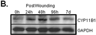
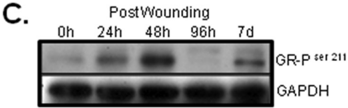
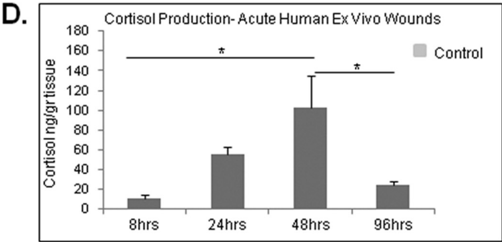
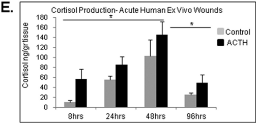
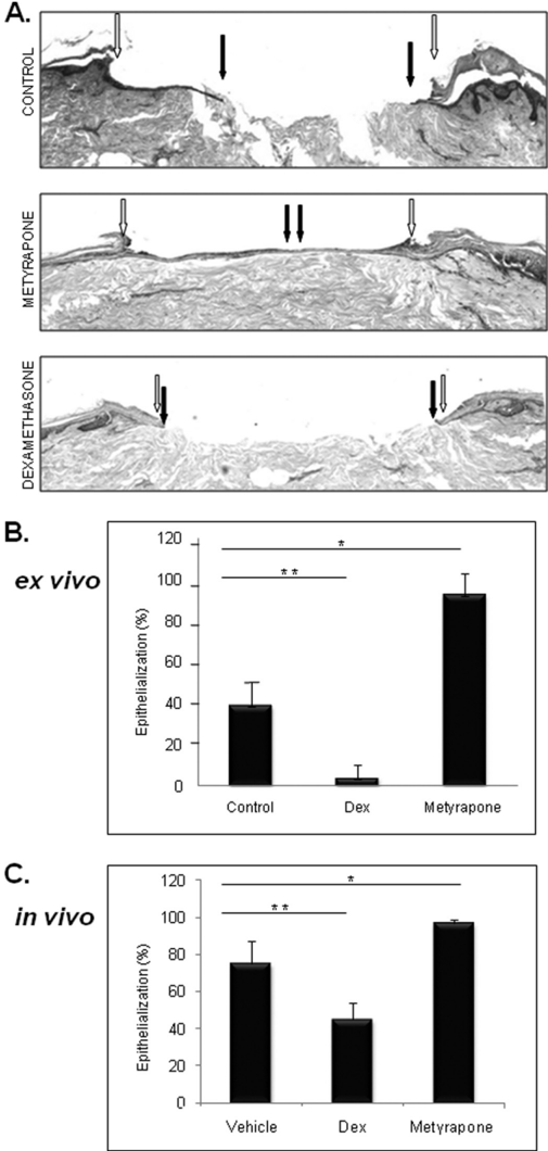
Glucocorticoids (GCs) are known inhibitors of wound healing. In this study we report the novel finding that both keratinocytes in vitro and epidermis in vivo synthesize cortisol and how this synthesis regulates wound healing. We show that epidermis expresses enzymes essential for cortisol synthesis, including steroid 11 β-hydroxylase (CYP11B1), and an enzyme that controls negative feedback mechanism, 11β-hydroxysteroid dehydrogenase 2 (11βHSD2). We also found that cortisol synthesis in keratinocytes and skin can be stimulated by ACTH and inhibited by metyrapone (CYP11B1 enzyme inhibitor). Interestingly, IL-1β, the first epidermal signal of tissue injury, induces the expression of CYP11B1 and increases cortisol production by keratinocytes. Additionally, we found induction of CYP11B1 increased production of cortisol and activation of GR pathway during wound healing ex vivo and in vivo using human and porcine wound models, respectively. Conversely, inhibition of cortisol synthesis during wound healing increases IL-1β production, suggesting that cortisol synthesis in epidermis may serve as a local negative feedback to proinflammatory cytokines. Local GCs synthesis, therefore, may provide control of the initial proinflammatory response, preventing excessive inflammation upon tissue injury. Inhibition of GC synthesis accelerated wound closure in vivo, providing the evidence that modulation of cortisol synthesis in epidermis may be an important regulatory mechanism during wound healing.
Overview
The claims and statistics of this paper haven’t been checked yet.
Run the check — free, no account needed. It reads the open-access text and checks the sources it cites.
Integrity signals
Registry
Cites
Statistics
Figures
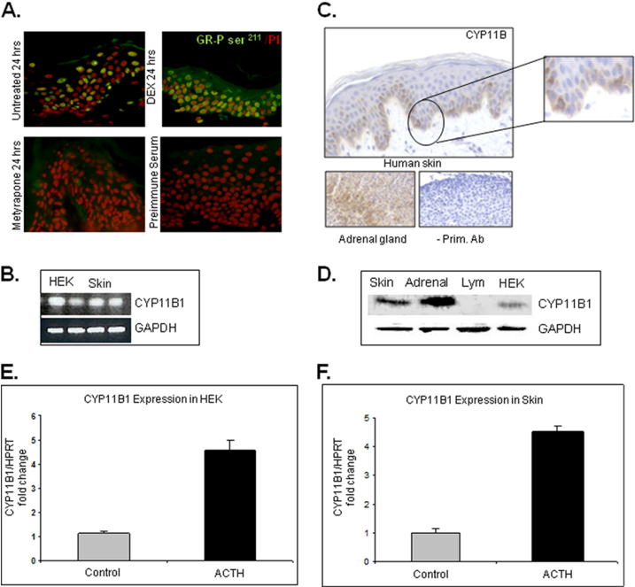
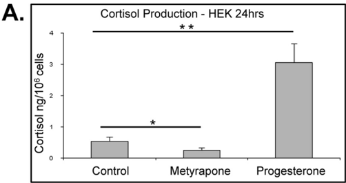
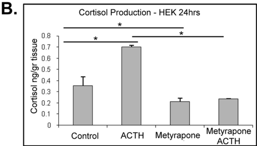
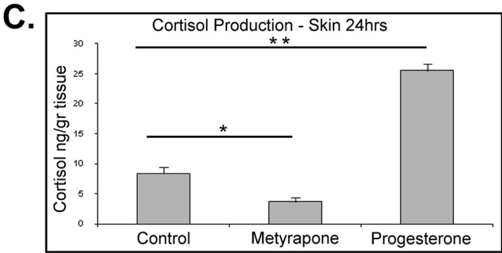
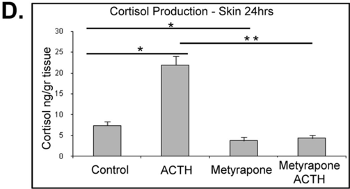
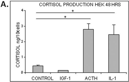
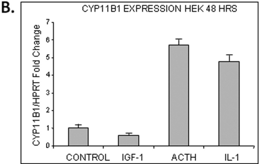
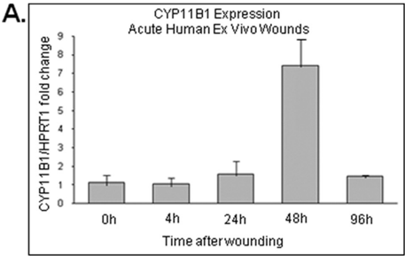
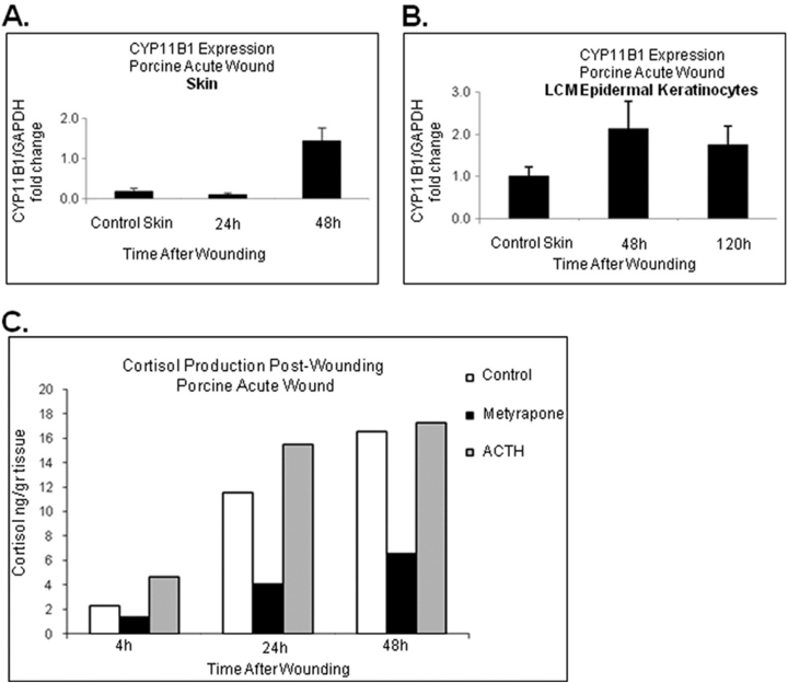
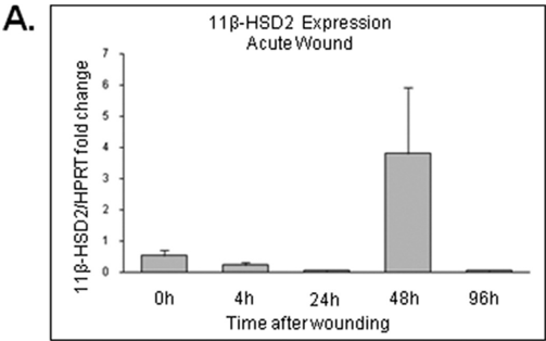
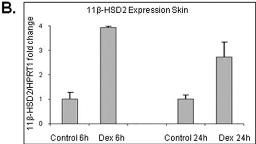
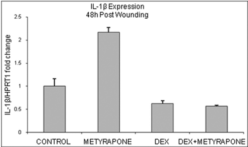
Verification
Claims the paper makes
The claims and statistics of this paper haven’t been checked yet.
Run the check — free, no account needed. It reads the open-access text and checks the sources it cites.
Claims other papers make about this one
Each is checked against this paper’s own text: does it say what the citing paper says it does?
In the citing paper
“Skin also expresses 11βHSD2 which works in the oxidative direction, converting cortisol to the inactive cortisone [23, 64–66, 81].”
From Steroidogenesis in the skin: Implications for local immune functions (2013) · cites this paper as [65]
In the citing paper
“In contrast, dexamethasone treatment of human skin was reported to increase 11βHSD2 expression at the mRNA level [65].”
From Steroidogenesis in the skin: Implications for local immune functions (2013) · cites this paper as [65]
Self-check
Percentages, sums and reported differences re-computed from the paper’s own numbers.
The self-check needs the paper’s full text.
Citations
Cites
Sapolsky, R. M., Romero, L. M., & Munck, A. U. (2000) · Endocrine Reviews · doi:10.1210/edrv.21.1.0389 · Cited by 7,541
PERSPECTIVE ARTICLE: Growth factors and cytokines in wound healing
Barrientos, S., Stojadinovic, O., Golinko, M. S., Brem, H., & Tomic‐Canic, M. (2008) · Wound Repair and Regeneration · doi:10.1111/j.1524-475x.2008.00410.x · Cited by 3,583
Antiinflammatory Action of Glucocorticoids — New Mechanisms for Old Drugs
Rhen, T., & Cidlowski, J. A. (2005) · New England Journal of Medicine · doi:10.1056/nejmra050541 · Cited by 3,149
Hypothalamic-pituitary-adrenal axis, neuroendocrine factors and stress
Tsigos, C., & Chrousos, G. P. (2002) · Journal of Psychosomatic Research · doi:10.1016/s0022-3999(02)00429-4 · Cited by 2,822
Cellular and molecular basis of wound healing in diabetes
Brem, H., & Tomic‐Canic, M. (2007) · Journal of Clinical Investigation · doi:10.1172/jci32169 · Cited by 1,863
Wound Macrophages Express TGF-α and Other Growth Factors in Vivo: Analysis by mRNA Phenotyping
Rappolee, D. A., Mark, D., Banda, M. J., & Werb, Z. (1988) · Science · doi:10.1126/science.3041594 · Cited by 1,105
Cloning and tissue distribution of the human 1 lβ-hydroxysteroid dehydrogenase type 2 enzyme
Albiston, A. L., Obeyesekere, V. R., Smith, R. E., & Krozowski, Z. S. (1994) · Molecular and Cellular Endocrinology · doi:10.1016/0303-7207(94)90176-7 · Cited by 712
Loots, M. A. M., Lamme, E. N., Zeegelaar, J., Mekkes, J. R., Bos, J. D., & Middelkoop, E. (1998) · Journal of Investigative Dermatology · doi:10.1046/j.1523-1747.1998.00381.x · Cited by 620
Interactions of cytokines, growth factors, and proteases in acute and chronic wounds
Mast, B. A., & Schultz, G. S. (1996) · Wound Repair and Regeneration · doi:10.1046/j.1524-475x.1996.40404.x · Cited by 598
Keratins and the Keratinocyte Activation Cycle
Freedberg, I. M., Tomic‐Canic, M., Komine, M., & Blumenberg, M. (2001) · Journal of Investigative Dermatology · doi:10.1046/j.1523-1747.2001.01327.x · Cited by 564
Research works printed but not linked: 6
- Pastar, 2008, Surg. Technol. Int., 17, 105
- Steinhoff, 2001, Curr. Opin. Allergy Clin. Immunol., 1, 469
- Zouboulis, 2000, Horm. Res., 54, 230
- Davis, 2008, Ostomy Wound Manage., 54, 16
- Kayes-Wandover, 2000, J. Clin. Endocrinol. Metab., 85, 2519
- Sapolsky, 2000, Endocr. Rev., 21, 55
Cited by
Sorted by citations
Epithelialization in Wound Healing: A Comprehensive Review
Pastar, I., Stojadinović, O., Yin, N. C., Ramirez, H. A., Nusbaum, A. G., Sawaya, A. P., Patel, S. B., Khalid, L., Isseroff, R. R., & Tomic‐Canic, M. (2014) · Advances in Wound Care · doi:10.1089/wound.2013.0473 · Cited by 1,604
“Inhibition of GC synthesis in keratinocytes resulted in increased expression of proinflammatory IL1-b implying that skin-specific cortisol synthesis serves as a local negative feedback to prevent excessive inflammation upon initial injury and serves to regulate epithelialization.(107) Another type of hormone, the cate…”
BackgroundMethodInfluential11β-hydroxysteroid dehydrogenases: Intracellular gate-keepers of tissue glucocorticoid action
Chapman, K., Holmes, M., & Seckl, J. (2013) · Physiological Reviews · doi:10.1152/physrev.00020.2012 · Cited by 887
“11 -HSD2 mRNA is also detectable by PCR in human skin (732), although whether this extends beyond known expression in the mineralocorticoid target cells of sweat glands (82) or if there is sufficient to influence glucocorticoid function elsewhere in the dermis/epidermis is unknown.”
BackgroundSlominski, A. T., Zmijewski, M. A., Skobowiat, C., Zbytek, B., Slominski, R. M., & Steketee, J. D. (2012) · Advances in anatomy, embryology and cell biology · doi:10.1007/978-3-642-19683-6 · Cited by 505
“, 2005e), IL-1 as well as by wound response (Vukelic et al., 2011) and high energy ultraviolet radiation (Skobowiat et al.”
BackgroundSlominski (2012) · doi:10.1007/978-3-642-19683-6_1 · Cited by 461
How UV Light Touches the Brain and Endocrine System Through Skin, and Why
Slominski, A. T., Zmijewski, M. A., Plonka, P. M., Szaflarski, J. P., & Paus, R. (2018) · Endocrinology · doi:10.1210/en.2017-03230 · Cited by 452
Metadata
- Authors
- Vukelic, S., Stojadinovic, O., Pastar, I., Rabach, M., Krzyzanowska, A., Lebrun, E., Davis, S. C., Resnik, S., Brem, H., & Tomic-Canic, M.
- Affiliations
- Lenox Hill Hospital (US)
- University of Miami (US)
- SUNY Downstate Health Sciences University (US)
- Hospital for Special Surgery (US)
- NYU Langone Health (US)
- Venue
- Journal of Biological Chemistry
- Publisher
- Elsevier BV
- Published
- 2011-03
- Volume
- 286
- Issue
- 12
- Pages
- 10265-10275
- ISSN
- 0021-9258 (ISSN-L)
- PMC
- PMC3060481
- OpenAlex
- W2080660266
- Cited by
- 193
- References
- 67
- Citations by year
- 2026: 6 · 2025: 12 · 2024: 7 · 2023: 6 · 2022: 12 · 2021: 11 · 2020: 18 · 2019: 9 · 2018: 11 · 2017: 18 · 2016: 17 · 2015: 8 · 2014: 15 · 2013: 19 · 2012: 13
- FWCI
- 18.81
- Type
- journal article
- Language
- en
- Open access
- hybrid
- doi.org
- Licence
- Locations
- Journal of Biological Chemistry (publishedVersion, cc-by)
- Europe PMC (PubMed Central) (publishedVersion)
- PubMed Central (publishedVersion)
- PubMed (publishedVersion)
- Funders
- National Institutes of Health
- National Institute of Nursing Research (R01NR008029, NR008029)
- National Institute of Diabetes and Digestive and Kidney Diseases (K24DK090135)
- Topics
- Health Sciences › Medicine › Surgery › Wound Healing and Treatments
- Health Sciences › Medicine › Endocrinology, Diabetes and Metabolism › Adrenal Hormones and Disorders
- Health Sciences › Medicine › Pharmacology (medical) › Inflammatory mediators and NSAID effects
- Keywords
- cortisol production, epidermis, keratinocytes, wound healing, IL-1β, tissue damage, CYP11B1, 11β-hydroxysteroid dehydrogenase type 2, glucocorticoid receptor, ACTH, metyrapone, negative feedback regulation, inflammatory cytokines
- MeSH terms
- Adrenocorticotropic Hormone, Animals, Enzyme Inhibitors, Epidermis, Hormones, Humans, Hydrocortisone, Inflammation, Metyrapone, Steroid 11-beta-Hydroxylase, Swine, Wound Healing, Keratinocytes, Interleukin-1beta
About these checks
Registry, retraction, Cites and statistics results are computed, not judged, with one exception: a printed reference that no index lists is read once by a model before it counts as a citation error. Claims are extracted by a model and kept only when their quote appears verbatim in the text; each cited source is then checked, with a model’s help, for whether it says what the claim relies on. Those verdicts can be wrong — read the claim’s quote and the cited source before relying on one.
Statistics and arithmetic are recomputed, not judged.
Confidence bands are shown only for fields calibrated on our annotated test set. None is calibrated yet, so no band is shown.
What counts as a claim ›How we measure confidence ›How we check this ›
Are you an author, or do you see a problem with this page? Request a review
