Cited by 137

The DOI matches its Crossref record; 12 of the 12 references it cites were found in a registry or an index.

Interactions of Pathological Hallmark Proteins

Oláh, Judit, Vincze, Orsolya, Virók, Dezső, Simon, Dóra, Bozsó, Zsolt, Tőkési, Natália, Horváth, István, Hlavanda, Emma, Kovács, János, Magyar, Anna, Szűcs, Mária, Orosz, Ferenc, Penke, Botond, Ovádi, Judit·2011·Journal of Biological Chemistry

Overview

The claims and statistics of this paper haven’t been checked yet.

Run the check — free, no account needed. It reads the open-access text and checks the sources it cites.

A few free checks a day · papers already verified open instantly · results are public

Integrity signals

Registry

Crossref · 10.1074/jbc.m111.243907

Cites

12 of 12 references verified0 citation errors · 0 not checkable

Statistics

Figures

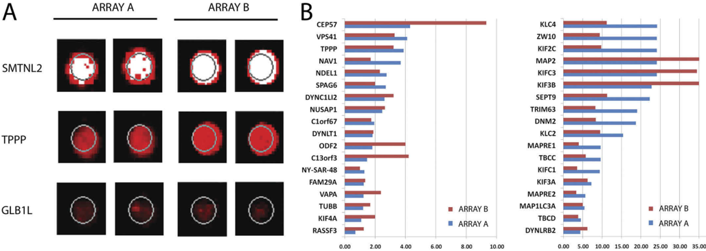
FIGURE1 Proteinarray-basedinteractomeanalysisofoligomericA 𝛃 42 . A , to explore the interacting protein partners of 𝛃 -amyloid, 10 𝛍 MA 𝛃 42 peptidewas hybridized onto the Protoarray 4.0 protein array. Duplicate spot images show a strong A 𝛃 42 binder smoothelin-like protein 2 ( SMTNL2 ), the TPPP/p25, and a weak A 𝛃 42 binder 𝛃 -galactosidase-1-like protein ( GLB1L ) on both applied protein arrays. B , the cellular localization of the A 𝛃 42 interacting proteins were characterized using the GO data base. Members of the microtubule GO cellular content category are shown, with their normalized signal intensities on both applied arrays.
FIGURE1 Proteinarray-basedinteractomeanalysisofoligomericA 𝛃 42 . A , to explore the interacting protein partners of 𝛃 -amyloid, 10 𝛍 MA 𝛃 42 peptidewas hybridized onto the Protoarray 4.0 protein array. Duplicate spot images show a strong A 𝛃 42 binder smoothelin-like protein 2 ( SMTNL2 ), the TPPP/p25, and a weak A 𝛃 42 binder 𝛃 -galactosidase-1-like protein ( GLB1L ) on both applied protein arrays. B , the cellular localization of the A 𝛃 42 interacting proteins were characterized using the GO data base. Members of the microtubule GO cellular content category are shown, with their normalized signal intensities on both applied arrays.CC-BY · From the PDF
Image 2
CC-BY · From the PDF
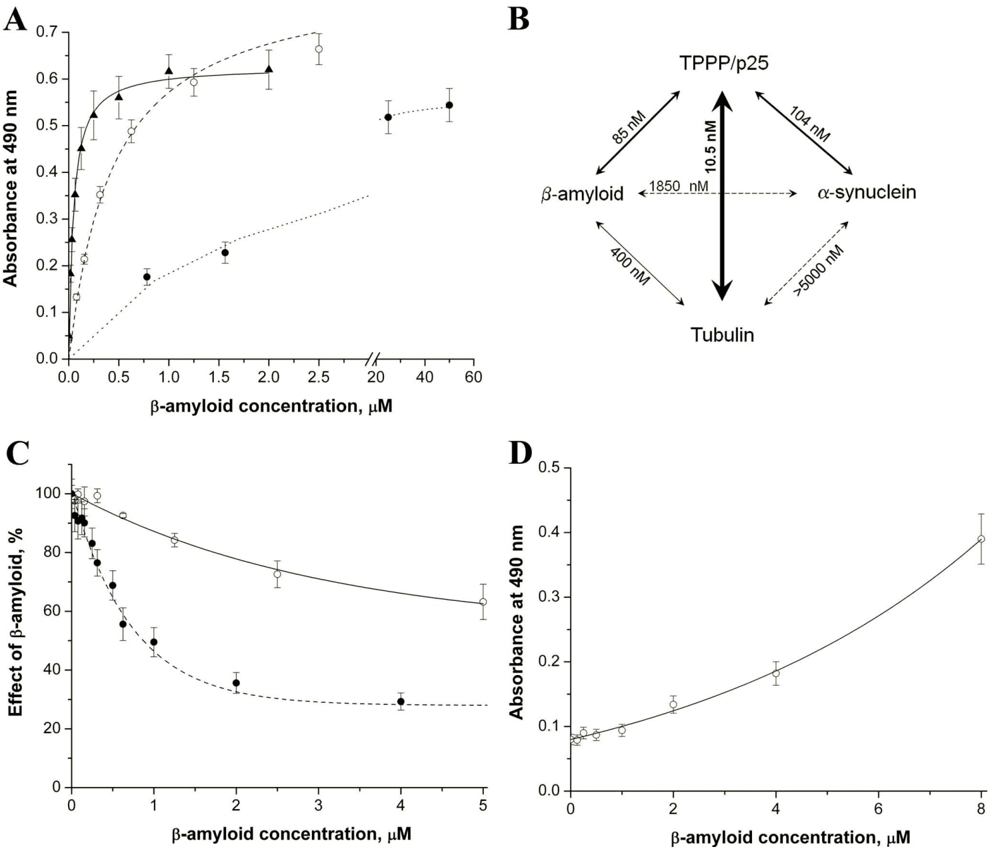
FIGURE3 TheinteractionofoligomericA 𝛃 42 withdifferentproteinsfollowedbyELISA. A , the plate was coated with 0.5 𝛍 g/well TPPP/p25 ( Œ ), tubulin ( E ), or 𝛂 -synuclein ( F ), then it was incubated with A 𝛃 42 at different concentrations. B , the apparent dissociation constants ( Kd ) characteristic for the interactions were evaluated by non-linear fitting of the hyperbolic saturation curves using the Microcal Origin 6.0 software. C , E , the plate was coated with TPPP/p25 then incubatedwithA 𝛃 42 . After incubation, tubulin was added in constant concentration (100 nM). After further incubation, anti-tubulin was added. F , the plate was coated with TPPP/p25 and incubated with A 𝛃 42 , 100 nM 𝛂 -synuclein was added, and then anti𝛂 -synuclein was added. The effect of the A 𝛃 42 wascalculated as the absorbance at a given A 𝛃 42 concentration divided by the absorbance without A 𝛃 42 . D , E , the plate was coated with 𝛂 -synuclein, then incubated with A 𝛃 42 . After incubation, tubulin was added in constant concentration (100 nM). After further incubation, anti-tubulin was added. The average of three-five independent experiments and the S.E. is shown.
FIGURE3 TheinteractionofoligomericA 𝛃 42 withdifferentproteinsfollowedbyELISA. A , the plate was coated with 0.5 𝛍 g/well TPPP/p25 ( Œ ), tubulin ( E ), or 𝛂 -synuclein ( F ), then it was incubated with A 𝛃 42 at different concentrations. B , the apparent dissociation constants ( Kd ) characteristic for the interactions were evaluated by non-linear fitting of the hyperbolic saturation curves using the Microcal Origin 6.0 software. C , E , the plate was coated with TPPP/p25 then incubatedwithA 𝛃 42 . After incubation, tubulin was added in constant concentration (100 nM). After further incubation, anti-tubulin was added. F , the plate was coated with TPPP/p25 and incubated with A 𝛃 42 , 100 nM 𝛂 -synuclein was added, and then anti𝛂 -synuclein was added. The effect of the A 𝛃 42 wascalculated as the absorbance at a given A 𝛃 42 concentration divided by the absorbance without A 𝛃 42 . D , E , the plate was coated with 𝛂 -synuclein, then incubated with A 𝛃 42 . After incubation, tubulin was added in constant concentration (100 nM). After further incubation, anti-tubulin was added. The average of three-five independent experiments and the S.E. is shown.CC-BY · From the PDF
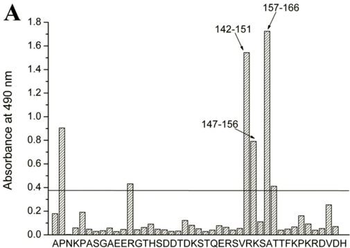
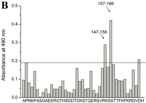
FIGURE4 MappingofpossiblebindingsitesbetweenA 𝛃 42 or 𝛂 -synuclein and TPPP/p25 by MULTIPIN peptide technology (Pepscan analysis). The amino acid sequence of the TPPP/p25 was used to synthesize a complete set of overlapping decapeptides covalently attached to the surfaces of derivative polyethylenepinsinaformatcompatiblewithstandardELISA.Theseoverlapping peptides covered the entire sequence of the protein. Pins were coated with 4 𝛍 M A 𝛃 42 ( A ) or 1.5 𝛍 M 𝛂 -synuclein ( B ), then anti-A 𝛃 or anti𝛂 -synuclein wasadded,respectively.Theabsorbancesofthepeptides(indicatedby letters of the first amino acid of the decapeptides ) are shown. The reaction of a pincoupled peptide was scored positive (significance level, indicated by a line ) when the ELISA absorption was significantly higher than the 2-fold average absorption of the peptides.
FIGURE4 MappingofpossiblebindingsitesbetweenA 𝛃 42 or 𝛂 -synuclein and TPPP/p25 by MULTIPIN peptide technology (Pepscan analysis). The amino acid sequence of the TPPP/p25 was used to synthesize a complete set of overlapping decapeptides covalently attached to the surfaces of derivative polyethylenepinsinaformatcompatiblewithstandardELISA.Theseoverlapping peptides covered the entire sequence of the protein. Pins were coated with 4 𝛍 M A 𝛃 42 ( A ) or 1.5 𝛍 M 𝛂 -synuclein ( B ), then anti-A 𝛃 or anti𝛂 -synuclein wasadded,respectively.Theabsorbancesofthepeptides(indicatedby letters of the first amino acid of the decapeptides ) are shown. The reaction of a pincoupled peptide was scored positive (significance level, indicated by a line ) when the ELISA absorption was significantly higher than the 2-fold average absorption of the peptides.CC-BY · From the PDF
Image 5
CC-BY · From the PDF
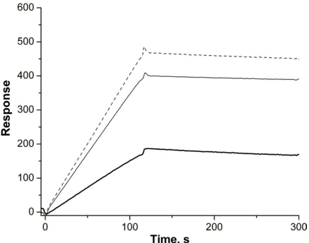
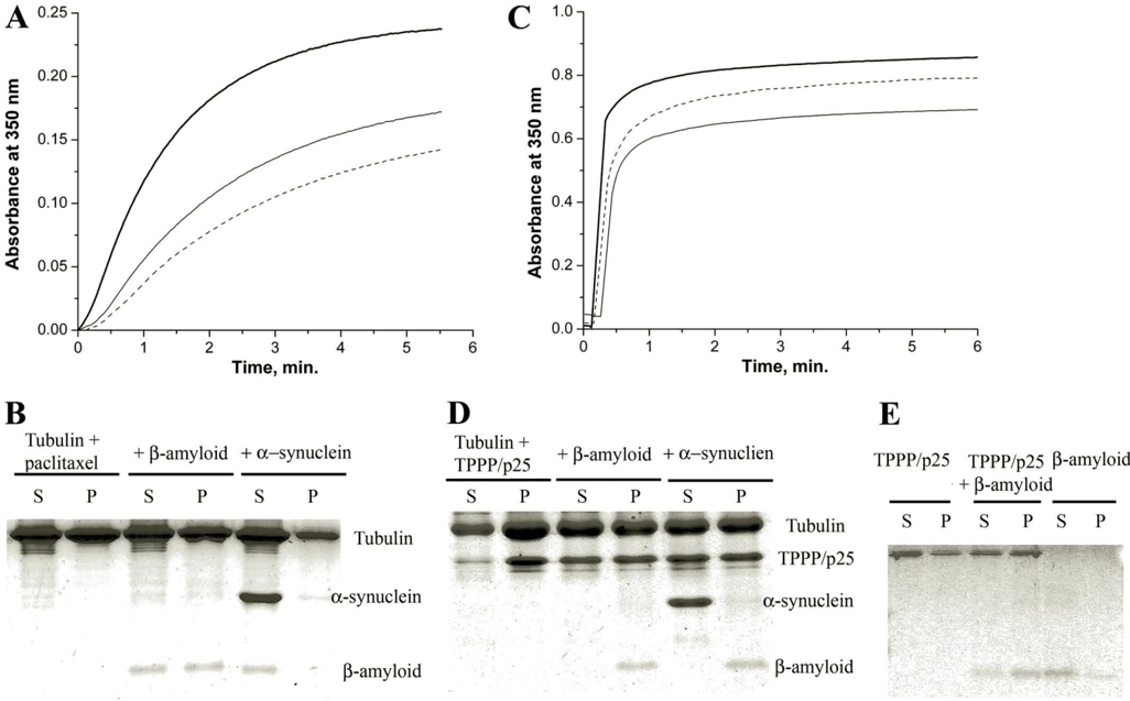
FIGURE 5 The effect of the interacting partners on tubulin assembly as determined via a turbidimetric assay followed by pelleting experiments. Tubulin polymerization was induced by paclitaxel ( A and B ) or by TPPP/p25 ( C and D ). For the turbidimetric assay ( A and C ), control ( bold line ), in the presence of A 𝛃 42 ( solid line ) or A 𝛃 42 and 𝛂 -synuclein ( dashedline ) are shown. For the pelleting experiments ( B and D ), at the quasi endpoints of the polymerization curves the assay mixtures were centrifuged at 15,000 × g for 15 min at 37 °C, and the pellet ( P ) and the supernatant ( S ) fractions were separated followed by SDS-PAGE analysis on Tris-Tricine three-layer gels. Tubulin alone, incubated with A 𝛃 42 or with both A 𝛃 42 and 𝛂 -synuclein as indicated. The concentration of tubulin was 15 𝛍 M ( A and B ) or 7 𝛍 M ( C ) or 10 𝛍 M ( D ) and the concentration of A 𝛃 42 was 15 𝛍 M ( A and B ) or 10 mM ( C ) or 25 𝛍 M ( D ), whereas that of the TPPP/p25 was 3 𝛍 M ( C ) or 10 𝛍 M( D ) and that of the 𝛂 -synuclein was 10 𝛍 M( A , B , and D ) or 5 𝛍 M( C ). E , for the pelleting experiment, 2 𝛍 MTPPP/p25 alone, TPPP/p25 incubated with 25 𝛍 MA 𝛃 42 , and A 𝛃 42 alone as indicated were used. Three-five independent experiments were performed; S.E. for turbidimetry and pelleting were ± 5 and ± 10%, respectively.
FIGURE 5 The effect of the interacting partners on tubulin assembly as determined via a turbidimetric assay followed by pelleting experiments. Tubulin polymerization was induced by paclitaxel ( A and B ) or by TPPP/p25 ( C and D ). For the turbidimetric assay ( A and C ), control ( bold line ), in the presence of A 𝛃 42 ( solid line ) or A 𝛃 42 and 𝛂 -synuclein ( dashedline ) are shown. For the pelleting experiments ( B and D ), at the quasi endpoints of the polymerization curves the assay mixtures were centrifuged at 15,000 × g for 15 min at 37 °C, and the pellet ( P ) and the supernatant ( S ) fractions were separated followed by SDS-PAGE analysis on Tris-Tricine three-layer gels. Tubulin alone, incubated with A 𝛃 42 or with both A 𝛃 42 and 𝛂 -synuclein as indicated. The concentration of tubulin was 15 𝛍 M ( A and B ) or 7 𝛍 M ( C ) or 10 𝛍 M ( D ) and the concentration of A 𝛃 42 was 15 𝛍 M ( A and B ) or 10 mM ( C ) or 25 𝛍 M ( D ), whereas that of the TPPP/p25 was 3 𝛍 M ( C ) or 10 𝛍 M( D ) and that of the 𝛂 -synuclein was 10 𝛍 M( A , B , and D ) or 5 𝛍 M( C ). E , for the pelleting experiment, 2 𝛍 MTPPP/p25 alone, TPPP/p25 incubated with 25 𝛍 MA 𝛃 42 , and A 𝛃 42 alone as indicated were used. Three-five independent experiments were performed; S.E. for turbidimetry and pelleting were ± 5 and ± 10%, respectively.CC-BY · From the PDF
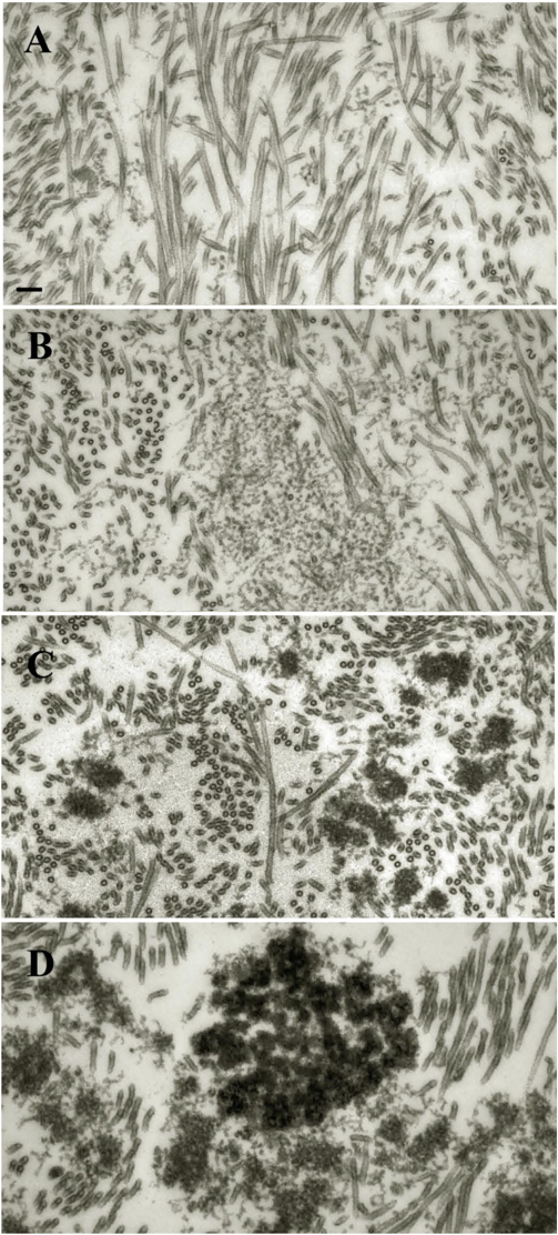
FIGURE6 ElectronmicroscopicanalysisoftheeffectofA 𝛃 42 oligomerson paclitaxel- or TPPP/p25-induced tubulin polymerization. Samples were prepared with 20 𝛍 Mpaclitaxel ( A and B ) or with 1.5 𝛍 MTPPP/p25 ( C and D ) in the presence of 10 𝛍 M A 𝛃 42 ( B and D ) or in its absence ( A and C ). The tubulin concentration was 10 𝛍 M ( A and B ) or 7 𝛍 M ( C and D ). Scale bar , 100 nm.
FIGURE6 ElectronmicroscopicanalysisoftheeffectofA 𝛃 42 oligomerson paclitaxel- or TPPP/p25-induced tubulin polymerization. Samples were prepared with 20 𝛍 Mpaclitaxel ( A and B ) or with 1.5 𝛍 MTPPP/p25 ( C and D ) in the presence of 10 𝛍 M A 𝛃 42 ( B and D ) or in its absence ( A and C ). The tubulin concentration was 10 𝛍 M ( A and B ) or 7 𝛍 M ( C and D ). Scale bar , 100 nm.CC-BY · From the PDF
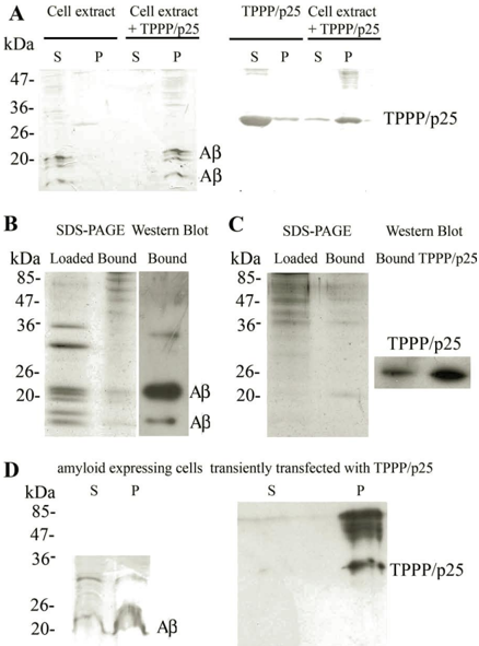
FIGURE7 InteractionofTPPP/p25andA 𝛃 atcell level. A , shownis a co-precipitation assay. Extract of amyloid expressing CHO7PA2 cells was incubated without or with 20 𝛍 M TPPP/p25 overnight at 4 °C, or 20 𝛍 M TPPP/p25 alone was incubated under the same conditions, and after centrifugation, the supernatant ( S ) and the pellet ( P ) fractions were subjected to Western blot to test the partition of A 𝛃 and TPPP/p25 as indicated. B , extract of amyloid expressing CHO7PA2 cells loaded to TPPP/p25 affinity column is shown. SDSPAGE analysis of the cell extract loaded to the column and the bound proteins. After elution, the bound proteins were subjected to Western blot using A 𝛃 antibody. C , extract of the SPM fraction loaded to A 𝛃 affinity column. SDS-PAGE analysis of the SPM extract loaded to the column and the bound proteins. After elution, the bound proteins and 0.9 ng of human recombinant TPPP/p25 were subjected to Western blot using TPPP/p25 antiserum. D , shown is a co-precipitation assay. Amyloid-expressing CHO7PA2 cells were transiently transfected with human recombinant TPPP/p25 and were lysed by sonication. After centrifugation, the supernatant and the pellet fractions were subjected to Western blot to test the partition of A 𝛃 and TPPP/p25 as indicated.
FIGURE7 InteractionofTPPP/p25andA 𝛃 atcell level. A , shownis a co-precipitation assay. Extract of amyloid expressing CHO7PA2 cells was incubated without or with 20 𝛍 M TPPP/p25 overnight at 4 °C, or 20 𝛍 M TPPP/p25 alone was incubated under the same conditions, and after centrifugation, the supernatant ( S ) and the pellet ( P ) fractions were subjected to Western blot to test the partition of A 𝛃 and TPPP/p25 as indicated. B , extract of amyloid expressing CHO7PA2 cells loaded to TPPP/p25 affinity column is shown. SDSPAGE analysis of the cell extract loaded to the column and the bound proteins. After elution, the bound proteins were subjected to Western blot using A 𝛃 antibody. C , extract of the SPM fraction loaded to A 𝛃 affinity column. SDS-PAGE analysis of the SPM extract loaded to the column and the bound proteins. After elution, the bound proteins and 0.9 ng of human recombinant TPPP/p25 were subjected to Western blot using TPPP/p25 antiserum. D , shown is a co-precipitation assay. Amyloid-expressing CHO7PA2 cells were transiently transfected with human recombinant TPPP/p25 and were lysed by sonication. After centrifugation, the supernatant and the pellet fractions were subjected to Western blot to test the partition of A 𝛃 and TPPP/p25 as indicated.CC-BY · From the PDF
Image 9
CC-BY · From the PDF
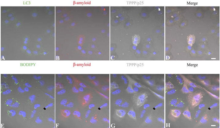
FIGURE8 RepresentativepicturesofintracellularA 𝛃 andTPPP/p25aggregationinCHO7PA2andCHO10cells. AmyloidexpressingCHO7PA2cells( A-D ) and TPPP/p25 expressing CHO10 cells ( E-H ) were transiently transfected with human recombinant TPPP/p25 and A 𝛃 42 , respectively, and immunostained for TPPP/p25 ( C and G , gray ), A 𝛃 ( B and F , red ), and the autophagic marker LC3 ( A , green ), whereas the general intracellular membrane marker (BODIPY 500/510) wasdetectedbyits own signal ( E , green ). Note the appearance of cytoplasmic aggregates in the case of TPPP/p25 and 𝛃 -amyloid co-expression on the merged pictures ( D and H ) that show no co-localization with either vacuole markers. Blue , DAPI. Scale bar , 10 𝛍 m.
FIGURE8 RepresentativepicturesofintracellularA 𝛃 andTPPP/p25aggregationinCHO7PA2andCHO10cells. AmyloidexpressingCHO7PA2cells( A-D ) and TPPP/p25 expressing CHO10 cells ( E-H ) were transiently transfected with human recombinant TPPP/p25 and A 𝛃 42 , respectively, and immunostained for TPPP/p25 ( C and G , gray ), A 𝛃 ( B and F , red ), and the autophagic marker LC3 ( A , green ), whereas the general intracellular membrane marker (BODIPY 500/510) wasdetectedbyits own signal ( E , green ). Note the appearance of cytoplasmic aggregates in the case of TPPP/p25 and 𝛃 -amyloid co-expression on the merged pictures ( D and H ) that show no co-localization with either vacuole markers. Blue , DAPI. Scale bar , 10 𝛍 m.CC-BY · From the PDF

Verification

Claims the paper makes

The claims and statistics of this paper haven’t been checked yet.

Run the check — free, no account needed. It reads the open-access text and checks the sources it cites.

A few free checks a day · papers already verified open instantly · results are public

Claims other papers make about this one

Each is checked against this paper’s own text: does it say what the citing paper says it does?

Self-check

Percentages, sums and reported differences re-computed from the paper’s own numbers.

The self-check needs the paper’s full text.

Citations

Cites

1 retracted

Show all 12 references

Research works printed but not linked: 47

  1. Hardy, 2002, Science, 297, 353, 10.1126/science.1072994 · DOI · Paper page
  2. Spillantini, 1998, Proc. Natl. Acad. Sci. U.S.A., 95, 6469, 10.1073/pnas.95.11.6469 · DOI · Paper page
  3. Irvine, 2008, Mol. Med., 14, 451, 10.2119/2007-00100.Irvine · DOI
  4. Avila, 2006, FEBS Lett., 580, 2922, 10.1016/j.febslet.2006.02.067 · DOI
  5. Hiltunen, 2009, J. Alzheimers Dis., 18, 401, 10.3233/JAD-2009-1154 · DOI
  6. Carrell, 2005, Trends Cell Biol., 15, 574, 10.1016/j.tcb.2005.09.005 · DOI
  7. Hartmann, 1997, Nat. Med., 3, 1016, 10.1038/nm0997-1016 · DOI
  8. Roychaudhuri, 2009, J. Biol. Chem., 284, 4749, 10.1074/jbc.R800036200 · DOI
  9. Shah, 2009, Neurobiol. Dis., 36, 11, 10.1016/j.nbd.2009.05.016 · DOI
  10. Verdier, 2005, J. Neurochem., 94, 617, 10.1111/j.1471-4159.2005.03158.x · DOI
  11. Crews, 2009, Neurotox. Res., 16, 306, 10.1007/s12640-009-9073-6 · DOI
  12. Lei, 2010, Int. J. Biochem. Cell Biol., 42, 1775, 10.1016/j.biocel.2010.07.016 · DOI
  13. Kazmierczak, 2008, Neurochem. Int., 53, 263, 10.1016/j.neuint.2008.08.004 · DOI
  14. Hlavanda, 2002, Biochemistry, 41, 8657, 10.1021/bi020140g · DOI
  15. Tirián, 2003, Proc. Natl. Acad. Sci. U.S.A., 100, 13976, 10.1073/pnas.2436331100 · DOI
  16. Lehotzky, 2010, Glia, 58, 157, 10.1002/glia.20909 · DOI
  17. Kovács, 2004, Neurobiol. Dis., 17, 155, 10.1016/j.nbd.2004.06.006 · DOI
  18. Orosz, 2004, Biol. Cell, 96, 701, 10.1016/j.biolcel.2004.08.002 · DOI
  19. Lindersson, 2005, J. Biol. Chem., 280, 5703, 10.1074/jbc.M410409200 · DOI
  20. Geysen, 1984, Proc. Natl. Acad. Sci. U.S.A., 81, 3998, 10.1073/pnas.81.13.3998 · DOI
  21. Krchnák, 1988, Int. J. Pept. Protein Res., 32, 415, 10.1111/j.1399-3011.1988.tb01276.x · DOI
  22. Bozso, 2010, Peptides, 31, 248, 10.1016/j.peptides.2009.12.001 · DOI
  23. Na, 1986, Biochemistry, 25, 6214, 10.1021/bi00368a057 · DOI
  24. Paik, 1997, Arch. Biochem. Biophys., 344, 325, 10.1006/abbi.1997.0207 · DOI
  25. Fábián, 2002, J. Pharmacol. Exp. Ther., 302, 774, 10.1124/jpet.102.036152 · DOI
  26. Bradford, 1976, Anal. Biochem., 72, 248, 10.1016/0003-2697(76)90527-3 · DOI · Paper page
  27. Virok, 2011, J. Proteome. Res., 10, 1538, 10.1021/pr1009096 · DOI
  28. Sherman, 2007, BMC Bioinformatics, 8, 426, 10.1186/1471-2105-8-426 · DOI
  29. Tokési, 2010, J. Biol. Chem., 285, 17896, 10.1074/jbc.M109.096578 · DOI
  30. Oyama, 2000, Biochim. Biophys. Acta, 1479, 91, 10.1016/S0167-4838(00)00057-1 · DOI
  31. Verdier, 2008, J. Pept. Sci., 14, 755, 10.1002/psc.998 · DOI
  32. Henning Jensen, 2001, Methods Mol. Med., 62, 61
  33. Koudinov, 1998, Clin. Chim. Acta, 270, 75, 10.1016/S0009-8981(97)00207-6 · DOI
  34. Wilhelmus, 2007, Mol. Neurobiol., 35, 203, 10.1007/s12035-007-0029-7 · DOI
  35. Duce, 2006, J. Mol. Biol., 361, 493, 10.1016/j.jmb.2006.06.038 · DOI
  36. Hlavanda, 2007, J. Biol. Chem., 282, 29531, 10.1074/jbc.M703466200 · DOI
  37. Vincze, 2006, Biochemistry, 45, 13818, 10.1021/bi061305e · DOI
  38. Lehotzky, 2004, J. Cell Sci., 117, 6249, 10.1242/jcs.01550 · DOI
  39. Reddy, 2009, Exp. Neurol., 218, 286, 10.1016/j.expneurol.2009.03.042 · DOI
  40. Ovádi, 2008, IUBMB Life, 60, 637, 10.1002/iub.112 · DOI
  41. Clinton, 2010, J. Neurosci., 30, 7281, 10.1523/JNEUROSCI.0490-10.2010 · DOI · Paper page
  42. Hughes, 1998, Proc. Natl. Acad. Sci. U.S.A., 95, 3275, 10.1073/pnas.95.6.3275 · DOI
  43. Golabek, 1996, J. Biol. Chem., 271, 10602, 10.1074/jbc.271.18.10602 · DOI
  44. Ahn, 2010, Proc. Natl. Acad. Sci. U.S.A., 107, 21812, 10.1073/pnas.1010373107 · DOI
  45. Martin, 2008, PLoS One, 3, e2750, 10.1371/journal.pone.0002750 · DOI
  46. Parihar, 2010, J. Alzheimers Dis., 22, 741, 10.3233/JAD-2010-101020 · DOI
  47. Palop, 2010, Nat. Neurosci., 13, 812, 10.1038/nn.2583 · DOI · Paper page

Cited by

Sorted by citations

See all 137

Metadata

Authors
Oláh, J., Vincze, O., Virók, D., Simon, D., Bozsó, Z., Tőkési, N., Horváth, I., Hlavanda, E., Kovács, J., Magyar, A., Szűcs, M., Orosz, F., Penke, B., & Ovádi, J.
Venue
Journal of Biological Chemistry
Publisher
Elsevier BV
Published
2011-09
Volume
286
Issue
39
Pages
34088-34100
ISSN
0021-9258
Cited by
137
References
12
Language
en

About these checks

Registry, retraction, Cites and statistics results are computed, not judged, with one exception: a printed reference that no index lists is read once by a model before it counts as a citation error. Claims are extracted by a model and kept only when their quote appears verbatim in the text; each cited source is then checked, with a model’s help, for whether it says what the claim relies on. Those verdicts can be wrong — read the claim’s quote and the cited source before relying on one.

Statistics and arithmetic are recomputed, not judged.

Confidence bands are shown only for fields calibrated on our annotated test set. None is calibrated yet, so no band is shown.

What counts as a claim ›How we measure confidence ›How we check this ›

Are you an author, or do you see a problem with this page? Request a review

How to cite

Cite CoChat for the checks on this page, and the paper for its own findings.

This page
CoChat. (2026). Citation check: Interactions of Pathological Hallmark Proteins. https://app.cochat.ai/papers/10.1074/jbc.m111.243907
The paper
Oláh, J., Vincze, O., Virók, D., Simon, D., Bozsó, Z., Tőkési, N., Horváth, I., Hlavanda, E., Kovács, J., Magyar, A., Szűcs, M., Orosz, F., Penke, B., & Ovádi, J. (2011). Interactions of Pathological Hallmark Proteins. Journal of Biological Chemistry. https://doi.org/10.1074/jbc.m111.243907