Cited by 42

The DOI matches its Crossref record.

Binding to Syntenin-1 Protein Defines a New Mode of Ubiquitin-based Interactions Regulated by Phosphorylation

Rajesh, Sundaresan, Bago, Ružica, Odintsova, Elena, Muratov, Gayrat, Baldwin, Gouri, Sridhar, Pooja, Rajesh, Sandya, Overduin, Michael, Berditchevski, Fedor·2011·Journal of Biological Chemistry

Overview

The claims and statistics of this paper haven’t been checked yet.

Run the check — free, no account needed. It reads the open-access text and checks the sources it cites.

A few free checks a day · papers already verified open instantly · results are public

Integrity signals

Registry

Crossref · 10.1074/jbc.m111.262402

Cites

Statistics

Figures

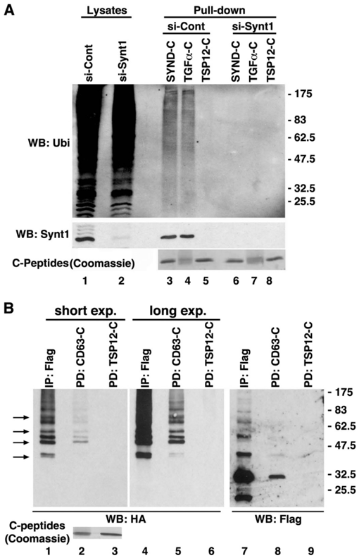
FIGURE 1 Syntenin-1 links its transmembrane partners to ubiquitylated proteins. A , lysates prepared from HeLa cells transfected with either the siRNAtargetingsyntenin-1orcontrolsiRNAwereincubatedwithbiotinylated peptides corresponding to the cytoplasmic tails of syndecan-2 ( SYND-C ), TGF 𝛂 ( TGF 𝛂 -C ), and Tspan-12-C ( TSP12-C ). Complexes were collected on NeutrAvidin-agarose and resolved on SDS-PAGE. Pulled-down ubiquitylated proteins interacting with the peptides were visualized with anti-Ub mAb (FK2). Shownareresults of the pulldown experiments. Lower panel showsefficiency of syntenin-1 knockdown. Note, knockdown of syntenin-1 markedly disrupted the interaction of the peptides with ubiquitylated proteins. WB , Western blotting. B , lysates prepared from HEK293T cells expressing FLAG-syntenin-1 and HA-Ub were incubated with either biotinylated CD63-C and Tspan12-C (control) peptides or anti-FLAG mAb. Protein complexes were collected on NeutrAvidin-agarose, resolved on SDS-PAGE, and subsequently probed with anti-HA and anti-FLAG polyclonal Abs in Western blotting. Arrows point to ubiquitylated proteins from the syntenin-1 immunoprecipitate ( IP ; lane 1 ) that are distinct from the ubiquitylated species of syntenin-1 (characteristic ladder, lane 7 ). Note that CD63-C interacts with both ubiquitylated syntenin-1 and the associated proteins. Two exposures are shown to compare the patterns of proteins precipitated with anti-FLAG mAb and CD63-C.
FIGURE 1 Syntenin-1 links its transmembrane partners to ubiquitylated proteins. A , lysates prepared from HeLa cells transfected with either the siRNAtargetingsyntenin-1orcontrolsiRNAwereincubatedwithbiotinylated peptides corresponding to the cytoplasmic tails of syndecan-2 ( SYND-C ), TGF 𝛂 ( TGF 𝛂 -C ), and Tspan-12-C ( TSP12-C ). Complexes were collected on NeutrAvidin-agarose and resolved on SDS-PAGE. Pulled-down ubiquitylated proteins interacting with the peptides were visualized with anti-Ub mAb (FK2). Shownareresults of the pulldown experiments. Lower panel showsefficiency of syntenin-1 knockdown. Note, knockdown of syntenin-1 markedly disrupted the interaction of the peptides with ubiquitylated proteins. WB , Western blotting. B , lysates prepared from HEK293T cells expressing FLAG-syntenin-1 and HA-Ub were incubated with either biotinylated CD63-C and Tspan12-C (control) peptides or anti-FLAG mAb. Protein complexes were collected on NeutrAvidin-agarose, resolved on SDS-PAGE, and subsequently probed with anti-HA and anti-FLAG polyclonal Abs in Western blotting. Arrows point to ubiquitylated proteins from the syntenin-1 immunoprecipitate ( IP ; lane 1 ) that are distinct from the ubiquitylated species of syntenin-1 (characteristic ladder, lane 7 ). Note that CD63-C interacts with both ubiquitylated syntenin-1 and the associated proteins. Two exposures are shown to compare the patterns of proteins precipitated with anti-FLAG mAb and CD63-C.CC-BY · From the PDF
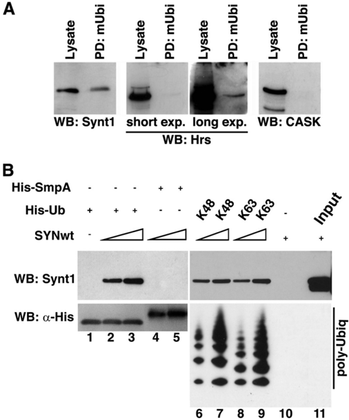
FIGURE 2 Syntenin-1 interacts with Ub. A , syntenin-1 is retained by immobilized mono-Ub from cellular lysates. Cellular lysates prepared from HeLa cells were incubated with mono-Ub covalently attached to agarose beads ( mUbi ); interacting proteins were eluted from the beads and equal volumes of the eluate (controlled by Western blotting with mAb to actin) were resolved on SDS-PAGE. Syntenin-1, Hrs, and CASK were detected by Western blotting ( WB ). B , upper , recombinant syntenin-1 (0.1 and 0.5 𝛍 g) was incubated with immobilized His-Ub or His-SmpA (negative control) and His-poly-Ub (2-7) chains (Lys 48 -linked) or His-poly-Ub (2-7) chains (Lys 63 -linked). Interactions between the proteins were visualized by Western blotting using anti-syntenin-1 mAb. Lower , loading of His-Ub species was controlled using anti-His HRP mAb antibody.
FIGURE 2 Syntenin-1 interacts with Ub. A , syntenin-1 is retained by immobilized mono-Ub from cellular lysates. Cellular lysates prepared from HeLa cells were incubated with mono-Ub covalently attached to agarose beads ( mUbi ); interacting proteins were eluted from the beads and equal volumes of the eluate (controlled by Western blotting with mAb to actin) were resolved on SDS-PAGE. Syntenin-1, Hrs, and CASK were detected by Western blotting ( WB ). B , upper , recombinant syntenin-1 (0.1 and 0.5 𝛍 g) was incubated with immobilized His-Ub or His-SmpA (negative control) and His-poly-Ub (2-7) chains (Lys 48 -linked) or His-poly-Ub (2-7) chains (Lys 63 -linked). Interactions between the proteins were visualized by Western blotting using anti-syntenin-1 mAb. Lower , loading of His-Ub species was controlled using anti-His HRP mAb antibody.CC-BY · From the PDF
Image 3
CC-BY · From the PDF
Image 4
CC-BY · From the PDF
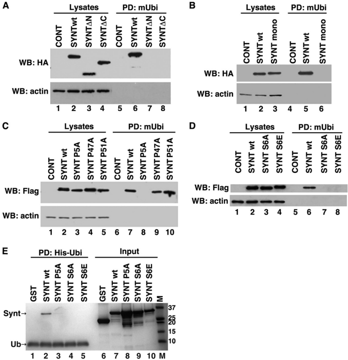
FIGURE 4 Both the N- and C-terminal regions are important for the interaction of syntenin-1 with Ub: role of dimerization. A , protein lysates prepared from HEK293T cells expressing syntenin-1WT-HA, syntenin𝚫 N-HA ( SYNT𝚫 N ), and syntenin𝚫 C-HA ( SYNT -𝚫 C ) were incubated with mUb covalently attached to agarose beads, and interacting proteins were resolved on SDS-PAGE. Syntenin-1 proteins were detected by Western blotting ( WB ) with anti-HA Ab. In the control experiments cells were transfected with pcDNA ( lanes 1 and 5 ). B , Protein lysates prepared from HEK293T cells expressing HA-tagged syntenin-1WT or dimerization mutant (F195A/L233A, referred to as SYNTmono ), were incubated with mUb covalently attached to agarose beads, and interacting proteins were resolved on SDS-PAGE. Syntenin-1 was detected by Western blotting with anti-HA Ab. C and D , protein lysates prepared from HEK293T cells expressing syntenin-1WT-FLAG, and various FLAG-tagged mutants of syntenin-1 were incubated with mUb covalently attached to agarose beads. Interacting proteins were resolved on SDS-PAGE. Syntenin-1 was detected by Western blotting with anti-FLAG Ab. In the control experiments cells were transfected with pcDNA ( lanes 1 and 5 for B and D ; lanes 1 and 4 for C ). E , recombinant wild-type syntenin-1 ( SYNTwt ) and syntenin-1 point mutants (P5A, SYNT P5A; S6A, SYNT S6A; and S6E, SYNT S6E) (0.5 𝛍 g each) were incubated with 30 𝛍 l of His-Ub-immobilized nickel-Sepharose beads in 50 mM NaPO4, pH 7.4, 500 mM NaCl, 0.5 mM tris(2-carboxyethyl)phosphine, 20 mM imidazole, 0.2% Triton X-100, 0.02% BSA. Bound proteins were eluted with 50 mM EDTA. The protein samples were resolved using SDS-PAGE and visualized with Coomassie Brilliant Blue staining. Note, additional low molecular mass protein bands correspond to proteolytic products of full-length syntenin. PD , pulldown.
FIGURE 4 Both the N- and C-terminal regions are important for the interaction of syntenin-1 with Ub: role of dimerization. A , protein lysates prepared from HEK293T cells expressing syntenin-1WT-HA, syntenin𝚫 N-HA ( SYNT𝚫 N ), and syntenin𝚫 C-HA ( SYNT -𝚫 C ) were incubated with mUb covalently attached to agarose beads, and interacting proteins were resolved on SDS-PAGE. Syntenin-1 proteins were detected by Western blotting ( WB ) with anti-HA Ab. In the control experiments cells were transfected with pcDNA ( lanes 1 and 5 ). B , Protein lysates prepared from HEK293T cells expressing HA-tagged syntenin-1WT or dimerization mutant (F195A/L233A, referred to as SYNTmono ), were incubated with mUb covalently attached to agarose beads, and interacting proteins were resolved on SDS-PAGE. Syntenin-1 was detected by Western blotting with anti-HA Ab. C and D , protein lysates prepared from HEK293T cells expressing syntenin-1WT-FLAG, and various FLAG-tagged mutants of syntenin-1 were incubated with mUb covalently attached to agarose beads. Interacting proteins were resolved on SDS-PAGE. Syntenin-1 was detected by Western blotting with anti-FLAG Ab. In the control experiments cells were transfected with pcDNA ( lanes 1 and 5 for B and D ; lanes 1 and 4 for C ). E , recombinant wild-type syntenin-1 ( SYNTwt ) and syntenin-1 point mutants (P5A, SYNT P5A; S6A, SYNT S6A; and S6E, SYNT S6E) (0.5 𝛍 g each) were incubated with 30 𝛍 l of His-Ub-immobilized nickel-Sepharose beads in 50 mM NaPO4, pH 7.4, 500 mM NaCl, 0.5 mM tris(2-carboxyethyl)phosphine, 20 mM imidazole, 0.2% Triton X-100, 0.02% BSA. Bound proteins were eluted with 50 mM EDTA. The protein samples were resolved using SDS-PAGE and visualized with Coomassie Brilliant Blue staining. Note, additional low molecular mass protein bands correspond to proteolytic products of full-length syntenin. PD , pulldown.CC-BY · From the PDF
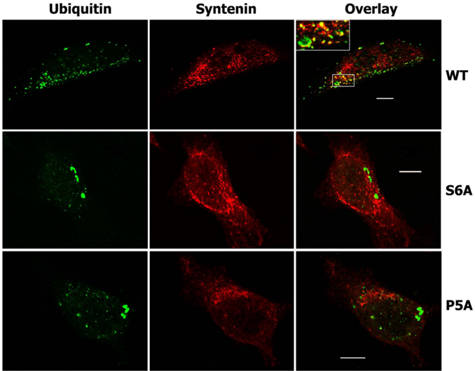
FIGURE 5 Co-localization of syntenin-1 and ubiquitylated proteins. HeLa cells expressing FLAG-tagged syntenin-1 WT or syntenin-1 S6A ( S6A ) and syntenin-1 P5A ( P5A ) mutants were fixed with 2% paraformaldehyde and permeabilized with 0.1% Triton X-100. Cells were co-stained with anti-FLAG rabbit pAb and anti-Ub mouse mAb (clone FK2). Staining was visualized with Alexa Fluor 488-conjugated goat anti-mouse antibody and labeled with Alexa Fluor 594-conjugated anti-rabbit antibody. Overlay panels and insets show partial co-localization of wild-type syntenin-1 ( red ) with ubiquitylated cellular proteins ( green ) and lack of co-localization in cells expressing mutants of syntenin-1 ( red ). Scale bars , 10 𝛍 m.
FIGURE 5 Co-localization of syntenin-1 and ubiquitylated proteins. HeLa cells expressing FLAG-tagged syntenin-1 WT or syntenin-1 S6A ( S6A ) and syntenin-1 P5A ( P5A ) mutants were fixed with 2% paraformaldehyde and permeabilized with 0.1% Triton X-100. Cells were co-stained with anti-FLAG rabbit pAb and anti-Ub mouse mAb (clone FK2). Staining was visualized with Alexa Fluor 488-conjugated goat anti-mouse antibody and labeled with Alexa Fluor 594-conjugated anti-rabbit antibody. Overlay panels and insets show partial co-localization of wild-type syntenin-1 ( red ) with ubiquitylated cellular proteins ( green ) and lack of co-localization in cells expressing mutants of syntenin-1 ( red ). Scale bars , 10 𝛍 m.CC-BY · From the PDF
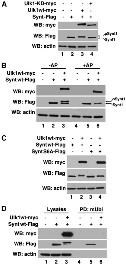
FIGURE 6 Ulk1 modifies syntenin via phosphorylation. A , protein lysates prepared from HEK293T cells expressing syntenin-1WT-FLAG, Ulk1-Myc, and the Ulk1-KD-Myc mutant were resolved by SDS-PAGE. B , samples prepared as aboveweresubjectedtotreatment with alkaline phosphatase ( AP ) for 40 min at 37 °C ( lanes 4 -6 ) and subsequently resolved on 10% SDS-PAGE. Note, alkalkine phosphatase treatment resulted in the loss of modification indicating that syntenin-1 is phosphorylated in the presence of Ulk1. C , protein lysates prepared from 293T cells expressing syntenin-1WT-FLAG, syntenin-1 S6A-FLAG mutant, and Ulk1-Myc were resolved on SDS-PAGE. D , protein lysates prepared from 293T cells expressing syntenin-1WT-FLAG and Ulk1Myc were incubated with immobilized monoubiquitin ( mUbi ), and proteins were resolved on SDS-PAGE. In the control experiments, cells were transfected with pcDNA ( lanes 1 and 4 ). Syntenin-1 and Ulk1 were detected by Western blotting ( WB ) with anti-FLAG and anti-Myc Abs, respectively ( A-D ). Actin was used as a loading control in all blots.
FIGURE 6 Ulk1 modifies syntenin via phosphorylation. A , protein lysates prepared from HEK293T cells expressing syntenin-1WT-FLAG, Ulk1-Myc, and the Ulk1-KD-Myc mutant were resolved by SDS-PAGE. B , samples prepared as aboveweresubjectedtotreatment with alkaline phosphatase ( AP ) for 40 min at 37 °C ( lanes 4 -6 ) and subsequently resolved on 10% SDS-PAGE. Note, alkalkine phosphatase treatment resulted in the loss of modification indicating that syntenin-1 is phosphorylated in the presence of Ulk1. C , protein lysates prepared from 293T cells expressing syntenin-1WT-FLAG, syntenin-1 S6A-FLAG mutant, and Ulk1-Myc were resolved on SDS-PAGE. D , protein lysates prepared from 293T cells expressing syntenin-1WT-FLAG and Ulk1Myc were incubated with immobilized monoubiquitin ( mUbi ), and proteins were resolved on SDS-PAGE. In the control experiments, cells were transfected with pcDNA ( lanes 1 and 4 ). Syntenin-1 and Ulk1 were detected by Western blotting ( WB ) with anti-FLAG and anti-Myc Abs, respectively ( A-D ). Actin was used as a loading control in all blots.CC-BY · From the PDF
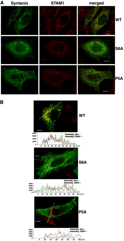
FIGURE 7 Role of Ub binding in cellular localization of syntenin-1. HeLa cells expressing FLAG-tagged syntenin-1 (WT or syntenin-1 S6A ( S6A ) and syntenin-1 P5A ( P5A ) mutants) were fixed with 2% paraformaldehyde and permeabilized with 0.1% Triton X-100. A , cells were co-stained with anti-FLAG mouse mAb (M2) and anti-STAM rabbit pAb. Staining was visualized with Alexa Fluor 488-conjugated goat anti-mouse antibody and labeled with Alexa Fluor 594-conjugated anti-rabbit antibody. Overlay panels show minimal co-localization syntenin-1 proteins ( green ) with STAM ( red ). B , cells were co-stainedwithanti-FLAGrabbitpAbandanti-CD63mousemAb(6H1).Staining was visualized with Alexa Fluor 594-conjugated goat anti-mouse antibody and labeled with Alexa Fluor 488-conjugated anti-rabbit antibody. Shownare only overlay panels . Co-localization profiles (along the lines on the images directed toward the arrows ) are presented as the fluorescence intensity distribution for each channel (shown underneath each of the panels). Note the differences in degree of syntenin-1/CD63 co-localization in cells expressing the wild-type syntenin-1 ( top ) and syntenin-1 mutants ( middle and bottom ). Scale bars , 10 𝛍 m.
FIGURE 7 Role of Ub binding in cellular localization of syntenin-1. HeLa cells expressing FLAG-tagged syntenin-1 (WT or syntenin-1 S6A ( S6A ) and syntenin-1 P5A ( P5A ) mutants) were fixed with 2% paraformaldehyde and permeabilized with 0.1% Triton X-100. A , cells were co-stained with anti-FLAG mouse mAb (M2) and anti-STAM rabbit pAb. Staining was visualized with Alexa Fluor 488-conjugated goat anti-mouse antibody and labeled with Alexa Fluor 594-conjugated anti-rabbit antibody. Overlay panels show minimal co-localization syntenin-1 proteins ( green ) with STAM ( red ). B , cells were co-stainedwithanti-FLAGrabbitpAbandanti-CD63mousemAb(6H1).Staining was visualized with Alexa Fluor 594-conjugated goat anti-mouse antibody and labeled with Alexa Fluor 488-conjugated anti-rabbit antibody. Shownare only overlay panels . Co-localization profiles (along the lines on the images directed toward the arrows ) are presented as the fluorescence intensity distribution for each channel (shown underneath each of the panels). Note the differences in degree of syntenin-1/CD63 co-localization in cells expressing the wild-type syntenin-1 ( top ) and syntenin-1 mutants ( middle and bottom ). Scale bars , 10 𝛍 m.CC-BY · From the PDF

Verification

Claims the paper makes

The claims and statistics of this paper haven’t been checked yet.

Run the check — free, no account needed. It reads the open-access text and checks the sources it cites.

A few free checks a day · papers already verified open instantly · results are public

Claims other papers make about this one

Each is checked against this paper’s own text: does it say what the citing paper says it does?

Self-check

Percentages, sums and reported differences re-computed from the paper’s own numbers.

The self-check needs the paper’s full text.

Citations

Cites

No references could be resolved yet.

Research works printed but not linked: 40

  1. Hemler, 2003, Annu. Rev. Cell Dev. Biol., 19, 397, 10.1146/annurev.cellbio.19.111301.153609 · DOI
  2. Berditchevski, 2007, Traffic, 8, 89, 10.1111/j.1600-0854.2006.00515.x · DOI
  3. Latysheva, 2006, Mol. Cell. Biol., 26, 7707, 10.1128/MCB.00849-06 · DOI
  4. Grootjans, 1997, Proc. Nat. Acad. Sci. U.S.A., 94, 13683, 10.1073/pnas.94.25.13683 · DOI
  5. Zimmermann, 2001, Mol. Biol. Cell, 12, 339, 10.1091/mbc.12.2.339 · DOI
  6. Beekman, 2008, J. Cell Sci., 121, 1349, 10.1242/jcs.026401 · DOI
  7. Boukerche, 2007, Cancer Res., 67, 1812, 10.1158/0008-5472.CAN-06-3875 · DOI
  8. Boukerche, 2008, Proc. Natl. Acad. Sci. U.S.A., 105, 15914, 10.1073/pnas.0808171105 · DOI
  9. Boukerche, 2005, Cancer Res., 65, 10901, 10.1158/0008-5472.CAN-05-1614 · DOI
  10. Hwangbo, 2011, J. Biol. Chem., 286, 33601, 10.1074/jbc.M110.206789 · DOI
  11. Tomoda, 2004, Genes Dev., 18, 541, 10.1101/gad.1151204 · DOI
  12. McClelland, 2009, Proc. Natl. Acad. Sci. U.S.A., 106, 20487, 10.1073/pnas.0811862106 · DOI
  13. Ureña, 1999, J. Cell Sci., 112, 773, 10.1242/jcs.112.6.773 · DOI
  14. Zimmermann, 2005, Dev. Cell, 9, 377, 10.1016/j.devcel.2005.07.011 · DOI
  15. Hurley, 2006, Annu. Rev. Biophys. Biomol. Struct., 35, 277, 10.1146/annurev.biophys.35.040405.102126 · DOI · Paper page
  16. Dikic, 2009, Nat. Rev. Mol. Cell Biol., 10, 659, 10.1038/nrm2767 · DOI
  17. Okumura, 2011, Mol. Cell. Biochem., 352, 163, 10.1007/s11010-011-0750-4 · DOI
  18. Sakamoto, 1999, Biochemistry, 38, 11634, 10.1021/bi990310y · DOI
  19. Berditchevski, 1999, J. Cell Biol., 146, 477, 10.1083/jcb.146.2.477 · DOI
  20. Hirano, 2006, Nat. Struct. Mol. Biol., 13, 272, 10.1038/nsmb1051 · DOI
  21. Hurley, 2006, Biochem. J., 399, 361, 10.1042/BJ20061138 · DOI
  22. Kang, 2003, Structure, 11, 459, 10.1016/S0969-2126(03)00052-2 · DOI
  23. Koroll, 2001, J. Biol. Chem., 276, 10646, 10.1074/jbc.M010647200 · DOI
  24. Ko, 2006, J. Neurosci., 26, 963, 10.1523/JNEUROSCI.4475-05.2006 · DOI
  25. Young, 2006, J. Cell Sci., 119, 3888, 10.1242/jcs.03172 · DOI · Paper page
  26. Hurley, 2010, Crit. Rev. Biochem. Mol. Biol., 45, 463, 10.3109/10409238.2010.502516 · DOI
  27. Mizuno, 2006, Traffic, 7, 1017, 10.1111/j.1600-0854.2006.00452.x · DOI
  28. Kang, 2003, Cell, 113, 621, 10.1016/S0092-8674(03)00362-3 · DOI
  29. Prag, 2003, Cell, 113, 609, 10.1016/S0092-8674(03)00364-7 · DOI
  30. Bilodeau, 2004, J. Biol. Chem., 279, 54808, 10.1074/jbc.M406654200 · DOI
  31. Komander, 2009, Nat. Rev. Mol. Cell Biol., 10, 550, 10.1038/nrm2731 · DOI · Paper page
  32. Drag, 2008, Biochem. J., 415, 367, 10.1042/BJ20080779 · DOI
  33. Reyes-Turcu, 2006, Cell, 124, 1197, 10.1016/j.cell.2006.02.038 · DOI
  34. Carvallo, 2010, J. Biol. Chem., 285, 29546, 10.1074/jbc.M110.155812 · DOI
  35. de Juan-Sanz, J., Zafra, F., Lopez-Corcuera, B., Aragon, C. (September 12, 2011) Traffic 10.1111/j.1600-0854.2011.01278.x. · DOI
  36. Martinez-Moczygemba, 2007, J. Leukoc. Biol., 81, 1137, 10.1189/jlb.0706465 · DOI
  37. Pelkmans, 2005, Nature, 436, 78, 10.1038/nature03571 · DOI
  38. Zhou, 2007, Proc. Nat. Acad. Sci. U.S.A., 104, 5842, 10.1073/pnas.0701402104 · DOI · Paper page
  39. Chan, 2007, J. Biol. Chem., 282, 25464, 10.1074/jbc.M703663200 · DOI · Paper page
  40. Ganley, 2009, J. Biol. Chem., 284, 12297, 10.1074/jbc.M900573200 · DOI · Paper page

Cited by

Sorted by citations

Metadata

Authors
Rajesh, S., Bago, R., Odintsova, E., Muratov, G., Baldwin, G., Sridhar, P., Rajesh, S., Overduin, M., & Berditchevski, F.
Venue
Journal of Biological Chemistry
Publisher
Elsevier BV
Published
2011-11
Volume
286
Issue
45
Pages
39606-39614
ISSN
0021-9258
Cited by
42
References
0
Language
en

About these checks

Registry, retraction, Cites and statistics results are computed, not judged, with one exception: a printed reference that no index lists is read once by a model before it counts as a citation error. Claims are extracted by a model and kept only when their quote appears verbatim in the text; each cited source is then checked, with a model’s help, for whether it says what the claim relies on. Those verdicts can be wrong — read the claim’s quote and the cited source before relying on one.

Statistics and arithmetic are recomputed, not judged.

Confidence bands are shown only for fields calibrated on our annotated test set. None is calibrated yet, so no band is shown.

What counts as a claim ›How we measure confidence ›How we check this ›

Are you an author, or do you see a problem with this page? Request a review