Cited by 55

The DOI matches its Crossref record; 6 of the 6 references it cites were found in a registry or an index.

p62 Plays a Protective Role in the Autophagic Degradation of Polyglutamine Protein Oligomers in Polyglutamine Disease Model Flies

Saitoh, Yuji, Fujikake, Nobuhiro, Okamoto, Yuma, Popiel, H. Akiko, Hatanaka, Yusuke, Ueyama, Morio, Suzuki, Mari, Gaumer, Sébastien, Murata, Miho, Wada, Keiji, Nagai, Yoshitaka·2015·Journal of Biological Chemistry

Overview

The claims and statistics of this paper haven’t been checked yet.

Run the check — free, no account needed. It reads the open-access text and checks the sources it cites.

A few free checks a day · papers already verified open instantly · results are public

Integrity signals

Registry

Crossref · 10.1074/jbc.m114.590281

Cites

6 of 6 references verified0 citation errors · 0 not checkable

Statistics

Figures

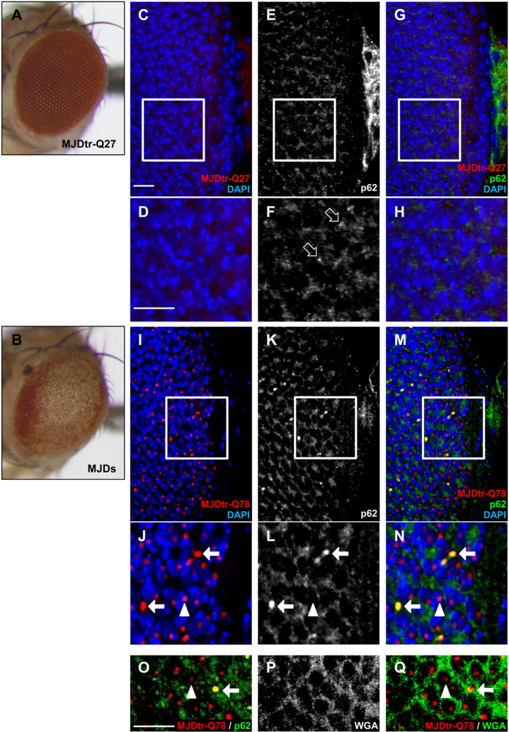
FIGURE1 p62co-localizes with cytoplasmic aggregates of the MJDtr-Q78 protein. A and B , light microscopic images of the external compound eyes of the control 1-day-old adult MJDtr-Q27 flies ( A ) and MJDtr-Q78S flies ( MJDs ) ( B ). C -N , confocal microscopic images of the larval eye discs of the control MJDtr-Q27 flies ( C -H ) and MJDtr-Q78Sflies ( I -Q ), stained with an anti-HA antibody to detect the MJDtr-Q27 or MJDtr-Q78 protein ( red ), an anti-Ref(2)P/p62 antibody ( white in E , F , K , and L and green in G , H , M , N , and O ), DAPI for nuclear staining ( blue ), and wheat germ agglutinin to define the nuclear membrane ( white in P and green in Q ). D , F , H , J , L , and N , high magnification images of the indicated areas of C , E , G , I , K , and M , respectively. The white arrows and arrowheads indicate cytoplasmic and nuclear MJDtr-Q78 protein aggregates, respectively, and the open arrows indicate punctate dotlike structures of p62. Bars , 10 𝛍 m. Fly genotypes were as follows: g mr-GAL4 / + ;; UAS-MJDtr-Q27 / + ( A and C -H ) and gmr-GAL4 / + ; UAS-MJDtr-Q78S / + ( B and I -Q ).
FIGURE1 p62co-localizes with cytoplasmic aggregates of the MJDtr-Q78 protein. A and B , light microscopic images of the external compound eyes of the control 1-day-old adult MJDtr-Q27 flies ( A ) and MJDtr-Q78S flies ( MJDs ) ( B ). C -N , confocal microscopic images of the larval eye discs of the control MJDtr-Q27 flies ( C -H ) and MJDtr-Q78Sflies ( I -Q ), stained with an anti-HA antibody to detect the MJDtr-Q27 or MJDtr-Q78 protein ( red ), an anti-Ref(2)P/p62 antibody ( white in E , F , K , and L and green in G , H , M , N , and O ), DAPI for nuclear staining ( blue ), and wheat germ agglutinin to define the nuclear membrane ( white in P and green in Q ). D , F , H , J , L , and N , high magnification images of the indicated areas of C , E , G , I , K , and M , respectively. The white arrows and arrowheads indicate cytoplasmic and nuclear MJDtr-Q78 protein aggregates, respectively, and the open arrows indicate punctate dotlike structures of p62. Bars , 10 𝛍 m. Fly genotypes were as follows: g mr-GAL4 / + ;; UAS-MJDtr-Q27 / + ( A and C -H ) and gmr-GAL4 / + ; UAS-MJDtr-Q78S / + ( B and I -Q ).CC-BY · From the PDF
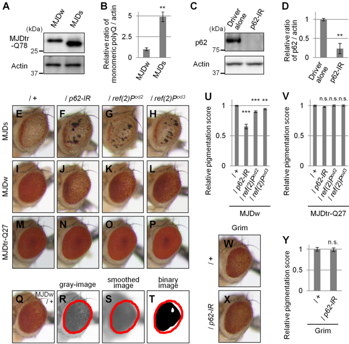
FIGURE 2 Loss of p62 function causes exacerbation of eye degeneration in MJDtr-Q78 flies. A and B , Western blot analysis of the monomeric MJDtr-Q78 protein in larval eye disc lysates of the MJDtr-Q78S flies ( MJDs ) and the MJDtr-Q78W ( MJDw ) flies expressing the MJDtr-Q78 protein under the gmr-GAL4 driver, using an anti-HA antibody to detect the MJDtr-Q78 protein. The expression level of actin was used as a protein-loading control. The graph shows the relative ratio of the MJDtr-Q78 protein to actin. The relative amount of each protein was measured by densitometric analysis of the bands in A . Data are presented as the mean ± S.E. (**, p < 0.01, versus the MJDw flies) ( n = 3). Fly genotypes were gmr-GAL4 / + ;; UAS-MJDtr-Q78W / + and gmr-GAL4 / + ; UAS-MJDtr-Q78S / + . C and D , knockdown efficiency of RNAi-mediated knockdown of ref(2)P , the Drosophila ortholog of the p62 gene. The p62 protein in lysates prepared from adult fly heads expressing an IR targeted to ref(2)P/p62 under the tub-GAL4 driver was detected with the anti-Ref(2)P/p62 antibody by Western blot analysis. The expression level of actin was used as a protein-loading control. The graph shows the relative ratio of p62 protein to actin. The relative amount of each protein was measured by densitometric analyses of the bands in C . Data are presented as the mean ± S.E. (**, p < 0.01, versus the control flies expressing the GAL4 protein alone) ( n = 3). Fly genotypes were tub-GAL4 / + and UAS-p62-IR / + ; tub-GAL4 / + . E -P , light microscopic images of the external compound eyes of 1-day-old adult flies of two different MJDtr-Q78 fly lines, MJDtr-Q78S flies ( E , MJDs ) and MJDtr-Q78W flies ( I , MJDw ), expressing p62-IR ( F and J ) or bearing p62 mutations, namely, ref(2)P od2 ( G and K ) and ref(2)P od3 ( H and L ), and the control MJDtr-Q27 flies ( M -P ). Note that the p62 mutantflies also possess a wild-type p62 allelle in trans tothemutantallele.Flygenotypeswereasfollows: gmr-GAL4 / + ; UAS-MJDtr-Q78S/ + ( E ); gmr-GAL4 / + ; UAS-MJDtr-Q78S / UAS-p62-IR ( F ); gmr-GAL4 / + ; UAS-MJDtr-Q78S / ref(2)P od2 ( G ); gmr-GAL4 / + ; UAS-MJDtr-Q78S / ref(2)P od3 ( H ); gmr-GAL4 / + ;; UAS-MJDtr-Q78W / + ( I ); gmr-GAL4 / + ; UAS-p62-IR / + ; UAS-MJDtrQ78W / + ( J ); gmr-GAL4 / + ; ref(2)P od2 / + ; UAS-MJDtr-Q78W / + ( K ); gmr-GAL4 / + ; ref(2)P od3/ + UAS-MJDtr-Q78W / + ( L ); gmr-GAL4 / + ;; UAS-MJDtr-Q27 / + ( M ); gmrGAL4 / + ; UAS-p62-IR / + ; UAS-MJDtr-Q27 / + ( N ); gmr-GAL4 / + ; ref(2)P od2 / + ; UAS-MJDtr-Q27 / + ( O ); and gmr-GAL4 / + ; ref(2)P od3 / + ; UAS-MJDtr-Q27 / + ( P ). Q -T , calculation of eye pigmentation score. Shown are light microscopic images of the external compound eye of an MJDtr-Q78W fly ( Q ; MJDw ), the grayscale images extracted from Q ( R ), the smoothed image of R ( S ), and the binary image of R ( T ). Red outlines show the region of interest defined to evaluate the eye pigmentation. Note that the images shown here are representative images to explain this procedure clearly. The fly genotype used was gmr-GAL4 / + ;; UASMJDtr-Q78W / + . U and V , quantitative imaging analyses of eye pigmentation in the 1-day-old adult MJDtr-Q78W flies ( U ) or the control 1-day-old adult MJDtr-Q27 flies ( V ) expressing p62-IR alone or bearing p62 mutations, namely ref(2)P od2 and ref(2)P od3 . More than four eye images were analyzed for each genotype. Data are presented as the mean ± S.E. (**, p < 0.01; ***, p < 0.001; n.s. , not significant, versus the MJDtr-Q78W flies in U and the MJDtr-Q27 flies in V , respectively). W and X , light microscopic images of the external compound eyes of 1-day-old adult flies expressing the Grim protein with or without p62 knockdown. Fly genotypes were as follows: gmr-GAL4 / + ; gmr-grim / + ( W ) and gmr-GAL4 / + ; gmr-grim/UAS-p62-IR ( X ). Y , quantitative imaging analyses of eye pigmentation in the 1-day-old adult grim flies with or without expressing p62-IR. More than five eye images were analyzed for each genotype. Data are presented as the mean ± S.E. ( error bars ) ( n.s. , not significant, versus the grim flies).
FIGURE 2 Loss of p62 function causes exacerbation of eye degeneration in MJDtr-Q78 flies. A and B , Western blot analysis of the monomeric MJDtr-Q78 protein in larval eye disc lysates of the MJDtr-Q78S flies ( MJDs ) and the MJDtr-Q78W ( MJDw ) flies expressing the MJDtr-Q78 protein under the gmr-GAL4 driver, using an anti-HA antibody to detect the MJDtr-Q78 protein. The expression level of actin was used as a protein-loading control. The graph shows the relative ratio of the MJDtr-Q78 protein to actin. The relative amount of each protein was measured by densitometric analysis of the bands in A . Data are presented as the mean ± S.E. (**, p < 0.01, versus the MJDw flies) ( n = 3). Fly genotypes were gmr-GAL4 / + ;; UAS-MJDtr-Q78W / + and gmr-GAL4 / + ; UAS-MJDtr-Q78S / + . C and D , knockdown efficiency of RNAi-mediated knockdown of ref(2)P , the Drosophila ortholog of the p62 gene. The p62 protein in lysates prepared from adult fly heads expressing an IR targeted to ref(2)P/p62 under the tub-GAL4 driver was detected with the anti-Ref(2)P/p62 antibody by Western blot analysis. The expression level of actin was used as a protein-loading control. The graph shows the relative ratio of p62 protein to actin. The relative amount of each protein was measured by densitometric analyses of the bands in C . Data are presented as the mean ± S.E. (**, p < 0.01, versus the control flies expressing the GAL4 protein alone) ( n = 3). Fly genotypes were tub-GAL4 / + and UAS-p62-IR / + ; tub-GAL4 / + . E -P , light microscopic images of the external compound eyes of 1-day-old adult flies of two different MJDtr-Q78 fly lines, MJDtr-Q78S flies ( E , MJDs ) and MJDtr-Q78W flies ( I , MJDw ), expressing p62-IR ( F and J ) or bearing p62 mutations, namely, ref(2)P od2 ( G and K ) and ref(2)P od3 ( H and L ), and the control MJDtr-Q27 flies ( M -P ). Note that the p62 mutantflies also possess a wild-type p62 allelle in trans tothemutantallele.Flygenotypeswereasfollows: gmr-GAL4 / + ; UAS-MJDtr-Q78S/ + ( E ); gmr-GAL4 / + ; UAS-MJDtr-Q78S / UAS-p62-IR ( F ); gmr-GAL4 / + ; UAS-MJDtr-Q78S / ref(2)P od2 ( G ); gmr-GAL4 / + ; UAS-MJDtr-Q78S / ref(2)P od3 ( H ); gmr-GAL4 / + ;; UAS-MJDtr-Q78W / + ( I ); gmr-GAL4 / + ; UAS-p62-IR / + ; UAS-MJDtrQ78W / + ( J ); gmr-GAL4 / + ; ref(2)P od2 / + ; UAS-MJDtr-Q78W / + ( K ); gmr-GAL4 / + ; ref(2)P od3/ + UAS-MJDtr-Q78W / + ( L ); gmr-GAL4 / + ;; UAS-MJDtr-Q27 / + ( M ); gmrGAL4 / + ; UAS-p62-IR / + ; UAS-MJDtr-Q27 / + ( N ); gmr-GAL4 / + ; ref(2)P od2 / + ; UAS-MJDtr-Q27 / + ( O ); and gmr-GAL4 / + ; ref(2)P od3 / + ; UAS-MJDtr-Q27 / + ( P ). Q -T , calculation of eye pigmentation score. Shown are light microscopic images of the external compound eye of an MJDtr-Q78W fly ( Q ; MJDw ), the grayscale images extracted from Q ( R ), the smoothed image of R ( S ), and the binary image of R ( T ). Red outlines show the region of interest defined to evaluate the eye pigmentation. Note that the images shown here are representative images to explain this procedure clearly. The fly genotype used was gmr-GAL4 / + ;; UASMJDtr-Q78W / + . U and V , quantitative imaging analyses of eye pigmentation in the 1-day-old adult MJDtr-Q78W flies ( U ) or the control 1-day-old adult MJDtr-Q27 flies ( V ) expressing p62-IR alone or bearing p62 mutations, namely ref(2)P od2 and ref(2)P od3 . More than four eye images were analyzed for each genotype. Data are presented as the mean ± S.E. (**, p < 0.01; ***, p < 0.001; n.s. , not significant, versus the MJDtr-Q78W flies in U and the MJDtr-Q27 flies in V , respectively). W and X , light microscopic images of the external compound eyes of 1-day-old adult flies expressing the Grim protein with or without p62 knockdown. Fly genotypes were as follows: gmr-GAL4 / + ; gmr-grim / + ( W ) and gmr-GAL4 / + ; gmr-grim/UAS-p62-IR ( X ). Y , quantitative imaging analyses of eye pigmentation in the 1-day-old adult grim flies with or without expressing p62-IR. More than five eye images were analyzed for each genotype. Data are presented as the mean ± S.E. ( error bars ) ( n.s. , not significant, versus the grim flies).CC-BY · From the PDF
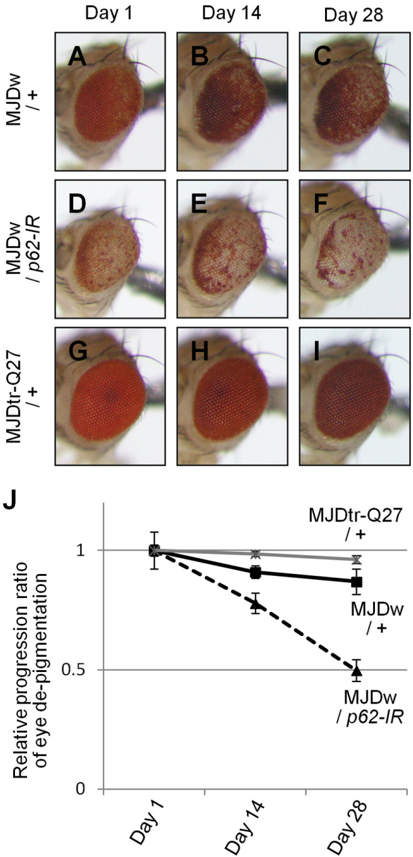
FIGURE3 Theeffectof p62 knockdownonage-relatedprogressionofeye degenerationintheMJDtr-Q78Wflies. A -F , light microscopic images of the external compound eyes of 1-, 14-, and 28-day-old adult flies expressing the MJDtr-Q78 protein alone ( A -C , MJDw/ + ) or co-expressing p62-IR ( D -F , MJDw/ p62-IR ) and the control MJDtr-Q27 flies ( G -I , MJDtr-Q27/ + ). J , relative progression ratio of eye depigmentation by performing quantitative imaging analyses of eye pigmentation in the MJDw flies with or without expressing p62-IR and the MJDtr-Q27 flies, respectively. More than five eye images were analyzed for each genotype. Data are presented as the mean ± S.E. ( error bars ). Fly genotypes were as follows: gmr-GAL4 / + ;; UAS-MJDtr-Q78W / + ( A -C ); gmr-GAL4 / + ; UAS-p62-IR / + ; UAS-MJDtr-Q78W / + ( D -F ); and gmr-GAL4 / + ;; UAS-MJDtr-Q27 / + ( G -I ).
FIGURE3 Theeffectof p62 knockdownonage-relatedprogressionofeye degenerationintheMJDtr-Q78Wflies. A -F , light microscopic images of the external compound eyes of 1-, 14-, and 28-day-old adult flies expressing the MJDtr-Q78 protein alone ( A -C , MJDw/ + ) or co-expressing p62-IR ( D -F , MJDw/ p62-IR ) and the control MJDtr-Q27 flies ( G -I , MJDtr-Q27/ + ). J , relative progression ratio of eye depigmentation by performing quantitative imaging analyses of eye pigmentation in the MJDw flies with or without expressing p62-IR and the MJDtr-Q27 flies, respectively. More than five eye images were analyzed for each genotype. Data are presented as the mean ± S.E. ( error bars ). Fly genotypes were as follows: gmr-GAL4 / + ;; UAS-MJDtr-Q78W / + ( A -C ); gmr-GAL4 / + ; UAS-p62-IR / + ; UAS-MJDtr-Q78W / + ( D -F ); and gmr-GAL4 / + ;; UAS-MJDtr-Q27 / + ( G -I ).CC-BY · From the PDF
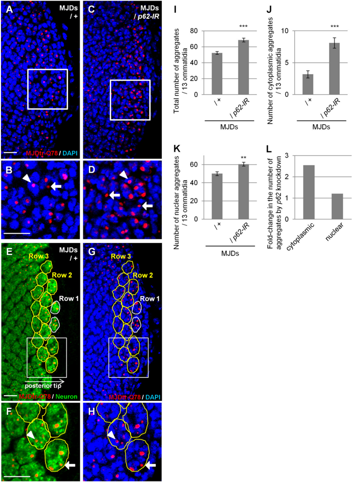
FIGURE 4 Loss of p62 function results in an increase in cytoplasmic MJDtr-Q78 protein aggregates. A -D , confocal microscopic images of the larval eye discs of flies expressing the MJDtr-Q78 protein alone ( A and B , MJDs ) or co-expressing p62-IR ( C and D ), stained with an anti-HA antibody to detect the MJDtr-Q78 protein ( red ) and DAPI for nuclear staining ( blue ). B and D , high magnification images of the indicated areas of A and C , respectively. The arrows and arrowheads indicate cytoplasmic and nuclear MJDtr-Q78 protein aggregates, respectively. Bars , 10 𝛍 m.Flygenotypeswereasfollows: gmr-GAL4 / + ; UAS-MJDtrQ78S / + ( A and B ) and gmr-GAL4 / + ; UAS-MJDtr-Q78S/UAS-p62-IR ( C and D ). E -H , quantitative analyses of MJDtr-Q78 protein aggregates in the eye discs. Confocal microscopic images of an MJDtr-Q78S fly (MJDs) larval eye disc stained with an anti-HA antibody to detect the MJDtr-Q78 protein ( red ) and with an anti-elav antibody to detect the photoreceptor neurons ( E and F , green ). Nuclei were stained with DAPI ( G and H , blue ). Thirteen ommatidia in row 2 and row 3 at the posterior tip of the eye discs were selected ( yellow outlines ). F and H , high magnification images of the indicated areas of E and G , respectively. The MJDtr-Q78proteinaggregateswerecountedaseithercytoplasmicaggregates( arrow ) or nuclear aggregates ( arrowhead ), dependingonwhethertheymerged with the nucleus or not. The fly genotype used was gmr-GAL4 / + ; UAS-MJDtr-Q78S / + . Bars , 10 𝛍 m. I -K , the number of MJDtr-Q78 protein aggregates in the eye discs of MJDtr-Q78S flies with or without expression of p62-IR. The total numbers of MJDtr-Q78 protein aggregates ( I ), cytoplasmic aggregates ( J ), and nuclear aggregates ( K ) are presented. More than five eye discs were analyzed for both genotypes. Data are presented as the mean ± S.E. ( error bars ) (**, p < 0.01; ***, p < 0.001, versus the MJDtr-Q78S flies). L , the -fold change in the number of cytoplasmic or nuclear MJDtr-Q78 protein aggregates by p62 knockdown.
FIGURE 4 Loss of p62 function results in an increase in cytoplasmic MJDtr-Q78 protein aggregates. A -D , confocal microscopic images of the larval eye discs of flies expressing the MJDtr-Q78 protein alone ( A and B , MJDs ) or co-expressing p62-IR ( C and D ), stained with an anti-HA antibody to detect the MJDtr-Q78 protein ( red ) and DAPI for nuclear staining ( blue ). B and D , high magnification images of the indicated areas of A and C , respectively. The arrows and arrowheads indicate cytoplasmic and nuclear MJDtr-Q78 protein aggregates, respectively. Bars , 10 𝛍 m.Flygenotypeswereasfollows: gmr-GAL4 / + ; UAS-MJDtrQ78S / + ( A and B ) and gmr-GAL4 / + ; UAS-MJDtr-Q78S/UAS-p62-IR ( C and D ). E -H , quantitative analyses of MJDtr-Q78 protein aggregates in the eye discs. Confocal microscopic images of an MJDtr-Q78S fly (MJDs) larval eye disc stained with an anti-HA antibody to detect the MJDtr-Q78 protein ( red ) and with an anti-elav antibody to detect the photoreceptor neurons ( E and F , green ). Nuclei were stained with DAPI ( G and H , blue ). Thirteen ommatidia in row 2 and row 3 at the posterior tip of the eye discs were selected ( yellow outlines ). F and H , high magnification images of the indicated areas of E and G , respectively. The MJDtr-Q78proteinaggregateswerecountedaseithercytoplasmicaggregates( arrow ) or nuclear aggregates ( arrowhead ), dependingonwhethertheymerged with the nucleus or not. The fly genotype used was gmr-GAL4 / + ; UAS-MJDtr-Q78S / + . Bars , 10 𝛍 m. I -K , the number of MJDtr-Q78 protein aggregates in the eye discs of MJDtr-Q78S flies with or without expression of p62-IR. The total numbers of MJDtr-Q78 protein aggregates ( I ), cytoplasmic aggregates ( J ), and nuclear aggregates ( K ) are presented. More than five eye discs were analyzed for both genotypes. Data are presented as the mean ± S.E. ( error bars ) (**, p < 0.01; ***, p < 0.001, versus the MJDtr-Q78S flies). L , the -fold change in the number of cytoplasmic or nuclear MJDtr-Q78 protein aggregates by p62 knockdown.CC-BY · From the PDF
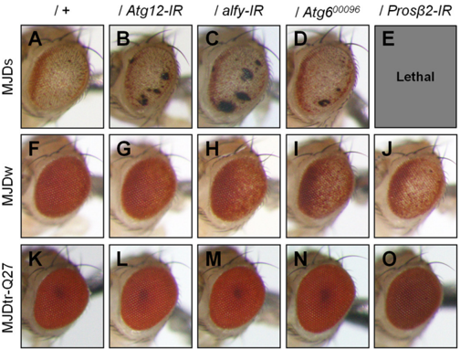
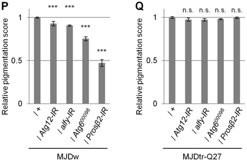
FIGURE 5 Loss of autophagic or proteasomal function causes exacerbationofeyedegenerationinMJDtr-Q78flies. A -O , light microscopic images of the external compound eyes of 1-day-old adult flies of two different MJDtrQ78 fly lines, MJDtr-Q78S flies ( A , MJDs ) and MJDtr-Q78W flies ( F , MJDw ); MJDtr-Q78 flies co-expressing Atg12-IR ( B and G ), alfy-IR ( C and H ), or the proteasome 𝛃 2 subunit (Pros 𝛃 2)-IR ( E and J ) or bearing an Atg6 mutation ( Atg6 00096 ) ( D and I ); and the control 1-day-old adult MJDtr-Q27 flies ( K -O ). Note that Pros 𝛃 2 knockdown showed a lethal phenotype in the MJDtr-Q78S flies. Fly genotypes were as follows: gmr-GAL4 / + ; UAS-MJDtr-Q78S / + ( A ); gmrGAL4 / + ; UAS-MJDtr-Q78S/UAS-Atg12-IR ( B ); gmr-GAL4 / + ; UAS-MJDtr-Q78S/ alfy-IR ( C ); gmr-GAL4 / + ; UAS-MJDtr-Q78S / + ; Atg6 00096 / + ( D ); gmr-GAL4 / + ; UAS-MJDtr-Q78S / + ; Pros 𝛃 2-IR / + ( E ); gmr-GAL4 / + ;; UAS-MJDtr-Q78W / + ( F ); gmr-GAL4 / + ; UAS-Atg12-IR / + ; UAS-MJDtr-Q78W / + ( G ); gmr-GAL4 / + ; UAS-alfyIR / + ; UAS-MJDtr-Q78W / + ( H ); gmr-GAL4 / + ;; UAS-MJDtr-Q78W/Atg6 00096 ( I ); gmr-GAL4 / + ;; UAS-MJDtr-Q78W/Pros 𝛃 2-IR ( J ); gmr-GAL4 / + ;; UAS-MJDtrQ27 / + ( K ); gmr-GAL4 / + ; UAS-Atg12-IR / + ; UAS-MJDtr-Q27 / + ( L ); gmr-GAL4 / + ; alfy-IR / + ; UAS-MJDtr-Q27 / + ( M ); gmr-GAL4 / + ;; UAS-MJDtr-Q27/Atg6 00096 ( N ); and gmr-GAL4 / + ;; UAS-MJDtr-Q27/Pros 𝛃 2-IR ( O ). P and Q , quantitative imaging analyses of eye pigmentation in the 1-day-old adult MJDtr-Q78W flies ( P ) or the control 1-day-old adult MJDtr-Q27 flies ( Q ) expressing Atg12-IR, alfy-IR, or Pros 𝛃 2-IR or bearing an Atg6 mutation. More than four eye images were analyzed for each genotype. Data are presented as the mean ± S.E. ( error bars ) (***, p < 0.001; n.s. , not significant, versus the MJDtr-Q78W flies in P and versus the MJDtr-Q27 flies in Q , respectively).
FIGURE 5 Loss of autophagic or proteasomal function causes exacerbationofeyedegenerationinMJDtr-Q78flies. A -O , light microscopic images of the external compound eyes of 1-day-old adult flies of two different MJDtrQ78 fly lines, MJDtr-Q78S flies ( A , MJDs ) and MJDtr-Q78W flies ( F , MJDw ); MJDtr-Q78 flies co-expressing Atg12-IR ( B and G ), alfy-IR ( C and H ), or the proteasome 𝛃 2 subunit (Pros 𝛃 2)-IR ( E and J ) or bearing an Atg6 mutation ( Atg6 00096 ) ( D and I ); and the control 1-day-old adult MJDtr-Q27 flies ( K -O ). Note that Pros 𝛃 2 knockdown showed a lethal phenotype in the MJDtr-Q78S flies. Fly genotypes were as follows: gmr-GAL4 / + ; UAS-MJDtr-Q78S / + ( A ); gmrGAL4 / + ; UAS-MJDtr-Q78S/UAS-Atg12-IR ( B ); gmr-GAL4 / + ; UAS-MJDtr-Q78S/ alfy-IR ( C ); gmr-GAL4 / + ; UAS-MJDtr-Q78S / + ; Atg6 00096 / + ( D ); gmr-GAL4 / + ; UAS-MJDtr-Q78S / + ; Pros 𝛃 2-IR / + ( E ); gmr-GAL4 / + ;; UAS-MJDtr-Q78W / + ( F ); gmr-GAL4 / + ; UAS-Atg12-IR / + ; UAS-MJDtr-Q78W / + ( G ); gmr-GAL4 / + ; UAS-alfyIR / + ; UAS-MJDtr-Q78W / + ( H ); gmr-GAL4 / + ;; UAS-MJDtr-Q78W/Atg6 00096 ( I ); gmr-GAL4 / + ;; UAS-MJDtr-Q78W/Pros 𝛃 2-IR ( J ); gmr-GAL4 / + ;; UAS-MJDtrQ27 / + ( K ); gmr-GAL4 / + ; UAS-Atg12-IR / + ; UAS-MJDtr-Q27 / + ( L ); gmr-GAL4 / + ; alfy-IR / + ; UAS-MJDtr-Q27 / + ( M ); gmr-GAL4 / + ;; UAS-MJDtr-Q27/Atg6 00096 ( N ); and gmr-GAL4 / + ;; UAS-MJDtr-Q27/Pros 𝛃 2-IR ( O ). P and Q , quantitative imaging analyses of eye pigmentation in the 1-day-old adult MJDtr-Q78W flies ( P ) or the control 1-day-old adult MJDtr-Q27 flies ( Q ) expressing Atg12-IR, alfy-IR, or Pros 𝛃 2-IR or bearing an Atg6 mutation. More than four eye images were analyzed for each genotype. Data are presented as the mean ± S.E. ( error bars ) (***, p < 0.001; n.s. , not significant, versus the MJDtr-Q78W flies in P and versus the MJDtr-Q27 flies in Q , respectively).CC-BY · From the PDF
Image 6
CC-BY · From the PDF
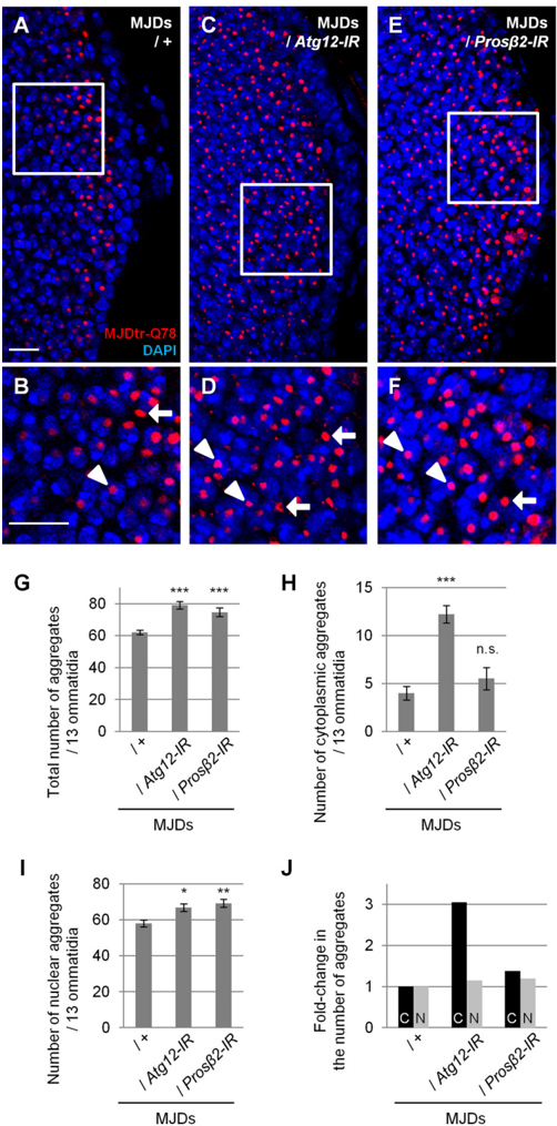
FIGURE 6 Loss of autophagic function results in an increase in cytoplasmic MJDtr-Q78 protein aggregates. A -F , confocal microscopic images of larval eye discs of flies expressing the MJDtr-Q78S protein alone ( A and B , MJDs ) or co-expressing either Atg12-IR ( C and D ) or the proteasome 𝛃 2 subunit ( Pros 𝛃 2 )-IR ( E and F ), stained with an anti-HA antibody to detect the MJDtr-Q78 protein ( red ) and DAPI for nuclear staining ( blue ). B , D , and F , high magnification images of the indicated areas of A , C , and E , respectively. The arrows and arrowheads indicate cytoplasmic and nuclear MJDtr-Q78 protein aggregates, respectively. Bars , 10 𝛍 m. Fly genotypes used were as follows: gmr-GAL4 / + ; UAS-MJDtr-Q78S / + ( A and B ); gmr-GAL4 / + ; UAS-MJDtr-Q78S/ UAS-Atg12-IR ( C and D ); and gmr-GAL4 / + ; UAS-MJDtr-Q78S / + ; Pros 𝛃 2-IR / + ( E and F ). G -I , number of MJDtr-Q78 protein aggregates in the eye discs of MJDtr-Q78S flies with or without expression of Atg12-IR or Pros 𝛃 2-IR. The total numbers of MJDtr-Q78 protein aggregates ( G ), cytoplasmic aggregates ( H ), and nuclear aggregates ( I ) are presented. More than five eye discs were analyzed for each genotype. Data are presented as the mean ± S.E. ( error bars ) (*, p < 0.05; **, < 0.01; ***, p < 0.001; n.s. , not significant, versus the MJDtr-Q78S flies). J , -fold change in the number of cytoplasmic ( C ) or nuclear ( N ) MJDtr-Q78 protein aggregates by knockdown of Atg12 or Pros 𝛃 2 .
FIGURE 6 Loss of autophagic function results in an increase in cytoplasmic MJDtr-Q78 protein aggregates. A -F , confocal microscopic images of larval eye discs of flies expressing the MJDtr-Q78S protein alone ( A and B , MJDs ) or co-expressing either Atg12-IR ( C and D ) or the proteasome 𝛃 2 subunit ( Pros 𝛃 2 )-IR ( E and F ), stained with an anti-HA antibody to detect the MJDtr-Q78 protein ( red ) and DAPI for nuclear staining ( blue ). B , D , and F , high magnification images of the indicated areas of A , C , and E , respectively. The arrows and arrowheads indicate cytoplasmic and nuclear MJDtr-Q78 protein aggregates, respectively. Bars , 10 𝛍 m. Fly genotypes used were as follows: gmr-GAL4 / + ; UAS-MJDtr-Q78S / + ( A and B ); gmr-GAL4 / + ; UAS-MJDtr-Q78S/ UAS-Atg12-IR ( C and D ); and gmr-GAL4 / + ; UAS-MJDtr-Q78S / + ; Pros 𝛃 2-IR / + ( E and F ). G -I , number of MJDtr-Q78 protein aggregates in the eye discs of MJDtr-Q78S flies with or without expression of Atg12-IR or Pros 𝛃 2-IR. The total numbers of MJDtr-Q78 protein aggregates ( G ), cytoplasmic aggregates ( H ), and nuclear aggregates ( I ) are presented. More than five eye discs were analyzed for each genotype. Data are presented as the mean ± S.E. ( error bars ) (*, p < 0.05; **, < 0.01; ***, p < 0.001; n.s. , not significant, versus the MJDtr-Q78S flies). J , -fold change in the number of cytoplasmic ( C ) or nuclear ( N ) MJDtr-Q78 protein aggregates by knockdown of Atg12 or Pros 𝛃 2 .CC-BY · From the PDF
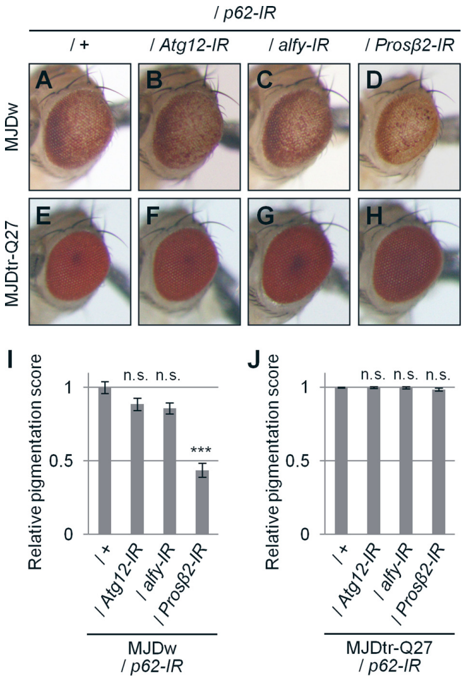
FIGURE 7 Knockdown of autophagy-related genes in addition to p62 knockdown does not cause additive exacerbation of eye degeneration in MJDtr-Q78W flies. A -H , light microscopic images of the external compoundeyesof1-day-oldadultMJDtr-Q78W( MJDw ) flies with p62 knockdown alone( A ) or together with Atg12 knockdown( B ), alfy knockdown( C ), or knockdown of Pros 𝛃 2 ( D ) and the control 1-day-old adult MJDtr-Q27 flies ( E -H ). Fly genotypes used were as follows: gmr-GAL4 / + ; UAS-p62-IR / + ; UAS-MJDtrQ78W / + ( A ); gmr-GAL4 / + ; UAS-p62-IR/UAS-Atg12-IR / + ; UAS-MJDtr-Q78W / + ( B ) gmr-GAL4 / + ; UAS-p62-IR/UAS-alfy-IR ; UAS-MJDtr-Q78W / + ( C ); gmr-GAL4 / + ; UAS-p62-IR / + ; UAS-MJDtr-Q78W/Pros 𝛃 2-IR ( D ); gmr-GAL4 / + ; UAS-p62-IR / + ; UAS-MJDtr-Q27 / + ( E ); gmr-GAL4 / + ; UAS-p62-IR/UAS-Atg12-IR ; UAS-MJDtrQ27 / + ( F ); gmr-GAL4 / + ; UAS-p62-IR/alfy-IR ; UAS-MJDtr-Q27 / + ( G ); and gmrGAL4 / + ; UAS-p62-IR / + ; UAS-MJDtr-Q27/Pros 𝛃 2-IR ( H ). I and J , quantitative imaging analyses of eye pigmentation in the 1-day-old adult MJDtr-Q78W flies ( I ) or the control 1-day-old adult MJDtr-Q27 flies ( J ) with p62 knockdown alone or together with Atg12 knockdown, alfy knockdown, or Pros 𝛃 2 knockdown. More than five eye images were analyzed for each genotype. Data are presented as the mean ± S.E. ( error bars ) (***, p < 0.001; n.s. , not significant, versus the MJDtr-Q78W flies with p62 knockdown in I and versus the MJDtrQ27 flies in J , respectively).
FIGURE 7 Knockdown of autophagy-related genes in addition to p62 knockdown does not cause additive exacerbation of eye degeneration in MJDtr-Q78W flies. A -H , light microscopic images of the external compoundeyesof1-day-oldadultMJDtr-Q78W( MJDw ) flies with p62 knockdown alone( A ) or together with Atg12 knockdown( B ), alfy knockdown( C ), or knockdown of Pros 𝛃 2 ( D ) and the control 1-day-old adult MJDtr-Q27 flies ( E -H ). Fly genotypes used were as follows: gmr-GAL4 / + ; UAS-p62-IR / + ; UAS-MJDtrQ78W / + ( A ); gmr-GAL4 / + ; UAS-p62-IR/UAS-Atg12-IR / + ; UAS-MJDtr-Q78W / + ( B ) gmr-GAL4 / + ; UAS-p62-IR/UAS-alfy-IR ; UAS-MJDtr-Q78W / + ( C ); gmr-GAL4 / + ; UAS-p62-IR / + ; UAS-MJDtr-Q78W/Pros 𝛃 2-IR ( D ); gmr-GAL4 / + ; UAS-p62-IR / + ; UAS-MJDtr-Q27 / + ( E ); gmr-GAL4 / + ; UAS-p62-IR/UAS-Atg12-IR ; UAS-MJDtrQ27 / + ( F ); gmr-GAL4 / + ; UAS-p62-IR/alfy-IR ; UAS-MJDtr-Q27 / + ( G ); and gmrGAL4 / + ; UAS-p62-IR / + ; UAS-MJDtr-Q27/Pros 𝛃 2-IR ( H ). I and J , quantitative imaging analyses of eye pigmentation in the 1-day-old adult MJDtr-Q78W flies ( I ) or the control 1-day-old adult MJDtr-Q27 flies ( J ) with p62 knockdown alone or together with Atg12 knockdown, alfy knockdown, or Pros 𝛃 2 knockdown. More than five eye images were analyzed for each genotype. Data are presented as the mean ± S.E. ( error bars ) (***, p < 0.001; n.s. , not significant, versus the MJDtr-Q78W flies with p62 knockdown in I and versus the MJDtrQ27 flies in J , respectively).CC-BY · From the PDF
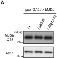
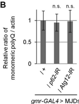
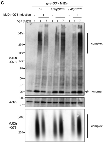
FIGURE 8 Loss of p62 function delays the degradation of the MJDtr-Q78 protein in vivo . A and B , Western blot analysis of the monomeric MJDtr-Q78 protein in larval eye disc lysates of the MJDtr-Q78S flies ( MJDs ) expressing the MJDtr-Q78proteinalone( + ) or co-expressing either p62-IR or Atg12-IR under the gmr-GAL4 driver, using an anti-HA antibody to detect the MJDtr-Q78 protein. The expression level of actin was used as a protein-loading control. The graph shows the ratio of the MJDtr-Q78 protein to actin. The relative amount of each protein was measured by densitometric analysis of the bands in A . Data are presented as the mean ± S.E. ( error bars ) ( n.s. , not significant, versus the MJDtr-Q78S flies) ( n = 3-4). Fly genotypes used were gmr-GAL4 / + ; UASMJDtr-Q78S / + , gmr-GAL4 / + ; UAS-MJDtr-Q78S/UAS-p62-IR , and gmr-GAL4 / + ; UAS-MJDtr-Q78S/UAS-Atg12-IR . C , Western blot analysis ( top and middle panels ) and SDS-AGE ( bottompanel ) of the MJDtr-Q78 protein in lysates prepared from 1-day-old and 7-day-old adult fly heads, expressing the MJDtr-Q78 protein alone ( + ) or bearing either the p62 mutation( ref(2)P od3 ) or Atg6 mutation ( Atg6 00096 ), under the gmr -GeneSwitch ( gmr-GS ) driver. MJDtr-Q78 protein expression was induced only during the larval stage by RU486 treatment (10 𝛍 g/ml) for the evaluation of MJDtr-Q78 protein turnover. In Western blot analysis, the monomeric MJDtr-Q78 protein ( monomer ) and its high molecular weight complexes ( complex ) were detected with an anti-HA antibody. The expression level of actin was used as a protein-loading control. The groups without MJDtr-Q78 protein induction ( -) were evaluated for leak MJDtr-Q78 protein expression. In SDS-AGE, the high molecular weight complexes of the MJDtr-Q78 protein were detected with an anti-HA antibody. Fly genotypes used were gmr-GS / + ; UAS-MJDtr-Q78S / + , gmr-GS / + ; UAS-MJDtr-Q78S/ ref(2)P od3 , and gmr-GS / + ; UAS-MJDtr-Q78S / + ; Atg6 00096 / + .
FIGURE 8 Loss of p62 function delays the degradation of the MJDtr-Q78 protein in vivo . A and B , Western blot analysis of the monomeric MJDtr-Q78 protein in larval eye disc lysates of the MJDtr-Q78S flies ( MJDs ) expressing the MJDtr-Q78proteinalone( + ) or co-expressing either p62-IR or Atg12-IR under the gmr-GAL4 driver, using an anti-HA antibody to detect the MJDtr-Q78 protein. The expression level of actin was used as a protein-loading control. The graph shows the ratio of the MJDtr-Q78 protein to actin. The relative amount of each protein was measured by densitometric analysis of the bands in A . Data are presented as the mean ± S.E. ( error bars ) ( n.s. , not significant, versus the MJDtr-Q78S flies) ( n = 3-4). Fly genotypes used were gmr-GAL4 / + ; UASMJDtr-Q78S / + , gmr-GAL4 / + ; UAS-MJDtr-Q78S/UAS-p62-IR , and gmr-GAL4 / + ; UAS-MJDtr-Q78S/UAS-Atg12-IR . C , Western blot analysis ( top and middle panels ) and SDS-AGE ( bottompanel ) of the MJDtr-Q78 protein in lysates prepared from 1-day-old and 7-day-old adult fly heads, expressing the MJDtr-Q78 protein alone ( + ) or bearing either the p62 mutation( ref(2)P od3 ) or Atg6 mutation ( Atg6 00096 ), under the gmr -GeneSwitch ( gmr-GS ) driver. MJDtr-Q78 protein expression was induced only during the larval stage by RU486 treatment (10 𝛍 g/ml) for the evaluation of MJDtr-Q78 protein turnover. In Western blot analysis, the monomeric MJDtr-Q78 protein ( monomer ) and its high molecular weight complexes ( complex ) were detected with an anti-HA antibody. The expression level of actin was used as a protein-loading control. The groups without MJDtr-Q78 protein induction ( -) were evaluated for leak MJDtr-Q78 protein expression. In SDS-AGE, the high molecular weight complexes of the MJDtr-Q78 protein were detected with an anti-HA antibody. Fly genotypes used were gmr-GS / + ; UAS-MJDtr-Q78S / + , gmr-GS / + ; UAS-MJDtr-Q78S/ ref(2)P od3 , and gmr-GS / + ; UAS-MJDtr-Q78S / + ; Atg6 00096 / + .CC-BY · From the PDF
Image 10
CC-BY · From the PDF
Image 11
CC-BY · From the PDF
Image 12
CC-BY · From the PDF
Image 13
CC-BY · From the PDF
Image 14
CC-BY · From the PDF
Image 15
CC-BY · From the PDF
Image 16
CC-BY · From the PDF
Image 17
CC-BY · From the PDF
Image 18
CC-BY · From the PDF
Image 19
CC-BY · From the PDF
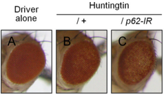
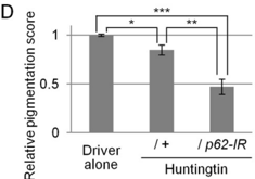
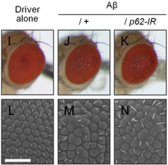
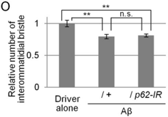
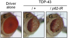
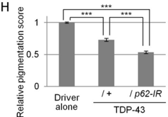
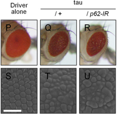
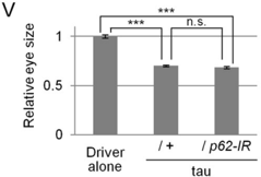
FIGURE 9 Effects of loss of p62 function in various neurodegenerative disease model flies. A -C , light microscopic images of the external compoundeyesof1-day-old adult flies expressing the mutant huntingtin protein with an expanded Gln-97 repeat with or without p62 knockdown ( B and C ) and the control flies expressing the GAL4 protein alone ( A ). Fly genotypes used were as follows: gmr-GAL4 / + ( A ); gmr-GAL4 / + ;; UAS-Httex1p97QP / + ( B ); gmr-GAL4 / + ; UAS-p62-IR / + ; UAS-Httex1p97QP / + ( C ). D , quantitative imaging analyses of eye pigmentation in A -C . More than five eye images were analyzed for each genotype. Data are presented as the mean ± S.E. ( error bars ) (*, p < 0.05; **, p < 0.01; ***, p < 0.001, versus the control flies expressing the GAL4proteinalone). E -G , light microscopic images of the external compound eyes of 7-day-old adult flies expressing the human TDP-43 protein with or without p62 knockdown ( F and G ), and the control flies expressing the GAL4 protein alone ( E ). Fly genotypes used were as follows: gmr-GAL4 / + ( E ); gmrGAL4 / + ; UAS -human TDP-43/ + ( F ); and gmr-GAL4 / + ; UAS-human TDP-43/ UAS-p62-IR ( G ). H , quantitative imaging analyses of eye pigmentation in E -G . More than five eye images were analyzed for each genotype. Data are presented as the mean ± S.E. (***, p < 0.001, versus the control flies expressing the GAL4 protein alone). I -N , light microscopic images ( I -K ) and SEM images ( L -N ) of the external compound eyes of 1-day-old adult flies expressing the mutant A 𝛃 protein with or without p62 knockdown ( J , K , M , and N ) and the control flies expressing the GAL4 protein alone ( I and L ). Bars , 50 𝛍 m. Fly genotypes used were as follows: gmr-GAL4 / + ( I and L ); gmr-GAL4 / + ;; UAS-A 𝛃 arc2 ( J and M ); and gmr-GAL4 / + ; UAS-p62-IR / + ; UAS-A 𝛃 arc2 / + ( K and N ). O , quantitative imaging analyses of the number of the interommatidial bristles in L -N . More than seven eye images were analyzed for each genotype. Data are presented as the mean ± S.E. (**, p < 0.01; n.s., not significant, versus the control flies expressing the GAL4 protein alone). P -U , light microscopic images ( P -R ) and SEM images ( S -U ) of the external compound eyes of 1-dayold adult flies expressing the mutant Tau protein with or without p62 knockdown ( Q , R , T , and U ) and the control flies expressing the GAL4 protein alone ( P and S ). Bars , 50 𝛍 m.Flygenotypesusedwereasfollows: gmr-GAL4 / + ( P and S ); gmr-GAL4 / + ;; UAS-R406Wtau / + ( Q and T ); and gmr-GAL4 / + ; UAS-p62-IR / + ; UAS-R406Wtau / + ( R and U ). V , quantitative imaging analyses of the eye size in
FIGURE 9 Effects of loss of p62 function in various neurodegenerative disease model flies. A -C , light microscopic images of the external compoundeyesof1-day-old adult flies expressing the mutant huntingtin protein with an expanded Gln-97 repeat with or without p62 knockdown ( B and C ) and the control flies expressing the GAL4 protein alone ( A ). Fly genotypes used were as follows: gmr-GAL4 / + ( A ); gmr-GAL4 / + ;; UAS-Httex1p97QP / + ( B ); gmr-GAL4 / + ; UAS-p62-IR / + ; UAS-Httex1p97QP / + ( C ). D , quantitative imaging analyses of eye pigmentation in A -C . More than five eye images were analyzed for each genotype. Data are presented as the mean ± S.E. ( error bars ) (*, p < 0.05; **, p < 0.01; ***, p < 0.001, versus the control flies expressing the GAL4proteinalone). E -G , light microscopic images of the external compound eyes of 7-day-old adult flies expressing the human TDP-43 protein with or without p62 knockdown ( F and G ), and the control flies expressing the GAL4 protein alone ( E ). Fly genotypes used were as follows: gmr-GAL4 / + ( E ); gmrGAL4 / + ; UAS -human TDP-43/ + ( F ); and gmr-GAL4 / + ; UAS-human TDP-43/ UAS-p62-IR ( G ). H , quantitative imaging analyses of eye pigmentation in E -G . More than five eye images were analyzed for each genotype. Data are presented as the mean ± S.E. (***, p < 0.001, versus the control flies expressing the GAL4 protein alone). I -N , light microscopic images ( I -K ) and SEM images ( L -N ) of the external compound eyes of 1-day-old adult flies expressing the mutant A 𝛃 protein with or without p62 knockdown ( J , K , M , and N ) and the control flies expressing the GAL4 protein alone ( I and L ). Bars , 50 𝛍 m. Fly genotypes used were as follows: gmr-GAL4 / + ( I and L ); gmr-GAL4 / + ;; UAS-A 𝛃 arc2 ( J and M ); and gmr-GAL4 / + ; UAS-p62-IR / + ; UAS-A 𝛃 arc2 / + ( K and N ). O , quantitative imaging analyses of the number of the interommatidial bristles in L -N . More than seven eye images were analyzed for each genotype. Data are presented as the mean ± S.E. (**, p < 0.01; n.s., not significant, versus the control flies expressing the GAL4 protein alone). P -U , light microscopic images ( P -R ) and SEM images ( S -U ) of the external compound eyes of 1-dayold adult flies expressing the mutant Tau protein with or without p62 knockdown ( Q , R , T , and U ) and the control flies expressing the GAL4 protein alone ( P and S ). Bars , 50 𝛍 m.Flygenotypesusedwereasfollows: gmr-GAL4 / + ( P and S ); gmr-GAL4 / + ;; UAS-R406Wtau / + ( Q and T ); and gmr-GAL4 / + ; UAS-p62-IR / + ; UAS-R406Wtau / + ( R and U ). V , quantitative imaging analyses of the eye size inCC-BY · From the PDF

Verification

Claims the paper makes

The claims and statistics of this paper haven’t been checked yet.

Run the check — free, no account needed. It reads the open-access text and checks the sources it cites.

A few free checks a day · papers already verified open instantly · results are public

Claims other papers make about this one

Each is checked against this paper’s own text: does it say what the citing paper says it does?

Self-check

Percentages, sums and reported differences re-computed from the paper’s own numbers.

The self-check needs the paper’s full text.

Citations

Cites

Research works printed but not linked: 39

  1. Gatchel, 2005, Diseases of unstable repeat expansion: mechanisms and common principles, Nat. Rev. Genet, 6, 743, 10.1038/nrg1691 · DOI
  2. Davies, 1997, Formation of neuronal intranuclear inclusions underlies the neurological dysfunction in mice transgenic for the HD mutation, Cell, 90, 537, 10.1016/S0092-8674(00)80513-9 · DOI
  3. Nagai, 2007, A toxic monomeric conformer of the polyglutamine protein, Nat. Struct. Mol. Biol, 14, 332, 10.1038/nsmb1215 · DOI
  4. Takahashi, 2008, Soluble polyglutamine oligomers formed prior to inclusion body formation are cytotoxic, Hum. Mol. Genet, 17, 345, 10.1093/hmg/ddm311 · DOI
  5. Verhoef, 2002, Aggregate formation inhibits proteasomal degradation of polyglutamine proteins, Hum. Mol. Genet, 11, 2689, 10.1093/hmg/11.22.2689 · DOI
  6. Holmberg, 2004, Inefficient degradation of truncated polyglutamine proteins by the proteasome, EMBO J, 23, 4307, 10.1038/sj.emboj.7600426 · DOI
  7. Venkatraman, 2004, Eukaryotic proteasomes cannot digest polyglutamine sequences and release them during degradation of polyglutamine-containing proteins, Mol. Cell, 14, 95, 10.1016/S1097-2765(04)00151-0 · DOI
  8. Ravikumar, 2002, Aggregate-prone proteins with polyglutamine and polyalanine expansions are degraded by autophagy, Hum. Mol. Genet, 11, 1107, 10.1093/hmg/11.9.1107 · DOI
  9. Johansen, 2011, Selective autophagy mediated by autophagic adapter proteins, Autophagy, 7, 279, 10.4161/auto.7.3.14487 · DOI
  10. Pankiv, 2007, p62/SQSTM1 binds directly to Atg8/LC3 to facilitate degradation of ubiquitinated protein aggregates by autophagy, J. Biol. Chem, 282, 24131, 10.1074/jbc.M702824200 · DOI · Paper page
  11. Kuusisto, 2001, Ubiquitin-binding protein p62 is present in neuronal and glial inclusions in human tauopathies and synucleinopathies, Neuroreport, 12, 2085, 10.1097/00001756-200107200-00009 · DOI
  12. Kuusisto, 2008, Use of p62/SQSTM1 antibodies for neuropathological diagnosis, Neuropathol. Appl. Neurobiol, 34, 169, 10.1111/j.1365-2990.2007.00884.x · DOI
  13. Yamaguchi, 1999, Ectopic expression of human p53 inhibits entry into S phase and induces apoptosis in the Drosophila eye imaginal disc, Oncogene, 18, 6767, 10.1038/sj.onc.1203113 · DOI
  14. Warrick, 1998, Expanded polyglutamine protein forms nuclear inclusions and causes neural degeneration in Drosophila, Cell, 93, 939, 10.1016/S0092-8674(00)81200-3 · DOI
  15. Steffan, 2004, SUMO modification of Huntingtin and Huntington's disease pathology, Science, 304, 100, 10.1126/science.1092194 · DOI
  16. Crowther, 2005, Intraneuronal Aβ, non-amyloid aggregates and neurodegeneration in a Drosophila model of Alzheimer's disease, Neuroscience, 132, 123, 10.1016/j.neuroscience.2004.12.025 · DOI
  17. Wittmann, 2001, Tauopathy in Drosophila: neurodegeneration without neurofibrillary tangles, Science, 293, 711, 10.1126/science.1062382 · DOI · Paper page
  18. Wyers, 1995, Localization of domains within the Drosophila Ref(2)P protein involved in the intracellular control of σ rhabdovirus multiplication, J. Virol, 69, 4463, 10.1128/jvi.69.7.4463-4470.1995 · DOI
  19. Ueyama, 2008, Functional analysis of proteoglycan galactosyltransferase II RNA interference mutant flies, J. Biol. Chem, 283, 6076, 10.1074/jbc.M709189200 · DOI
  20. Menzies, 2014, Calpain inhibition mediates autophagy-dependent protection against polyglutamine toxicity, Cell Death Differ
  21. Weiss, 2008, Sensitive biochemical aggregate detection reveals aggregation onset before symptom development in cellular and murine models of Huntington's disease, J. Neurochem, 104, 846, 10.1111/j.1471-4159.2007.05032.x · DOI
  22. Legleiter, 2010, Mutant huntingtin fragments form oligomers in a polyglutamine length-dependent manner in vitro in vivo, J. Biol. Chem, 285, 14777, 10.1074/jbc.M109.093708 · DOI
  23. Sontag, 2012, Detection of mutant huntingtin aggregation conformers and modulation of SDS-soluble fibrillar oligomers by small molecules, J. Huntingtons Dis, 1, 127
  24. Bjørkøy, 2005, p62/SQSTM1 forms protein aggregates degraded by autophagy and has a protective effect on huntingtin-induced cell death, J. Cell Biol, 171, 603, 10.1083/jcb.200507002 · DOI · Paper page
  25. Filimonenko, 2010, The selective macroautophagic degradation of aggregated proteins requires the PI3P-binding protein Alfy, Mol. Cell, 38, 265, 10.1016/j.molcel.2010.04.007 · DOI
  26. Roman, 2002, Conditional expression of UAS-transgenes in the adult eye with a new gene-switch vector system, Genesis, 34, 127, 10.1002/gene.10133 · DOI
  27. Korolchuk, 2009, Autophagy inhibition compromises degradation of ubiquitin-proteasome pathway substrates, Mol. Cell, 33, 517, 10.1016/j.molcel.2009.01.021 · DOI · Paper page
  28. Nagaoka, 2004, Increased expression of p62 in expanded polyglutamine-expressing cells and its association with polyglutamine inclusions, J. Neurochem, 91, 57, 10.1111/j.1471-4159.2004.02692.x · DOI
  29. Matsumoto, 2011, Serine 403 phosphorylation of p62/SQSTM1 regulates selective autophagic clearance of ubiquitinated proteins, Mol. Cell, 44, 279, 10.1016/j.molcel.2011.07.039 · DOI
  30. Doi, 2013, p62/SQSTM1 differentially removes the toxic mutant androgen receptor via autophagy and inclusion formation in a spinal and bulbar muscular atrophy mouse model, J. Neurosci, 33, 7710, 10.1523/JNEUROSCI.3021-12.2013 · DOI
  31. Schaffar, 2004, Cellular toxicity of polyglutamine expansion proteins: mechanism of transcription factor deactivation, Mol. Cell, 15, 95, 10.1016/j.molcel.2004.06.029 · DOI
  32. Kayed, 2003, Common structure of soluble amyloid oligomers implies common mechanism of pathogenesis, Science, 300, 486, 10.1126/science.1079469 · DOI · Paper page
  33. Martin, 2014, Autophagy in Huntington disease and huntingtin in autophagy, Trends Neurosci
  34. Brady, 2011, Regulation of TDP-43 aggregation by phosphorylation and p62/SQSTM1, J. Neurochem, 116, 248, 10.1111/j.1471-4159.2010.07098.x · DOI · Paper page
  35. Scotter, 2014, Differential roles of the ubiquitin proteasome system and autophagy in the clearance of soluble and aggregated TDP-43 species, J. Cell Sci, 127, 1263
  36. Shimura, 2004, CHIP-Hsc70 complex ubiquitinates phosphorylated Tau and enhances cell survival, J. Biol. Chem, 279, 4869, 10.1074/jbc.M305838200 · DOI
  37. Lee, 2013, Tau degradation: the ubiquitin-proteasome system versus the autophagy-lysosome system, Prog. Neurobiol, 105, 49, 10.1016/j.pneurobio.2013.03.001 · DOI · Paper page
  38. Iwata, 2000, Identification of the major Aβ1–42-degrading catabolic pathway in brain parenchyma: suppression leads to biochemical and pathological deposition, Nat. Med, 6, 143, 10.1038/72237 · DOI · Paper page
  39. Qiu, 1998, Insulin-degrading enzyme regulates extracellular levels of amyloid β-protein by degradation, J. Biol. Chem, 273, 32730, 10.1074/jbc.273.49.32730 · DOI · Paper page

Cited by

Sorted by citations

See all 55

Metadata

Authors
Saitoh, Y., Fujikake, N., Okamoto, Y., Popiel, H. A., Hatanaka, Y., Ueyama, M., Suzuki, M., Gaumer, S., Murata, M., Wada, K., & Nagai, Y.
Venue
Journal of Biological Chemistry
Publisher
Elsevier BV
Published
2015-01
Volume
290
Issue
3
Pages
1442-1453
ISSN
0021-9258
Cited by
55
References
6
Language
en

About these checks

Registry, retraction, Cites and statistics results are computed, not judged, with one exception: a printed reference that no index lists is read once by a model before it counts as a citation error. Claims are extracted by a model and kept only when their quote appears verbatim in the text; each cited source is then checked, with a model’s help, for whether it says what the claim relies on. Those verdicts can be wrong — read the claim’s quote and the cited source before relying on one.

Statistics and arithmetic are recomputed, not judged.

Confidence bands are shown only for fields calibrated on our annotated test set. None is calibrated yet, so no band is shown.

What counts as a claim ›How we measure confidence ›How we check this ›

Are you an author, or do you see a problem with this page? Request a review