

The DOI matches its Crossref record; 8 of the 8 references it cites were found in a registry or an index.
Endoplasmic reticulum stress responses in mouse models of Alzheimer's disease: Overexpression paradigm versus knockin paradigm
Abstract
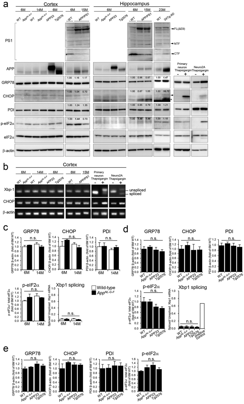
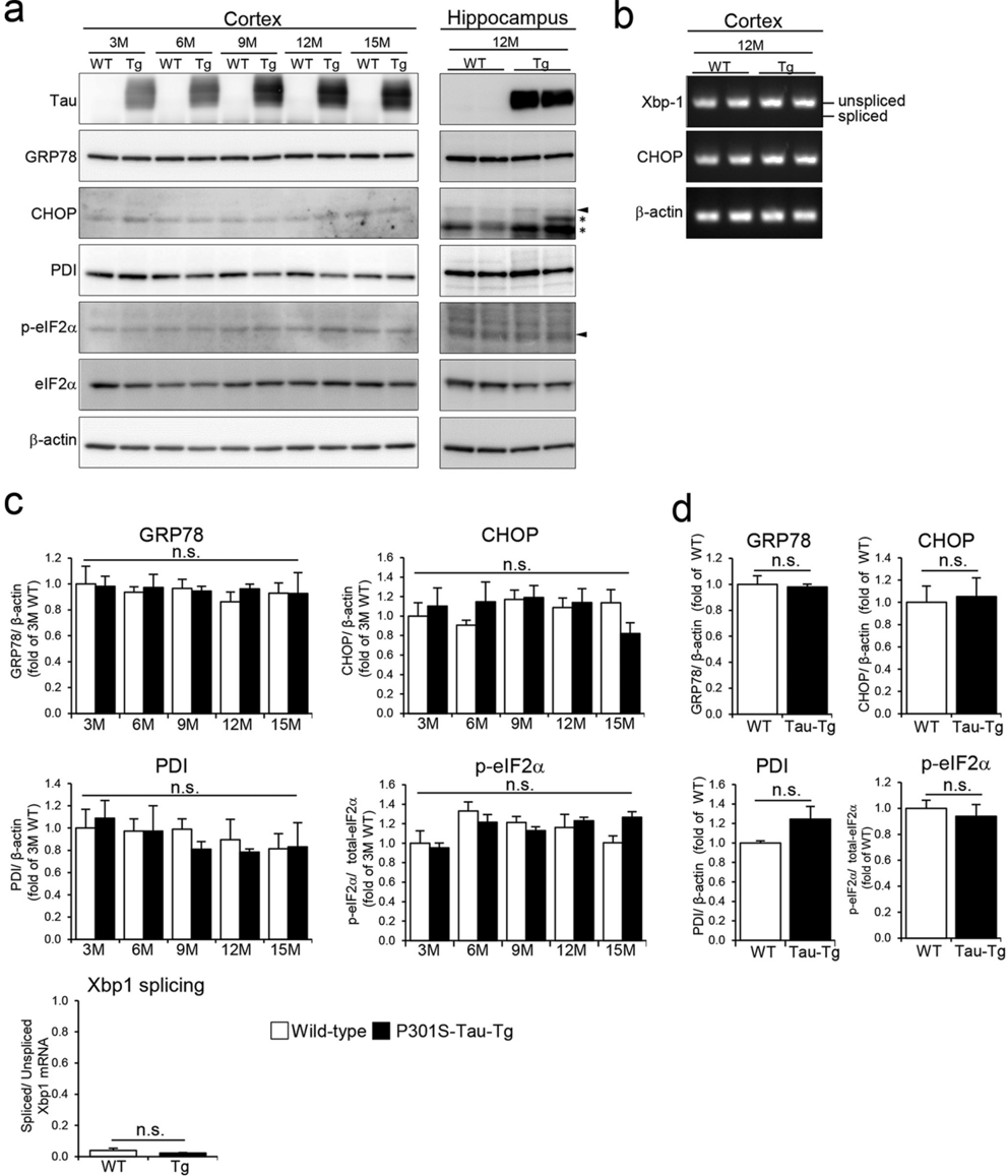
Endoplasmic reticulum (ER) stress is believed to play an important role in the etiology of Alzheimer's disease (AD). The accumulation of misfolded proteins and perturbation of intracellular calcium homeostasis are thought to underlie the induction of ER stress, resulting in neuronal dysfunction and cell death. Several reports have shown an increased ER stress response in amyloid precursor protein (APP) and presenilin1 (PS1) double-transgenic (Tg) AD mouse models. However, whether the ER stress observed in these mouse models is actually caused by AD pathology remains unclear. APP and PS1 contain one and nine transmembrane domains, respectively, for which it has been postulated that overexpressed membrane proteins can become wedged in a misfolded configuration in ER membranes, thereby inducing nonspecific ER stress. Here, we used an App-knockin (KI) AD mouse model that accumulates amyloid-β (Aβ) peptide without overexpressing APP to investigate whether the ER stress response is heightened because of Aβ pathology. Thorough examinations indicated that no ER stress responses arose in App-KI or single APP-Tg mice. These results suggest that PS1 overexpression or mutation induced a nonspecific ER stress response that was independent of Aβ pathology in the double-Tg mice. Moreover, we observed no ER stress in a mouse model of tauopathy (P301S-Tau-Tg mice) at various ages, suggesting that ER stress is also not essential in tau pathology–induced neurodegeneration. We conclude that the…
Overview
The claims and statistics of this paper haven’t been checked yet.
Run the check — free, no account needed. It reads the open-access text and checks the sources it cites.
Integrity signals
Registry
Cites
Statistics
Figures
Verification
Claims the paper makes
The claims and statistics of this paper haven’t been checked yet.
Run the check — free, no account needed. It reads the open-access text and checks the sources it cites.
Claims other papers make about this one
Each is checked against this paper’s own text: does it say what the citing paper says it does?
In the citing paper
“Utilizing KI mice rather than transgenic overexpression mice is beneficial for modeling diseased conditions while avoiding overexpression artifacts such as the destruction of endogenous loci, disturbances in cellular protein localization/interactions, and non-specific endoplasmic reticulum stress/calcium mobilization.6,7,8,9,10,11,12”
From Human MAPT knockin mouse models of frontotemporal dementia for the neurodegenerative research community (2025) · cites this paper as [8]
Self-check
Percentages, sums and reported differences re-computed from the paper’s own numbers.
The self-check needs the paper’s full text.
Citations
Cites
Synapse Loss and Microglial Activation Precede Tangles in a P301S Tauopathy Mouse Model
Yoshiyama, Y., Higuchi, M., Zhang, B., Huang, S.-M., Iwata, N., Saido, T. C., Maeda, J., Suhara, T., Trojanowski, J. Q., & Lee, V. M.-Y. (2007) · Neuron · doi:10.1016/j.neuron.2007.01.010 · Cited by 2,208
Self-citationTau-mediated neurodegeneration in Alzheimer’s disease and related disorders
Ballatore, C., Lee, V. M.-Y., & Trojanowski, J. Q. (2007) · Nature Reviews Neuroscience · doi:10.1038/nrn2194 · Cited by 2,174
Jankowsky, J. L., Fadale, D. J., Anderson, J., Xu, G. M., Gonzales, V., Jenkins, N. A., Copeland, N. G., Lee, M. K., Younkin, L. H., Wagner, S. L., et al. (2004) · Human Molecular Genetics · doi:10.1093/hmg/ddh019 · Cited by 1,575
Two amyloid precursor protein transgenic mouse models with Alzheimer disease-like pathology
Sturchler-Pierrat, C., Abramowski, D., Duke, M., Wiederhold, K.-H., Mistl, C., Rothacher, S., Ledermann, B., Bürki, K., Frey, P., Paganetti, P. A., et al. (1997) · Proceedings of the National Academy of Sciences · doi:10.1073/pnas.94.24.13287 · Cited by 1,507
Single App knock-in mouse models of Alzheimer’s disease
Saito, T., Matsuba, Y., Mihira, N., Takano, J., Nilsson, P., Itohara, S., Iwata, N., & Saido, T. C. (2014) · Nature Neuroscience · doi:10.1038/nn.3697 · Cited by 1,366
Self-citationA Molecular Web: Endoplasmic Reticulum Stress, Inflammation, and Oxidative Stress
Chaudhari, N., Talwar, P., Parimisetty, A., Lefebvre d’Hellencourt, C., & Ravanan, P. (2014) · Frontiers in Cellular Neuroscience · doi:10.3389/fncel.2014.00213 · Cited by 600
Chang, R. C. C., Wong, A. K. Y., Ng, H.-K., & Hugon, J. (2002) · NeuroReport · doi:10.1097/00001756-200212200-00011 · Cited by 289
Calpain Activation in Alzheimer's Model Mice Is an Artifact of APP and Presenilin Overexpression
Saito, T., Matsuba, Y., Yamazaki, N., Hashimoto, S., & Saido, T. C. (2016) · The Journal of Neuroscience · doi:10.1523/jneurosci.1907-16.2016 · Cited by 121
Self-citation
Research works printed but not linked: 33
- Hardy, 2002, The amyloid hypothesis of Alzheimer's disease: progress and problems on the road to therapeutics, Science, 297, 353, 10.1126/science.1072994 · DOI · Paper page
- Sasaguri, 2017, APP mouse models for Alzheimer's disease preclinical studies, EMBO J, 36, 2473, 10.15252/embj.201797397 · DOI · Paper page
- Higuchi, 2012, Mechanistic involvement of the calpain-calpastatin system in Alzheimer neuropathology, FASEB J, 26, 1204, 10.1096/fj.11-187740 · DOI
- Hitomi, 2004, Involvement of caspase-4 in endoplasmic reticulum stress-induced apoptosis and Aβ-induced cell death, J. Cell Biol, 165, 347, 10.1083/jcb.200310015 · DOI · Paper page
- Kang, 2013, Treadmill exercise represses neuronal cell death and inflammation during Aβ-induced ER stress by regulating unfolded protein response in aged presenilin 2 mutant mice, Apoptosis, 18, 1332, 10.1007/s10495-013-0884-9 · DOI
- Costa, 2013, Inhibition of mitochondrial cytochrome c oxidase potentiates Aβ-induced ER stress and cell death in cortical neurons, Mol. Cell. Neurosci, 52, 1, 10.1016/j.mcn.2012.09.005 · DOI
- Cornejo, 2013, The unfolded protein response in Alzheimer's disease, Semin. Immunopathol, 35, 277, 10.1007/s00281-013-0373-9 · DOI
- Endres, 2013, ER-stress in Alzheimer's disease: turning the scale?, Am. J. Neurodegener. Dis., 2, 247
- Barbero-Camps, 2014, Endoplasmic reticulum stress mediates amyloid β neurotoxicity via mitochondrial cholesterol trafficking, Am. J. Pathol, 184, 2066, 10.1016/j.ajpath.2014.03.014 · DOI
- Ron, 2007, Signal integration in the endoplasmic reticulum unfolded protein response, Nat. Rev. Mol. Cell Biol, 8, 519, 10.1038/nrm2199 · DOI · Paper page
- O'Connor, 2008, Phosphorylation of the translation initiation factor eIF2α increases BACE1 levels and promotes amyloidogenesis, Neuron, 60, 988, 10.1016/j.neuron.2008.10.047 · DOI · Paper page
- Reinhardt, 2014, Unfolded protein response signaling by transcription factor XBP-1 regulates ADAM10 and is affected in Alzheimer's disease, FASEB J, 28, 978, 10.1096/fj.13-234864 · DOI
- Soejima, 2013, Intracellular accumulation of toxic turn amyloid-β is associated with endoplasmic reticulum stress in Alzheimer's disease, Curr. Alzheimer Res., 10, 11
- Lee, 2010, Induction of the unfolded protein response and cell death pathway in Alzheimer's disease, but not in aged Tg2576 mice, Exp. Mol. Med., 42, 386, 10.3858/emm.2010.42.5.040 · DOI
- Tomita, 1999, C terminus of presenilin is required for overproduction of amyloidogenic Aβ42 through stabilization and endoproteolysis of presenilin, J. Neurosci, 19, 10627, 10.1523/JNEUROSCI.19-24-10627.1999 · DOI
- Sato, 2008, The C-terminal PAL motif and transmembrane domain 9 of presenilin 1 are involved in the formation of the catalytic pore of the γ-secretase, J. Neurosci, 28, 6264, 10.1523/JNEUROSCI.1163-08.2008 · DOI
- Annaert, 1999, Presenilin 1 controls γ-secretase processing of amyloid precursor protein in pre-Golgi compartments of hippocampal neurons, J. Cell Biol, 147, 277, 10.1083/jcb.147.2.277 · DOI · Paper page
- Area-Gomez, 2009, Presenilins are enriched in endoplasmic reticulum membranes associated with mitochondria, Am. J. Pathol, 175, 1810, 10.2353/ajpath.2009.090219 · DOI · Paper page
- Honarnejad, 2012, Presenilins: role in calcium homeostasis, Int. J. Biochem. Cell Biol, 44, 1983, 10.1016/j.biocel.2012.07.019 · DOI
- Zhang, 2010, Role of presenilins in neuronal calcium homeostasis, J. Neurosci, 30, 8566, 10.1523/JNEUROSCI.1554-10.2010 · DOI
- Green, 2008, SERCA pump activity is physiologically regulated by presenilin and regulates amyloid β production, J. Cell Biol, 181, 1107, 10.1083/jcb.200706171 · DOI · Paper page
- Guo, 1996, Alzheimer's PS-1 mutation perturbs calcium homeostasis and sensitizes PC12 cells to death induced by amyloid β-peptide, Neuroreport, 8, 379, 10.1097/00001756-199612200-00074 · DOI · Paper page
- Stutzmann, 2004, Dysregulated IP3 signaling in cortical neurons of knock-in mice expressing an Alzheimer's-linked mutation in presenilin1 results in exaggerated Ca2+ signals and altered membrane excitability, J. Neurosci, 24, 508, 10.1523/JNEUROSCI.4386-03.2004 · DOI · Paper page
- Chan, 2000, Presenilin-1 mutations increase levels of ryanodine receptors and calcium release in PC12 cells and cortical neurons, J. Biol. Chem., 275, 18195, 10.1074/jbc.M000040200 · DOI · Paper page
- Stutzmann, 2006, Enhanced ryanodine receptor recruitment contributes to Ca2+ disruptions in young, adult, and aged Alzheimer's disease mice, J. Neurosci, 26, 5180, 10.1523/JNEUROSCI.0739-06.2006 · DOI · Paper page
- Rogers, 1995, Use of thapsigargin to study Ca2+ homeostasis in cardiac cells, Biosci. Rep., 15, 341, 10.1007/BF01788366 · DOI · Paper page
- Katayama, 1999, Presenilin-1 mutations downregulate the signalling pathway of the unfolded-protein response, Nat. Cell Biol, 1, 479, 10.1038/70265 · DOI · Paper page
- Katayama, 2001, Disturbed activation of endoplasmic reticulum stress transducers by familial Alzheimer's disease-linked presenilin-1 mutations, J. Biol. Chem., 276, 43446, 10.1074/jbc.M104096200 · DOI
- Sato, 2000, Upregulation of BiP and CHOP by the unfolded-protein response is independent of presenilin expression, Nat. Cell Biol, 2, 863, 10.1038/35046500 · DOI
- Terro, 2002, Neurons overexpressing mutant presenilin-1 are more sensitive to apoptosis induced by endoplasmic reticulum-Golgi stress, J. Neurosci. Res., 69, 530, 10.1002/jnr.10312 · DOI
- Nijholt, 2011, Endoplasmic reticulum stress activates autophagy but not the proteasome in neuronal cells: implications for Alzheimer's disease, Cell Death Differ, 18, 1071, 10.1038/cdd.2010.176 · DOI
- Hsiao, 1996, Correlative memory deficits, Aβ elevation, and amyloid plaques in transgenic mice, Science, 274, 99, 10.1126/science.274.5284.99 · DOI · Paper page
- Hiroi, 2006, Bisphenol A binds to protein disulfide isomerase and inhibits its enzymatic and hormone-binding activities, Endocrinology, 147, 2773, 10.1210/en.2005-1235 · DOI
Cited by
ER stress and UPR in Alzheimer’s disease: mechanisms, pathogenesis, treatments
Ajoolabady, A., Lindholm, D., Ren, J., & Pratico, D. (2022) · Cell Death & Disease · doi:10.1038/s41419-022-05153-5 · Cited by 261
Recent advances in the modeling of Alzheimer’s disease
Sasaguri, H., Hashimoto, S., Watamura, N., Sato, K., Takamura, R., Nagata, K., Tsubuki, S., Ohshima, T., Yoshiki, A., Sato, K., et al. (2022) · Frontiers in Neuroscience · doi:10.3389/fnins.2022.807473 · Cited by 157
Self-citationCritical review: involvement of endoplasmic reticulum stress in the aetiology of Alzheimer's disease
Hashimoto, S., & Saido, T. C. (2018) · Open Biology · doi:10.1098/rsob.180024 · Cited by 129
Self-citationTau binding protein CAPON induces tau aggregation and neurodegeneration
Hashimoto, S., Matsuba, Y., Kamano, N., Mihira, N., Sahara, N., Takano, J., Muramatsu, S.-I., Saido, T. C., & Saito, T. (2019) · Nature Communications · doi:10.1038/s41467-019-10278-x · Cited by 111
Self-citationER stress is not elevated in the 5XFAD mouse model of Alzheimer's disease
Sadleir, K. R., Popovic, J., & Vassar, R. (2018) · Journal of Biological Chemistry · doi:10.1074/jbc.ra118.005769 · Cited by 60
Metadata
- Authors
- Hashimoto, S., Ishii, A., Kamano, N., Watamura, N., Saito, T., Ohshima, T., Yokosuka, M., & Saido, T. C.
- Venue
- Journal of Biological Chemistry
- Publisher
- Elsevier BV
- Published
- 2018-03
- Volume
- 293
- Issue
- 9
- Pages
- 3118-3125
- ISSN
- 0021-9258
- Cited by
- 76
- References
- 8
- Language
- en
- Licence
- www.elsevier.com/tdm/userlicense/1.0/ (tdm)
- creativecommons.org/licenses/by/4.0/ (vor, 912 d)
- Funders
- Ministry of Education, Culture, Sports, Science and Technology (Grant-in-aid for young scientists (B) 15K19036, Grant-in-aid for scientific research (B) 26290019)
- Japan Agency for Medical Research and Development (JP15km0908001)
About these checks
Registry, retraction, Cites and statistics results are computed, not judged, with one exception: a printed reference that no index lists is read once by a model before it counts as a citation error. Claims are extracted by a model and kept only when their quote appears verbatim in the text; each cited source is then checked, with a model’s help, for whether it says what the claim relies on. Those verdicts can be wrong — read the claim’s quote and the cited source before relying on one.
Statistics and arithmetic are recomputed, not judged.
Confidence bands are shown only for fields calibrated on our annotated test set. None is calibrated yet, so no band is shown.
What counts as a claim ›How we measure confidence ›How we check this ›
Are you an author, or do you see a problem with this page? Request a review
How to cite
Cite CoChat for the checks on this page, and the paper for its own findings.
- This page
- CoChat. (2026). Citation check: Endoplasmic reticulum stress responses in mouse models of Alzheimer's disease: Overexpression paradigm versus knockin paradigm. https://app.cochat.ai/papers/10.1074/jbc.m117.811315
- The paper
- Hashimoto, S., Ishii, A., Kamano, N., Watamura, N., Saito, T., Ohshima, T., Yokosuka, M., & Saido, T. C. (2018). Endoplasmic reticulum stress responses in mouse models of Alzheimer's disease: Overexpression paradigm versus knockin paradigm. Journal of Biological Chemistry. https://doi.org/10.1074/jbc.m117.811315
Data sources
Bibliographic data: Semantic Scholar, OpenAlex, Crossref.
Contains information from the Semantic Scholar Academic Graph Datasets, made available under the ODC Attribution License (ODC-BY 1.0).
Semantic Scholar’s data is described in: Kinney, R., et al. (2023). The Semantic Scholar Open Data Platform. arXiv:2301.10140.
