Cited by 141

The DOI matches its Crossref record; 1 of the 1 references it cites were found in a registry or an index.

Purity and yield of melanoma exosomes are dependent on isolation method

La Shu, Shin, Yang, Yunchen, Allen, Cheryl L., Hurley, Edward, Tung, Kaity H., Minderman, Hans, Wu, Yun, Ernstoff, Marc S.·2020·Journal of Extracellular Vesicles

Abstract

Both exosomes and soluble factors have been implicated in the generation of an immunosuppressive tumour microenvironment. Determining the contribution of each requires stringent control of purity of the isolated analytes. The present study compares several conventional exosome isolation methods for the presence of co‐enriched soluble factors while isolating exosomes from human melanoma‐derived cell lines. The resultant preparations were analysed by multiplex bead array analysis for cytokine profiles, and by electron microscopy and nanotracking analysis for exosome size distribution and concentration. It is demonstrated that the amount and repertoire of soluble factors in exosome preparations is dependent upon the isolation method used. A combination of ultrafiltration and size exclusion chromatography yielded up to 58‐fold more exosomes than ultracentrifugation, up to 836‐fold lower concentrations of co‐purified soluble factors when adjusted for exosome yield, and a greater than two‐fold increase in PD‐L1 expressing exosomes. Mechanistically, in context of the immunomodulatory effects of exosomes, the exosome isolation method should be carefully considered in order to limit any effects due instead to co‐eluted soluble factors.

Overview

The claims and statistics of this paper haven’t been checked yet.

Run the check — free, no account needed. It reads the open-access text and checks the sources it cites.

A few free checks a day · papers already verified open instantly · results are public

Integrity signals

Registry

Crossref · 10.1080/20013078.2019.1692401

Cites

1 of 1 references verified0 citation errors · 0 not checkable

Statistics

Figures

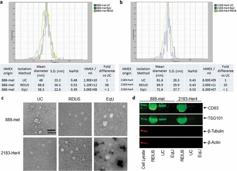
Figure 1. HMEX size distribution, concentration, morphology and protein characteristics in 888-mel and 2183-Her4 when various exosome isolation methods are used. 15 mL of supernatant was isolated from 16.5 × 10⁶ cells and was processed for exosome isolation using various methods. UC: Ultracentrifugation/REIUS: Rapid Exosome Isolation using Ultrafiltration and Size Exclusion Chromatography/EqU: ExoQuick Ultra. (a) HMEX size distribution in 888-mel and (b) 2183-Her4 is consistent regardless of exosome isolation method used. Concentrations of HMEX vary depending on the method of exosome isolation chosen. Exosome polydispersity index (NePdi) is the ratio of standard deviation over mean exosome size based on NTA (Zetaview). (c) Transmission Electron Microscopy images of isolated exosomes with negative staining by Uranyless. Circular morphology and the absence of internal staining indicate intact, compartmentalized vesicles. Size is consistent with results from Zetaview. (d) Protein characteristics of exosomes using western blotting technique. Exosomes isolated using different techniques were all concentrated to a 200 µl final volume, of which 50 µl was loaded for western blotting for comparison of yield. CD63 is an exosome-enriched marker and TSG-101 is an endosomal pathway marker specific for exosomes. β-tubulin and β-actin are cytoplasmic markers. The absence of β-tubulin and β-actin compared to cell lysate sample confirms the presence of purified exosomes with minimal cytoplasmic contaminants. CD63 forms a single fused band at higher protein concentrations but forms two distinct bands when loaded at lower protein concentrations in western blot (Supplementary Figure 7).
Figure 1. HMEX size distribution, concentration, morphology and protein characteristics in 888-mel and 2183-Her4 when various exosome isolation methods are used. 15 mL of supernatant was isolated from 16.5 × 10⁶ cells and was processed for exosome isolation using various methods. UC: Ultracentrifugation/REIUS: Rapid Exosome Isolation using Ultrafiltration and Size Exclusion Chromatography/EqU: ExoQuick Ultra. (a) HMEX size distribution in 888-mel and (b) 2183-Her4 is consistent regardless of exosome isolation method used. Concentrations of HMEX vary depending on the method of exosome isolation chosen. Exosome polydispersity index (NePdi) is the ratio of standard deviation over mean exosome size based on NTA (Zetaview). (c) Transmission Electron Microscopy images of isolated exosomes with negative staining by Uranyless. Circular morphology and the absence of internal staining indicate intact, compartmentalized vesicles. Size is consistent with results from Zetaview. (d) Protein characteristics of exosomes using western blotting technique. Exosomes isolated using different techniques were all concentrated to a 200 µl final volume, of which 50 µl was loaded for western blotting for comparison of yield. CD63 is an exosome-enriched marker and TSG-101 is an endosomal pathway marker specific for exosomes. β-tubulin and β-actin are cytoplasmic markers. The absence of β-tubulin and β-actin compared to cell lysate sample confirms the presence of purified exosomes with minimal cytoplasmic contaminants. CD63 forms a single fused band at higher protein concentrations but forms two distinct bands when loaded at lower protein concentrations in western blot (Supplementary Figure 7).CC BY-NC · PubMed Central
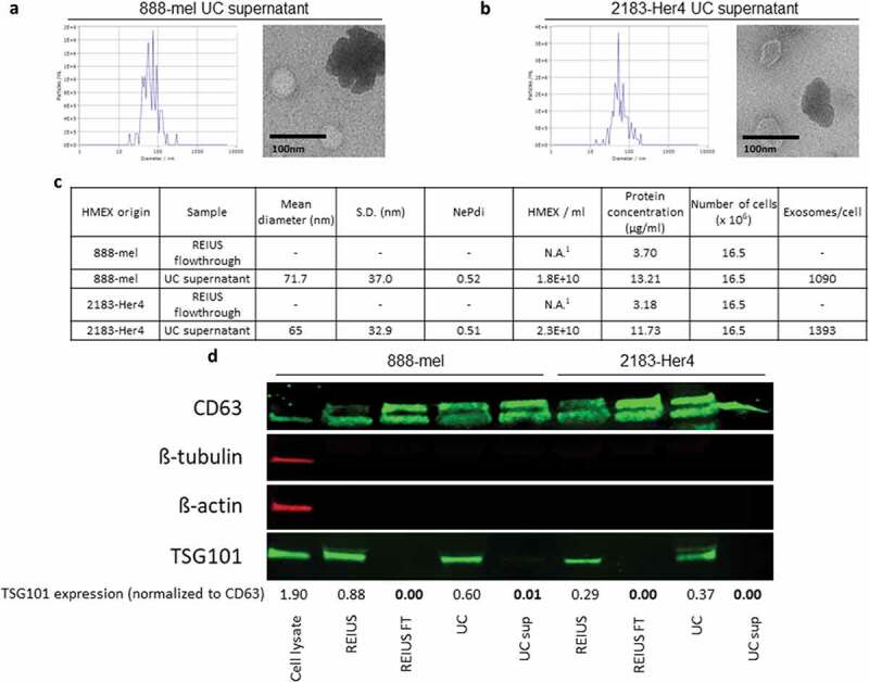
Figure 2. Significant proportion of exosomes remain in supernatant after UC. 15 ml of UC supernatant was concentrated using 3kD MWCO ultrafiltration columns to examine for the presence of exosomes in supernatant. UC supernatant size distribution and TEM images from both (a) 888-mel and (b) 2183-Her4. Significant protein aggregation is observed. (c) Physical characteristics of exosomes. UC supernatant has exosomes consistent in size to the UC-isolated exosome fraction. REIUS FT was concentrated using 3kD MWCO ultrafiltration columns to examine for the presence of exosomes. REIUS FT has no observable particles based on NTA. 1: below detection limit. (d) Protein characteristics of exosomes using western blotting technique. CD63 is an exosome-enriched marker and TSG-101 is an endosomal pathway marker specific for exosomes. β-tubulin and β-actin are cytoplasmic markers. The absence of β-tubulin and β-actin compared to cell lysate sample confirms the presence of purified exosomes with minimal cytoplasmic contaminants. For consistency of comparison, 20 µg of sample was loaded per well for western blotting.
Figure 2. Significant proportion of exosomes remain in supernatant after UC. 15 ml of UC supernatant was concentrated using 3kD MWCO ultrafiltration columns to examine for the presence of exosomes in supernatant. UC supernatant size distribution and TEM images from both (a) 888-mel and (b) 2183-Her4. Significant protein aggregation is observed. (c) Physical characteristics of exosomes. UC supernatant has exosomes consistent in size to the UC-isolated exosome fraction. REIUS FT was concentrated using 3kD MWCO ultrafiltration columns to examine for the presence of exosomes. REIUS FT has no observable particles based on NTA. 1: below detection limit. (d) Protein characteristics of exosomes using western blotting technique. CD63 is an exosome-enriched marker and TSG-101 is an endosomal pathway marker specific for exosomes. β-tubulin and β-actin are cytoplasmic markers. The absence of β-tubulin and β-actin compared to cell lysate sample confirms the presence of purified exosomes with minimal cytoplasmic contaminants. For consistency of comparison, 20 µg of sample was loaded per well for western blotting.CC BY-NC · PubMed Central
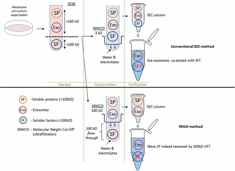
Figure 3. Schematic representation comparing exosome isolation strategies using SEC method. In conventional SEC, melanoma cell culture supernatant is harvested and is directly applied to SEC columns. In some cases, when concentration is required, low (3 kD) MWCO ultrafiltration is used to reduce excess water and electrolytes to load the correct volume onto SEC (100 μL). It is assumed that soluble biologics (e.g. cytokines) that remain are an integral part of exosomes. For REIUS method, 100 kD MWCO ultrafiltration is used to reduce soluble factors that are under 100 kD, followed by SEC. This method is proposed to reduce soluble factors more efficiently than SEC alone. For both methods, presence of non-exosomal soluble factors was examined in detail. Portions of this figure were made using templates from Motifolio Scientific Illustration Toolkits (www.motifolio.com).
Figure 3. Schematic representation comparing exosome isolation strategies using SEC method. In conventional SEC, melanoma cell culture supernatant is harvested and is directly applied to SEC columns. In some cases, when concentration is required, low (3 kD) MWCO ultrafiltration is used to reduce excess water and electrolytes to load the correct volume onto SEC (100 μL). It is assumed that soluble biologics (e.g. cytokines) that remain are an integral part of exosomes. For REIUS method, 100 kD MWCO ultrafiltration is used to reduce soluble factors that are under 100 kD, followed by SEC. This method is proposed to reduce soluble factors more efficiently than SEC alone. For both methods, presence of non-exosomal soluble factors was examined in detail. Portions of this figure were made using templates from Motifolio Scientific Illustration Toolkits (www.motifolio.com).CC BY-NC · PubMed Central
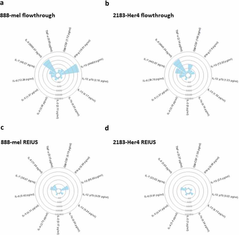
Figure 4. 100 kD MWCO ultrafiltration elimininates substantial cytokine contamination from cell culture supernatant. Circular cytokine plot overview of the 13-plex luminex assay result displaying cytokines that were reduced through 100 kD MWCO ultrafiltration (flow-through) and cytokines present with exosomes after REIUS method. Soluble factors detected in (a) 888-mel and (b) 2183-Her 4 supernatants from 100 kD MWCO UF flow-through that contains non-exosome associated soluble factors. Soluble factors detected in HMEX isolated using REIUS method from (c) 888-mel and (d) 2183-Her4 cell line. Individual data points used to generate the plot is included in supplementary data (supplementary table 2) (n = at least 6).
Figure 4. 100 kD MWCO ultrafiltration elimininates substantial cytokine contamination from cell culture supernatant. Circular cytokine plot overview of the 13-plex luminex assay result displaying cytokines that were reduced through 100 kD MWCO ultrafiltration (flow-through) and cytokines present with exosomes after REIUS method. Soluble factors detected in (a) 888-mel and (b) 2183-Her 4 supernatants from 100 kD MWCO UF flow-through that contains non-exosome associated soluble factors. Soluble factors detected in HMEX isolated using REIUS method from (c) 888-mel and (d) 2183-Her4 cell line. Individual data points used to generate the plot is included in supplementary data (supplementary table 2) (n = at least 6).CC BY-NC · PubMed Central
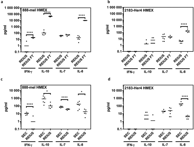
Figure 5. Determining efficiency of REIUS method in removing cytokines. (a,b) comparison between REIUS and REIUS flow-through (REIUS FT). (c,d) direct comparison between SEC alone and REIUS method and the presence of representative soluble factors (IFN-γ, IL-7, IL-8 and IL-10) in either 888-mel or 2183-Her4 supernatant isolates. REIUS method is capable of removing more soluble factors than SEC alone (n = at least 6).
Figure 5. Determining efficiency of REIUS method in removing cytokines. (a,b) comparison between REIUS and REIUS flow-through (REIUS FT). (c,d) direct comparison between SEC alone and REIUS method and the presence of representative soluble factors (IFN-γ, IL-7, IL-8 and IL-10) in either 888-mel or 2183-Her4 supernatant isolates. REIUS method is capable of removing more soluble factors than SEC alone (n = at least 6).CC BY-NC · PubMed Central
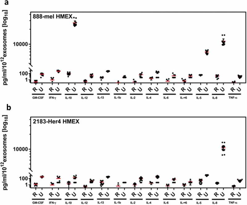
Figure 6. Ratio of soluble cytokine concentration to the number of exosomes isolated. When controlled for number of exosomes, cytokine profile per 1012 exosomes were up to 836 fold lower in REIUS compared to UC. A decrease was observed in both (a) 888-mel HMEX and (b) 2183-Her4 HMEX (n = at least 6).
Figure 6. Ratio of soluble cytokine concentration to the number of exosomes isolated. When controlled for number of exosomes, cytokine profile per 1012 exosomes were up to 836 fold lower in REIUS compared to UC. A decrease was observed in both (a) 888-mel HMEX and (b) 2183-Her4 HMEX (n = at least 6).CC BY-NC · PubMed Central
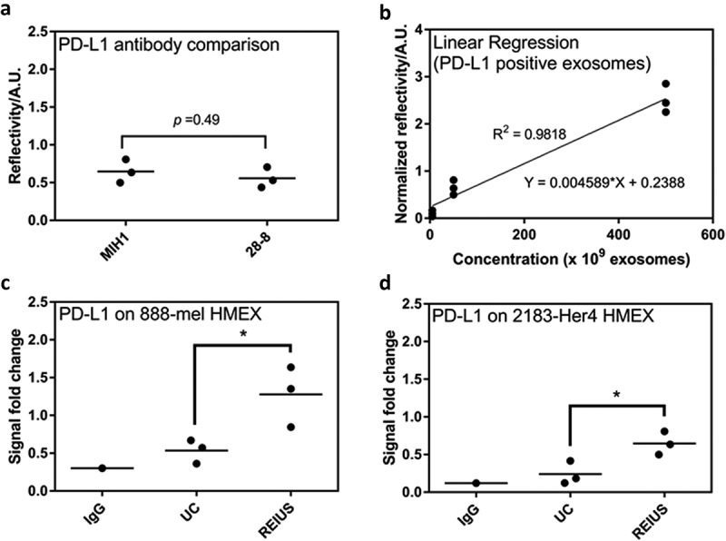
Figure 7. 15 ml of supernatant was isolated from 16.5 × 10⁶ cells and was processed for exosome isolation using various methods. Real-time assay measurement results for Compact Surface Plasmon Resonance Biosensor (SPR) detection of PD-L1. (a) The clone used, MIH1, is as effective as 28–8 for use in SPR assay when normalized by number of exosomes (1 × 10¹⁰). (b) Linear regression analysis of PD-L1 positive exosomes using MIH1 clone (R2 = 0.9818). When normalized by number of exosomes (1 × 10¹⁰), PD-L1 expression of exosomes in (c) 888-mel HMEX and (d) 2183-Her4 HMEX is as shown (n = at least 6).
Figure 7. 15 ml of supernatant was isolated from 16.5 × 10⁶ cells and was processed for exosome isolation using various methods. Real-time assay measurement results for Compact Surface Plasmon Resonance Biosensor (SPR) detection of PD-L1. (a) The clone used, MIH1, is as effective as 28–8 for use in SPR assay when normalized by number of exosomes (1 × 10¹⁰). (b) Linear regression analysis of PD-L1 positive exosomes using MIH1 clone (R2 = 0.9818). When normalized by number of exosomes (1 × 10¹⁰), PD-L1 expression of exosomes in (c) 888-mel HMEX and (d) 2183-Her4 HMEX is as shown (n = at least 6).CC BY-NC · PubMed Central

Verification

Claims the paper makes

The claims and statistics of this paper haven’t been checked yet.

Run the check — free, no account needed. It reads the open-access text and checks the sources it cites.

A few free checks a day · papers already verified open instantly · results are public

Claims other papers make about this one

Each is checked against this paper’s own text: does it say what the citing paper says it does?

Self-check

Percentages, sums and reported differences re-computed from the paper’s own numbers.

The self-check needs the paper’s full text.

Citations

Cites

Research works printed but not linked: 62

  1. 10.1038/nrc2621 · DOI
  2. 10.1080/00015458.2016.1242214 · DOI
  3. 10.1038/nrc.2017.6 · DOI · Paper page
  4. 10.21873/cgp.20015 · DOI
  5. 10.1093/infdis/jiu676 · DOI
  6. 10.1038/s41598-018-27190-x · DOI
  7. 10.4049/jimmunol.0900970 · DOI
  8. 10.1007/978-3-319-67577-0_6 · DOI
  9. Yoshimura A, 2017, Is the exosome a potential target for cancer immunotherapy?, Ann Transl Med, 5, 117, 10.21037/atm.2017.01.47 · DOI
  10. 10.3389/fcell.2018.00018 · DOI
  11. 10.1111/pcmr.12564 · DOI
  12. 10.1038/s41586-018-0392-8 · DOI · Paper page
  13. 10.15171/apb.2016.041 · DOI
  14. 10.1038/srep08913 · DOI
  15. 10.7150/ijbs.7.651 · DOI · Paper page
  16. 10.3402/jev.v2i0.19861 · DOI · Paper page
  17. Whiteside TL, 2017, Extracellular vesicles isolation and their biomarker potential: are we ready for testing?, Ann Transl Med, 5, 54, 10.21037/atm.2017.01.62 · DOI
  18. 10.1038/s41598-018-31323-7 · DOI
  19. 10.1021/acssensors.8b00230 · DOI
  20. Ansel N‐TCKM, 2016, Improved exosome isolation by sucrose gradient fractionation of ultracentrifuged crude exosome pellets, Protoc Exch
  21. 10.1016/j.jim.2014.06.007 · DOI
  22. NolanJ DugganE.Cell‐specific EV tetraspanin expression.Proceedings of the 34th congress of the International Society for Advancement of Cytometry;2019 Vancouver BC Canada p369. (#475/B337).
  23. Ringuette Goulet C, 2018, Exosomes induce fibroblast differentiation into cancer‐associated fibroblasts through TGFbeta signaling, Mol Cancer Res, 16, 1196, 10.1158/1541-7786.MCR-17-0784 · DOI
  24. 10.1007/978-3-642-78771-3_11 · DOI
  25. Castelli C, 1994, Expression of interleukin 1 alpha, interleukin 6, and tumor necrosis factor alpha genes in human melanoma clones is associated with that of mutated N‐RAS oncogene, Cancer Res, 54, 4785
  26. 10.1084/jem.20051848 · DOI
  27. 10.1186/s12967-017-1374-6 · DOI
  28. Lane RE, 2019, Optimizing size exclusion chromatography for extracellular vesicle enrichment and proteomic analysis from clinically relevant samples, Proteomics, 10.1002/pmic.201800156 · DOI
  29. 10.1158/1078-0432.CCR-16-1821 · DOI
  30. 10.7150/thno.18133 · DOI
  31. 10.3402/jev.v5.32945 · DOI
  32. Gheinani AH, 2018, Improved isolation strategies to increase the yield and purity of human urinary exosomes for biomarker discovery, Sci Rep, 8, 3945, 10.1038/s41598-018-22142-x · DOI
  33. 10.3892/mmr.2013.1634 · DOI
  34. 10.1152/ajprenal.00434.2006 · DOI
  35. 10.1016/j.chroma.2014.10.026 · DOI
  36. 10.1039/C7NR08360B · DOI
  37. 10.1371/journal.pone.0204276 · DOI
  38. 10.1038/s41598-017-15717-7 · DOI
  39. 10.1186/s13075-017-1228-x · DOI
  40. 10.1016/j.nano.2015.01.003 · DOI
  41. 10.1158/1078-0432.CCR-07-4843 · DOI · Paper page
  42. 10.1038/modpathol.2011.5 · DOI
  43. 10.1182/blood-2013-10-530469 · DOI
  44. Bar‐Eli M, 1999, Role of interleukin‐8 in tumor growth and metastasis of human melanoma, Pathobiology, 67, 12, 10.1159/000028045 · DOI
  45. Luca M, 1997, Expression of interleukin‐8 by human melanoma cells up‐regulates MMP‐2 activity and increases tumor growth and metastasis, Am J Pathol, 151, 1105
  46. 10.18632/oncotarget.13569 · DOI
  47. 10.1002/pmic.200900351 · DOI
  48. 10.1016/j.nano.2017.03.011 · DOI
  49. 10.1158/1078-0432.1010.11.3 · DOI
  50. Boing AN, 2014, Single‐step isolation of extracellular vesicles by size‐exclusion chromatography, J Extracell Vesicles, 3
  51. 10.1038/srep23978 · DOI
  52. 10.1007/978-1-4939-7253-1_8 · DOI
  53. 10.1038/srep33935 · DOI
  54. 10.1016/j.jmb.2015.09.019 · DOI
  55. 10.1074/mcp.M900152-MCP200 · DOI
  56. 10.1038/s41598-017-10646-x · DOI
  57. 10.1016/j.ymeth.2012.01.002 · DOI
  58. 10.1007/978-1-4939-2550-6_15 · DOI
  59. 10.3402/jev.v5.31655 · DOI
  60. 10.1016/j.ymeth.2015.02.019 · DOI
  61. 10.1016/j.clinbiochem.2013.10.020 · DOI
  62. 10.1007/s00216-018-1052-4 · DOI

Cited by

Sorted by citations

See all 141

Metadata

Authors
La Shu, S., Yang, Y., Allen, C. L., Hurley, E., Tung, K. H., Minderman, H., Wu, Y., & Ernstoff, M. S.
Venue
Journal of Extracellular Vesicles
Publisher
Wiley
Published
2019-11-20
Volume
9
Issue
1
ISSN
2001-3078
Cited by
141
References
1
Language
en
Funders
  • National Cancer Institute (5R33CA19124, S10OD018048)
  • National Institutes of Health (1R50CA211108)
  • Roswell Park Comprehensive Cancer Center (P30CA016056)
  • Roswell Park Cancer Institute (P30CA016056)

About these checks

Registry, retraction, Cites and statistics results are computed, not judged, with one exception: a printed reference that no index lists is read once by a model before it counts as a citation error. Claims are extracted by a model and kept only when their quote appears verbatim in the text; each cited source is then checked, with a model’s help, for whether it says what the claim relies on. Those verdicts can be wrong — read the claim’s quote and the cited source before relying on one.

Statistics and arithmetic are recomputed, not judged.

Confidence bands are shown only for fields calibrated on our annotated test set. None is calibrated yet, so no band is shown.

What counts as a claim ›How we measure confidence ›How we check this ›

Are you an author, or do you see a problem with this page? Request a review