

The DOI matches its Crossref record; 1 of the 1 references it cites were found in a registry or an index.
Epidermal Growth Factor and Membrane Trafficking
Abstract
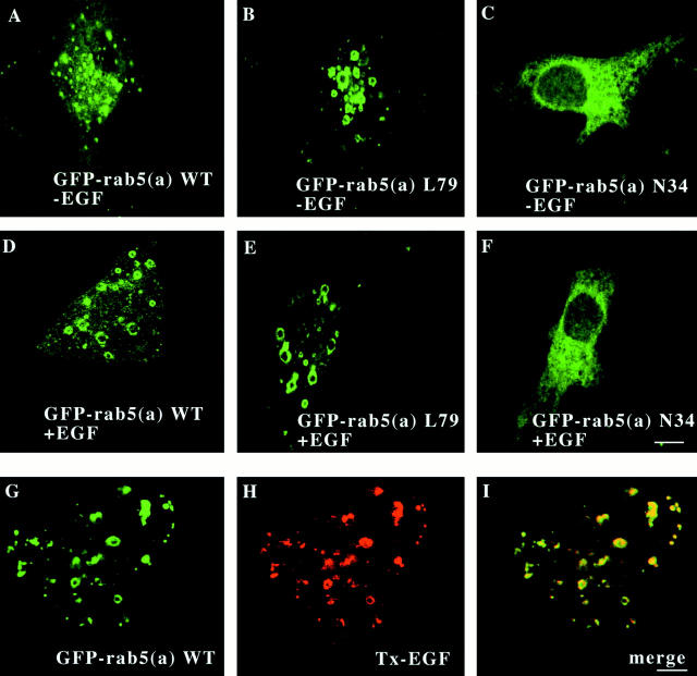
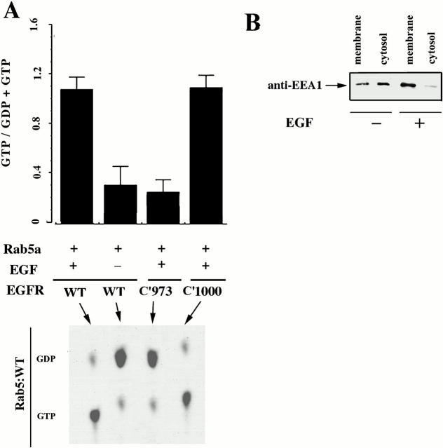
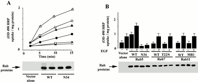
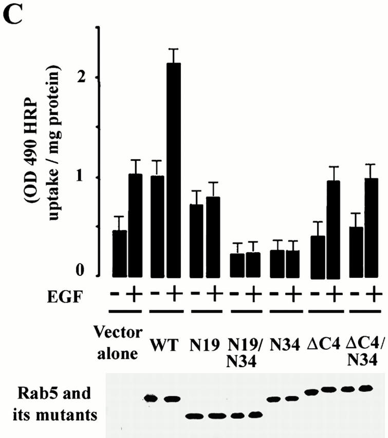
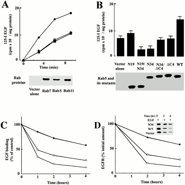
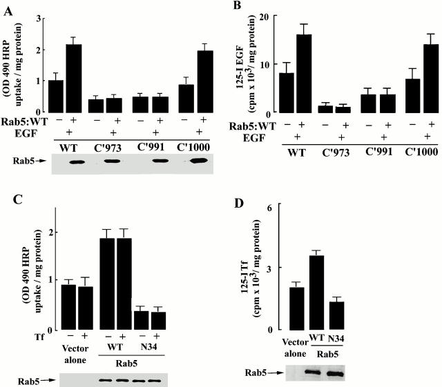
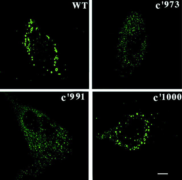
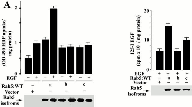
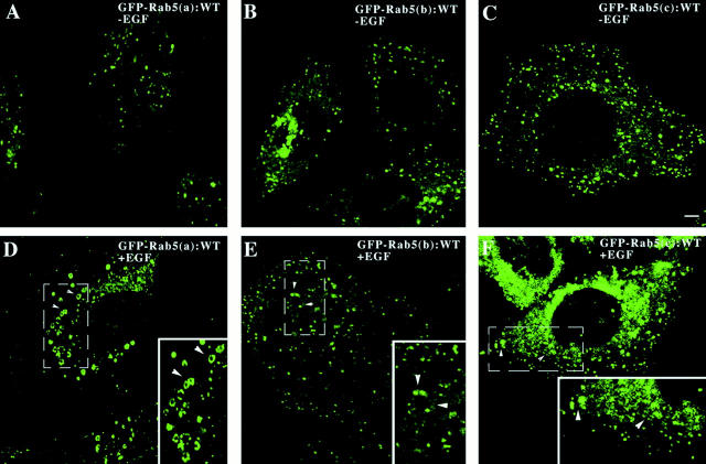
Activated epidermal growth factor receptors recruit various intracellular proteins leading to signal generation and endocytic trafficking. Although activated receptors are rapidly internalized into the endocytic compartment and subsequently degraded in lysosomes, the linkage between signaling and endocytosis is not well understood. Here we show that EGF stimulation of NR6 cells induces a specific, rapid and transient activation of Rab5a. EGF also enhanced translocation of the Rab5 effector, early endosomal autoantigen 1 (EEA1), from cytosol to membrane. The activation of endocytosis, fluid phase and receptor mediated, by EGF was enhanced by Rab5a expression, but not by Rab5b, Rab5c, or Rab5a truncated at the NH2 and/or COOH terminus. Dominant negative Rab5a (Rab5:N34) blocked EGF-stimulated receptor-mediated and fluid-phase endocytosis. EGF activation of Rab5a function was dependent on tyrosine residues in the COOH-terminal domain of the EGF receptor (EGFR). Removal of the entire COOH terminus by truncation (c'973 and c'991) abrogated ligand-induced Rab5a activation of endocytosis. A “kinase-dead” EGFR failed to stimulate Rab5a function. However, another EGF receptor mutant (c'1000), with the kinase domain intact and a single autophosphorylation site effectively signaled Rab5 activation. These results indicate that EGFR and Rab5a are linked via a cascade that results in the activation of Rab5a and that appears essential for internalization. The results point to an interdependent relationship between receptor activation, signal generation and endocytosis.
Overview
The claims and statistics of this paper haven’t been checked yet.
Run the check — free, no account needed. It reads the open-access text and checks the sources it cites.
Integrity signals
Registry
Cites
Statistics
Figures
Verification
Claims the paper makes
The claims and statistics of this paper haven’t been checked yet.
Run the check — free, no account needed. It reads the open-access text and checks the sources it cites.
Claims other papers make about this one
Each is checked against this paper’s own text: does it say what the citing paper says it does?
The source backs part of the claim, not all of it — or backs it for a different population, condition or comparison.
In the citing paper
“We found that, when overexpressed, SNX17 is found in large endosomal structures similar to those seen in cells overexpressing rab-5a, which the authors concluded were abnormally enlarged endosomes formed because further transport was blocked (21).”
From Sorting Motifs in the Intracellular Domain of the Low Density Lipoprotein Receptor Interact with a Novel Domain of Sorting Nexin-17 (2004) · cites this paper as [21]
Experimental — accuracy not yet measuredThe cited source says what the claim attributes to it.
In the citing paper
“Prolonged overexpression of SNX17-FLAG in CHO cells resulted in the formation of large abnormal vesicular structures (Fig. 9C, panels a and d) reminiscent of the disrupted endosomes seen in cells seen in cells overexpressing Rab-5 (21) or sorting nexin-1 (20).”
From Sorting Motifs in the Intracellular Domain of the Low Density Lipoprotein Receptor Interact with a Novel Domain of Sorting Nexin-17 (2004) · cites this paper as [21]
Self-check
Percentages, sums and reported differences re-computed from the paper’s own numbers.
The self-check needs the paper’s full text.
Citations
Cites
McBride, H. M., Rybin, V., Murphy, C., Giner, A., Teasdale, R., & Zerial, M. (1999) · Cell · doi:10.1016/s0092-8674(00)81966-2 · Cited by 520
Research works printed but not linked: 61
- Alvarez-Dominguez, 1998, Interferon-gamma selectively induces Rab5a synthesis and processing in mononuclear cells, J. Biol. Chem, 273, 33901, 10.1074/jbc.273.51.33901 · DOI
- Alvarez-Dominguez, 1999, Increased expression of Rab5a correlates directly with accelerated maturation of Listeria monocytogenes phagosomes, J. Biol. Chem, 274, 11459, 10.1074/jbc.274.17.11459 · DOI
- Barbieri, 1998, Evidence for a symmetrical requirement for Rab5:GTP in in vitro endosome–endosome fusion, J. Biol. Chem, 273, 25850, 10.1074/jbc.273.40.25850 · DOI
- Barbieri, 1998, Protein kinase B/akt and rab5 mediate Ras activation of endocytosis, J. Biol. Chem, 273, 19367, 10.1074/jbc.273.31.19367 · DOI
- Beguinot, 1984, Down-regulation of the epidermal growth factor receptor in KB cells is due to receptor internalization and subsequent degradation in lysosomes, Proc. Natl. Acad. Sci. USA., 81, 2384, 10.1073/pnas.81.8.2384 · DOI
- Benmerah, 1995, The tyrosine kinase substrate eps15 is constitutively associated with the plasma membrane adaptor AP-2, J. Cell Biol, 131, 1831, 10.1083/jcb.131.6.1831 · DOI
- Bucci, 1992, The small GTPase rab5 functions as a regulatory factor in the early endocytic pathway, Cell., 70, 715, 10.1016/0092-8674(92)90306-W · DOI
- Bunemann, 1999, G-protein coupled receptor kinases as modulators of G-protein signalling, J. Physiol., 517, 5, 10.1111/j.1469-7793.1999.0005z.x · DOI
- Carter, 1993, Multiple GTP-binding proteins participate in clathrin-coated vesicle-mediated endocytosis, J. Cell Biol, 120, 37, 10.1083/jcb.120.1.37 · DOI
- Chen, 1994, Cell movement elicited by epidermal growth factor receptor requires kinase and autophosphorylation but is separable from mitogenesis, J. Cell Biol, 124, 547, 10.1083/jcb.124.4.547 · DOI
- Chiariello, 1999, The small GTPases Rab5a, Rab5b and Rab5c are differentially phosphorylated in vitro, FEBS Lett, 453, 20, 10.1016/S0014-5793(99)00686-9 · DOI
- D'Souza-Schorey, 1995, A regulatory role for ARF6 in receptor-mediated endocytosis, Science., 267, 1175, 10.1126/science.7855600 · DOI
- Fischer von Mollard, 1994, Localization of Rab5 to synaptic vesicles identifies endosomal intermediate in synaptic vesicle recycling pathway, Eur. J. Cell Biol., 65, 319
- Goldstein, 1985, Receptor-mediated endocytosisconcepts emerging from the LDL receptor system, Annu. Rev. Cell. Biol, 1, 1, 10.1146/annurev.cb.01.110185.000245 · DOI · Paper page
- Grimes, 1996, Endocytosis of activated TrkAevidence that nerve growth factor induces formation of signaling endosomes, J. Neurosci., 16, 7950, 10.1523/JNEUROSCI.16-24-07950.1996 · DOI
- Gruenberg, 1995, Membrane transport in the endocytic pathway, Curr. Opin. Cell Biol, 7, 552, 10.1016/0955-0674(95)80013-1 · DOI
- Hama, 1999, Vps9p is a guanine nucleotide exchange factor involved in vesicle-mediated vacuolar protein transport, J. Biol. Chem., 274, 15284, 10.1074/jbc.274.21.15284 · DOI
- Hanover, 1984, Kinetics of transit of transferrin and epidermal growth factor through clathrin-coated membranes, Cell., 39, 283, 10.1016/0092-8674(84)90006-0 · DOI
- Haugh, 1999, Effect of epidermal growth factor receptor internalization on regulation of the phospholipase C-gamma1 signaling pathway, J. Biol. Chem, 274, 8958, 10.1074/jbc.274.13.8958 · DOI
- Heuser, 1993, Proton pumps populate the contractile vacuoles of Dictyostelium amoebae, J. Cell Biol, 121, 1311, 10.1083/jcb.121.6.1311 · DOI
- Hopkins, 1983, Internalization and processing of transferrin and the transferrin receptor in human carcinoma A-431 cells, J. Cell Biol, 97, 508, 10.1083/jcb.97.2.508 · DOI
- Hopkins, 1985, Receptor-mediated endocytosis of transferrin and epidermal growth factor receptorsa comparison of constitutive and ligand-induced uptake, J. Cell Sci, 3, 173, 10.1242/jcs.1985.Supplement_3.17 · DOI
- Horiuchi, 1997, A novel Rab5 GDP/GTP exchange factor complexed to Rabaptin-5 links nucleotide exchange to effector recruitment and function, Cell., 90, 1149, 10.1016/S0092-8674(00)80380-3 · DOI
- Iyer, 1999, The transcriptional program in the response of human fibroblasts to serum, Science., 283, 83, 10.1126/science.283.5398.83 · DOI
- Lamaze, 1993, Recruitment of epidermal growth factor and transferrin receptors into coated pits in vitrodiffering biochemical requirements, Mol. Biol. Cell., 4, 715, 10.1091/mbc.4.7.715 · DOI
- Li, 1993, Structure–function relationship of the small GTPase rab5, J. Biol. Chem, 268, 24475, 10.1016/S0021-9258(20)80550-X · DOI
- Li, 1994, Structural features of the GTP-binding defective Rab5 mutants required for their inhibitory activity on endocytosis, J. Biol. Chem, 269, 14631, 10.1016/S0021-9258(17)36671-1 · DOI
- Li, 1995, Evidence for phosphatidylinositol 3-kinase as a regulator of endocytosis via activation of Rab5, Proc. Natl. Acad. Sci. USA, 92, 10207, 10.1073/pnas.92.22.10207 · DOI
- Li, 1997, Uncoupling of membrane ruffling and pinocytosis during Ras signal transduction, J. Biol. Chem, 272, 10337, 10.1074/jbc.272.16.10337 · DOI · Paper page
- Lohi, 1998, EAST, an epidermal growth factor receptor- and Eps15-associated protein with Src homology 3 and tyrosine-based activation motif domains, J. Biol. Chem, 273, 21408, 10.1074/jbc.273.33.21408 · DOI
- Lutcke, 1995, Co-operative regulation of endocytosis by three Rab5 isoforms, FEBS Lett, 366, 65, 10.1016/0014-5793(95)00477-Q · DOI
- Luttrell, 1999, Regulation of tyrosine kinase cascades by G-protein-coupled receptors, Curr. Opin. Cell Biol, 11, 177, 10.1016/S0955-0674(99)80023-4 · DOI
- McLauchlan, 1998, A novel role for Rab5-GDI in ligand sequestration into clathrin-coated pits, Curr. Biol, 8, 34, 10.1016/S0960-9822(98)70018-1 · DOI
- Moghal, 1999, Multiple positive and negative regulators of signaling by the EGF-receptor, Curr. Opin. Cell. Biol, 11, 190, 10.1016/S0955-0674(99)80025-8 · DOI
- Olayioye, 1998, ErbB-1 and ErbB-2 acquire distinct signaling properties dependent upon their dimerization partner, Mol. Cell. Biol, 18, 5042, 10.1128/MCB.18.9.5042 · DOI
- Premont, 1999, beta2-Adrenergic receptor regulation by GIT1, a G protein-coupled receptor kinase-associated ADP ribosylation factor GTPase-activating protein, Proc. Natl. Acad. Sci. USA., 95, 14082, 10.1073/pnas.95.24.14082 · DOI
- Rizzo, 1999, Phospholipase D and its product, phosphatidic acid, mediate agonist-dependent Raf-1 translocation to the plasma membrane and the activation of the mitogen-activated protein kinase pathway, J. Biol. Chem, 274, 1131, 10.1074/jbc.274.2.1131 · DOI
- Roberts, 1999, Endosome fusion in living cells overexpressing GFP-rab5, J. Cell Sci, 112, 3667, 10.1242/jcs.112.21.3667 · DOI
- Simonsen, 1999, The Rab5 effector EEA1 interacts directly with syntaxin-6, J. Biol. Chem, 274, 28857, 10.1074/jbc.274.41.28857 · DOI
- Singer-Kruger, 1994, Role of three rab5-like GTPases, Ypt51p, Ypt52p, and Ypt53p, in the endocytic and vacuolar protein sorting pathways of yeast, J. Cell Biol, 125, 283, 10.1083/jcb.125.2.283 · DOI
- Sogaard, 1994, A rab protein is required for the assembly of SNARE complexes in the docking of transport vesicles, Cell., 78, 937, 10.1016/0092-8674(94)90270-4 · DOI
- Sorkin, 1998, Endocytosis and intracellular sorting of receptor tyrosine kinases, Front. Biosci, 3, 729, 10.2741/A316 · DOI
- Sorkin, 1993, Interaction of activated EGF receptors with coated pit adaptins, Science., 261, 612, 10.1126/science.8342026 · DOI
- Sorkin, 1991, Multiple autophosphorylation site mutations of the epidermal growth factor receptor. Analysis of kinase activity and endocytosis, J. Biol. Chem, 266, 8355, 10.1016/S0021-9258(18)92983-2 · DOI
- Sorkin, 1993, Pool of ligand-bound platelet-derived growth factor γ-receptors remain activated and tyrosine-phosphorylated after internalization, J. Cell Physiol, 156, 373, 10.1002/jcp.1041560221 · DOI
- Sorkina, 1999, Clathrin, adaptors and eps15 in endosomes containing activated epidermal growth factor receptors, J. Cell Sci, 112, 317, 10.1242/jcs.112.3.317 · DOI · Paper page
- Stenmark, 1999, FYVE-finger proteins—effectors of an inositol lipid, J. Cell Sci., 112, 4175, 10.1242/jcs.112.23.4175 · DOI
- Stenmark, 1994, Inhibition of rab5 GTPase activity stimulates membrane fusion in endocytosis, EMBO (Eur. Mol. Biol. Organ.) J, 13, 1287, 10.1002/j.1460-2075.1994.tb06381.x · DOI · Paper page
- Torrisi, 1999, Eps15 is recruited to the plasma membrane upon epidermal growth factor receptor activation and localizes to components of the endocytic pathway during receptor internalization, Mol. Biol. Cell., 10, 417, 10.1091/mbc.10.2.417 · DOI
- Ullrich, 1994, Membrane association of Rab5 mediated by GDP-dissociation inhibitor and accompanied by GDP/GTP exchange, Nature., 368, 157, 10.1038/368157a0 · DOI
- van Delft, 1997, Association and colocalization of Eps15 with adaptor protein-2 and clathrin, J. Cell Biol, 136, 811, 10.1083/jcb.136.4.811 · DOI
- Vickery, 1999, Distinct dynamin-dependent and independent mechanisms target structurally homologous dopamine receptors to different endocytic membranes, J. Cell Biol, 144, 31, 10.1083/jcb.144.1.31 · DOI
- Vieira, 1996, Control of EGF receptor signaling by clathrin-mediated endocytosis, Science., 27, 2086, 10.1126/science.274.5295.2086 · DOI · Paper page
- Wang, 1996, Requirement for the adapter protein GRB2 in EGF receptor endocytosis, Science., 272, 1935, 10.1126/science.272.5270.1935 · DOI
- Warren, 1997, Saturation of the endocytic pathway for the transferrin receptor does not affect the endocytosis of the epidermal growth factor receptor, J. Biol. Chem, 272, 2116, 10.1074/jbc.272.4.2116 · DOI · Paper page
- Waterman, 1999, The C-terminus of the kinase-defective neuregulin receptor ErbB-3 confers mitogenic superiority and dictates endocytic routing, EMBO (Eur. Mol. Biol. Organ.) J, 18, 3348, 10.1093/emboj/18.12.3348 · DOI
- Wells, 1999, Molecules in focus EGF receptor, Int. J. Biochem. Cell Biol, 31, 637, 10.1016/S1357-2725(99)00015-1 · DOI
- Wells, 1990, Ligand-induced transformation by a noninternalizing epidermal growth factor receptor, Science., 247, 962, 10.1126/science.2305263 · DOI
- West, 1989, Distinct endocytotic pathways in epidermal growth factor–stimulated human carcinoma A431 cells, J. Cell Biol, 109, 2731, 10.1083/jcb.109.6.2731 · DOI
- Wilde, 1999, EGF receptor signaling stimulates SRC kinase phosphorylation of clathrin, influencing clathrin redistribution and EGF uptake, Cell., 96, 677, 10.1016/S0092-8674(00)80578-4 · DOI
- Xiao, 1997, The tuberous sclerosis 2 gene product, tuberin, functions as a Rab5GTPase activating protein (GAP) in modulating endocytosis, J. Biol. Chem., 272, 6097, 10.1074/jbc.272.10.6097 · DOI
Cited by
Sorted by citations
Rab GTPases as coordinators of vesicle traffic
Stenmark, H. (2009) · Nature Reviews Molecular Cell Biology · doi:10.1038/nrm2728 · Cited by 3,413
Rab Proteins and the Compartmentalization of the Endosomal System
Wandinger-Ness, A., & Zerial, M. (2014) · Cold Spring Harbor Perspectives in Biology · doi:10.1101/cshperspect.a022616 · Cited by 586
Visualization of self-delivering hydrophobically modified siRNA cellular internalization
Ly, S., Navaroli, D. M., Didiot, M.-C., Cardia, J., Pandarinathan, L., Alterman, J. F., Fogarty, K., Standley, C., Lifshitz, L. M., Bellve, K. D., et al. (2017) · Nucleic Acids Research · doi:10.1093/nar/gkw1005 · Cited by 132
Burden, J. J., Sun, X.-M., García, A. B. G., & Soutar, A. K. (2004) · Journal of Biological Chemistry · doi:10.1074/jbc.m313689200 · Cited by 82
Metadata
- Authors
- Barbieri, M. A., Roberts, R. L., Gumusboga, A., Highfield, H., Alvarez-Dominguez, C., Wells, A., & Stahl, P. D.
- Affiliations
- aDepartment of Cell Biology and Physiology, Washington University School of Medicine, St. Louis, Missouri 63110
- bCentro de Biologia Molecular SEVERO OCHOA, 28033 Madrid, Spain
- cDepartment of Pathology, University of Pittsburgh, Pittsburgh, Pennsylvania 15261
- Venue
- The Journal of Cell Biology
- Publisher
- Rockefeller University Press
- Published
- 2000-10-30
- Volume
- 151
- Issue
- 3
- Pages
- 539-550
- ISSN
- 0021-9525
- 1540-8140
- PMC
- PMC2185585
- Cited by
- 204
- References
- 1
- Language
- en
About these checks
Registry, retraction, Cites and statistics results are computed, not judged, with one exception: a printed reference that no index lists is read once by a model before it counts as a citation error. Claims are extracted by a model and kept only when their quote appears verbatim in the text; each cited source is then checked, with a model’s help, for whether it says what the claim relies on. Those verdicts can be wrong — read the claim’s quote and the cited source before relying on one.
Statistics and arithmetic are recomputed, not judged.
Confidence bands are shown only for fields calibrated on our annotated test set. None is calibrated yet, so no band is shown.
What counts as a claim ›How we measure confidence ›How we check this ›
Are you an author, or do you see a problem with this page? Request a review
