

The DOI matches its Crossref record; 34 of the 34 references it cites were found in a registry or an index.
XBP1
Open accessAbstract
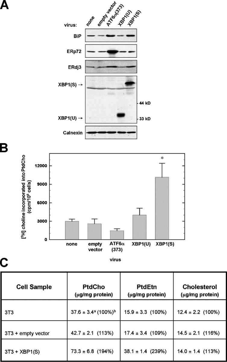
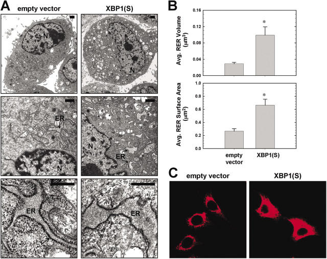
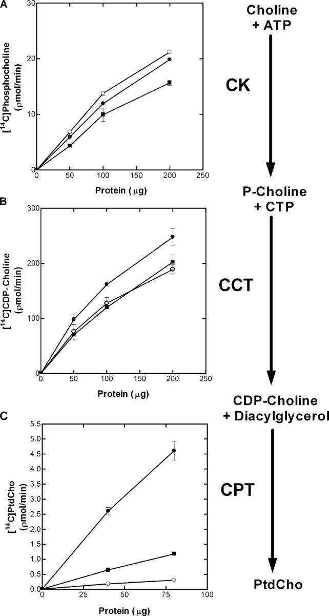
When the protein folding capacity of the endoplasmic reticulum (ER) is challenged, the unfolded protein response (UPR) maintains ER homeostasis by regulating protein synthesis and enhancing expression of resident ER proteins that facilitate protein maturation and degradation. Here, we report that enforced expression of XBP1(S), the active form of the XBP1 transcription factor generated by UPR-mediated splicing of XBP1 mRNA, is sufficient to induce synthesis of phosphatidylcholine, the primary phospholipid of the ER membrane. Cells overexpressing XBP1(S) exhibit elevated levels of membrane phospholipids, increased surface area and volume of rough ER, and enhanced activity of the cytidine diphosphocholine pathway of phosphatidylcholine biosynthesis. These data suggest that XBP1(S) links the mammalian UPR to phospholipid biosynthesis and ER biogenesis.
Overview
The claims and statistics of this paper haven’t been checked yet.
Free: it reads the open-access text and checks the first 5 claims that cite a source we can check. Create a free account to check every claim.
Integrity signals
Registry
Cites
Statistics
Figures


Verification
Claims the paper makes
The claims and statistics of this paper haven’t been checked yet.
Free: it reads the open-access text and checks the first 5 claims that cite a source we can check. Create a free account to check every claim.
Claims other papers make about this one
Each is checked against this paper’s own text: does it say what the citing paper says it does?
In the citing paper
“The increase in lysosomal proteins is consistent with the observations which found that the UPR remodels the lysosome as part of a pro‐survival response (Ron & Hampton, 2004; Sriburi et al, 2004; Brewer et al, 2008; Elfrink et al, 2013).”
From Differential dynamics of the mammalian mRNA and protein expression response to misfolding stress (2016) · cites this paper as Sriburi et al, 2004
Self-check
Percentages, sums and reported differences re-computed from the paper’s own numbers.
The self-check needs the paper’s full text.
Citations
Cites
A RAPID METHOD OF TOTAL LIPID EXTRACTION AND PURIFICATION
Bligh, E. G., & Dyer, W. J. (1959) · Canadian Journal of Biochemistry and Physiology · doi:10.1139/o59-099 · Cited by 54,751
Control of inositol 1,4,5-trisphosphate-induced Ca2+ release by cytosolic Ca2+
Bootman, M. D., Missiaen, L., Parys, J. B., De Smedt, H., & Casteels, R. (1995) · Biochemical Journal · doi:10.1042/bj3060445 · Cited by 5,120
Yoshida, H., Matsui, T., Yamamoto, A., Okada, T., & Mori, K. (2001) · Cell · doi:10.1016/s0092-8674(01)00611-0 · Cited by 4,039
Self-citationMutation of the mouse klotho gene leads to a syndrome resembling ageing
Kuro-o, M., Matsumura, Y., Aizawa, H., Kawaguchi, H., Suga, T., Utsugi, T., Ohyama, Y., Kurabayashi, M., Kaname, T., Kume, E., et al. (1997) · Nature · doi:10.1038/36285 · Cited by 3,837
IRE1 couples endoplasmic reticulum load to secretory capacity by processing the XBP-1 mRNA
Calfon, M., Zeng, H., Urano, F., Till, J. H., Hubbard, S. R., Harding, H. P., Clark, S. G., & Ron, D. (2002) · Nature · doi:10.1038/415092a · Cited by 2,820
Haze, K., Yoshida, H., Yanagi, H., Yura, T., & Mori, K. (1999) · Molecular Biology of the Cell · doi:10.1091/mbc.10.11.3787 · Cited by 2,069
Self-citationLee, A.-H., Iwakoshi, N. N., & Glimcher, L. H. (2003) · Molecular and Cellular Biology · doi:10.1128/mcb.23.21.7448-7459.2003 · Cited by 2,037
Principles and Techniques of Electron Microscopy
Mandal, A. K., Nordquist, J. A., & Hayat, M. A. (1981) · Transactions of the American Microscopical Society · doi:10.2307/3225561 · Cited by 1,433
A trip to the ER: coping with stress
Rutkowski, D. T., & Kaufman, R. J. (2004) · Trends in Cell Biology · doi:10.1016/j.tcb.2003.11.001 · Cited by 1,355
Shaffer, A. L., Shapiro-Shelef, M., Iwakoshi, N. N., Lee, A.-H., Qian, S.-B., Zhao, H., Yu, X., Yang, L., Tan, B. K., Rosenwald, A., et al. (2004) · Immunity · doi:10.1016/j.immuni.2004.06.010 · Cited by 1,054
Research works printed but not linked: 4
- 1959, Can. J. Med. Sci., 37, 911
- 2001, Biochem. J., 355, 19, 10.1042/bj3550019 · doi:10.1042/bj3550019 · Paper page
- 2003, Nat. Immunol., 4, 321
- 2001, Prog. Nucleic Acid Res. Mol. Biol., 65, 361
Cited by
Signal integration in the endoplasmic reticulum unfolded protein response
Ron, D., & Walter, P. (2007) · Nature Reviews Molecular Cell Biology · doi:10.1038/nrm2199 · Cited by 6,296
THE MAMMALIAN UNFOLDED PROTEIN RESPONSE
Schröder, M., & Kaufman, R. J. (2005) · Annual Review of Biochemistry · doi:10.1146/annurev.biochem.73.011303.074134 · Cited by 3,026
Mechanisms, regulation and functions of the unfolded protein response
Hetz, C., Zhang, K., & Kaufman, R. J. (2020) · Nature Reviews Molecular Cell Biology · doi:10.1038/s41580-020-0250-z · Cited by 2,666
Endoplasmic reticulum stress in liver disease
Malhi, H., & Kaufman, R. J. (2011) · Journal of Hepatology · doi:10.1016/j.jhep.2010.11.005 · Cited by 1,173
Yoshida, H. (2007) · The FEBS Journal · doi:10.1111/j.1742-4658.2007.05639.x · Cited by 1,086
Metadata
- Authors
- Sriburi, R., Jackowski, S., Mori, K., & Brewer, J. W.
- Affiliations
- Loyola University Chicago (US)
- St. Jude Children's Research Hospital (US)
- Kyoto University (JP)
- Venue
- The Journal of Cell Biology
- Publisher
- Rockefeller University Press
- Published
- 2004-10-04
- Volume
- 167
- Issue
- 1
- Pages
- 35-41
- ISSN
- 0021-9525 (ISSN-L)
- 1540-8140
- PMC
- PMC2172532
- OpenAlex
- W2005129842
- Cited by
- 653
- References
- 33
- Citations by year
- 2026: 19 · 2025: 18 · 2024: 28 · 2023: 26 · 2022: 29 · 2021: 30 · 2020: 32 · 2019: 39 · 2018: 31 · 2017: 34 · 2016: 22 · 2015: 32 · 2014: 34 · 2013: 27 · 2012: 37
- FWCI
- 9.57
- Type
- journal article
- Language
- en
- Open access
- bronze
- doi.org
- Locations
- The Journal of Cell Biology (publishedVersion)
- PubMed Central (publishedVersion, cc-by-nc-sa)
- PubMed (publishedVersion)
- Funders
- American Lebanese Syrian Associated Charities (GM45737, CA 21765)
- National Institutes of Health (CA21765))
- National Cancer Institute (CA21765, P30CA021765)
- National Institute of General Medical Sciences (R01GM061970, R01GM045737, GM61970, GM45737)
- Topics
- Life Sciences › Biochemistry, Genetics and Molecular Biology › Cell Biology › Endoplasmic Reticulum Stress and Disease
- Life Sciences › Biochemistry, Genetics and Molecular Biology › Cell Biology › Cellular transport and secretion
- Life Sciences › Neuroscience › Sensory Systems › Ion Channels and Receptors
- Keywords
- XBP1, unfolded protein response, endoplasmic reticulum, phosphatidylcholine synthesis, XBP1 splicing, ER homeostasis
- MeSH terms
- Regulatory Factor X Transcription Factors, X-Box Binding Protein 1, Animals, DNA-Binding Proteins, Dose-Response Relationship, Drug, Endoplasmic Reticulum, Fibroblasts, Microscopy, Fluorescence, Models, Biological, Nuclear Proteins, Phosphatidylcholines, Phospholipids, Plasmids, RNA Splicing, RNA, Messenger, Time Factors, Transcription Factors, Blotting, Northern, Transcriptional Activation, 3T3 Cells, Protein Folding, Protein Isoforms, Reverse Transcriptase Polymerase Chain Reaction, NIH 3T3 Cells, Mice
About these checks
Registry, retraction, Cites and statistics results are computed, not judged, with one exception: a printed reference that no index lists is read once by a model before it counts as a citation error. Claims are extracted by a model and kept only when their quote appears verbatim in the text; each cited source is then checked, with a model’s help, for whether it says what the claim relies on. Those verdicts can be wrong — read the claim’s quote and the cited source before relying on one.
Statistics and arithmetic are recomputed, not judged.
Confidence bands are shown only for fields calibrated on our annotated test set. None is calibrated yet, so no band is shown.
What counts as a claim ›How we measure confidence ›How we check this ›
Are you an author, or do you see a problem with this page? Request a review
How to cite
Cite CoChat for the checks on this page, and the paper for its own findings.
- This page
- CoChat. (2026). Citation check: XBP1. https://app.cochat.ai/papers/10.1083/jcb.200406136
- The paper
- Sriburi, R., Jackowski, S., Mori, K., & Brewer, J. W. (2004). XBP1. The Journal of Cell Biology. https://doi.org/10.1083/jcb.200406136
Data sources
Bibliographic data: Semantic Scholar, OpenAlex, Crossref.
Contains information from the Semantic Scholar Academic Graph Datasets, made available under the ODC Attribution License (ODC-BY 1.0).
Semantic Scholar’s data is described in: Kinney, R., et al. (2023). The Semantic Scholar Open Data Platform. arXiv:2301.10140.
