

The DOI matches its Crossref record; 65 of the 66 references it cites were found in a registry or an index.
Quantitative analysis of the native presynaptic cytomatrix by cryoelectron tomography
Open accessAbstract
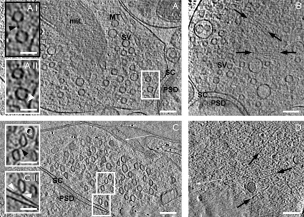
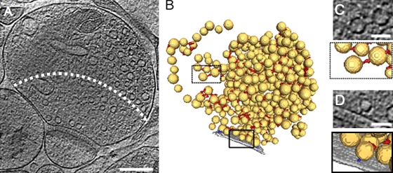
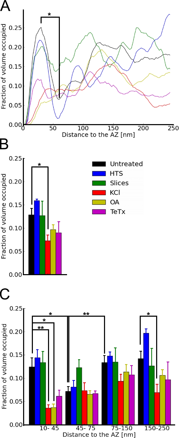
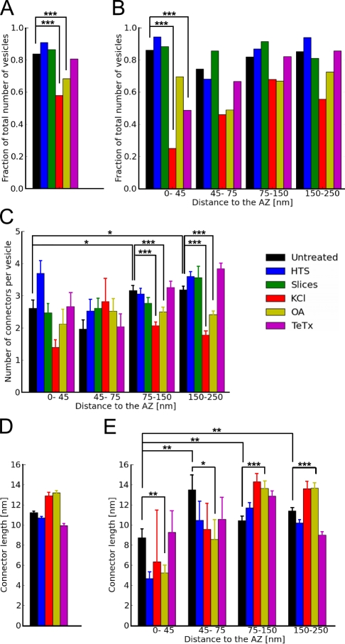
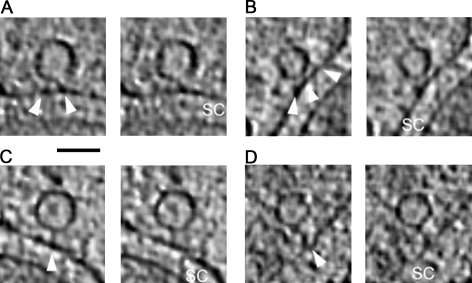
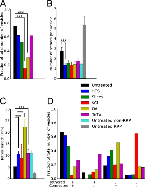
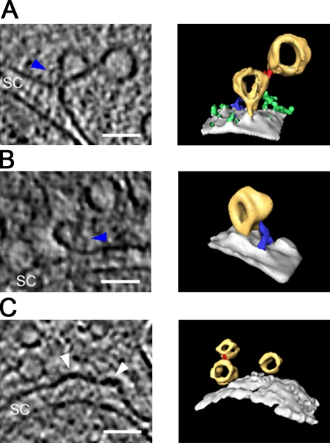
The presynaptic terminal contains a complex network of filaments whose precise organization and functions are not yet understood. The cryoelectron tomography experiments reported in this study indicate that these structures play a prominent role in synaptic vesicle release. Docked synaptic vesicles did not make membrane to membrane contact with the active zone but were instead linked to it by tethers of different length. Our observations are consistent with an exocytosis model in which vesicles are first anchored by long (>5 nm) tethers that give way to multiple short tethers once vesicles enter the readily releasable pool. The formation of short tethers was inhibited by tetanus toxin, indicating that it depends on soluble N-ethyl-maleimide sensitive fusion protein attachment protein receptor complex assembly. Vesicles were extensively interlinked via a set of connectors that underwent profound rearrangements upon synaptic stimulation and okadaic acid treatment, suggesting a role of these connectors in synaptic vesicle mobilization and neurotransmitter release.
Overview
The claims and statistics of this paper haven’t been checked yet.
Run the check — free, no account needed. It reads the open-access text and checks the sources it cites.
Integrity signals
Registry
Cites
Statistics
Figures
Verification
Claims the paper makes
The claims and statistics of this paper haven’t been checked yet.
Run the check — free, no account needed. It reads the open-access text and checks the sources it cites.
Claims other papers make about this one
Each is checked against this paper’s own text: does it say what the citing paper says it does?
The cited source doesn’t support the claim as written.
In the citing paper
“In cryo-EM studies of unfixed and unstained synapse preparations, however, no dense projections are detectable. The only structures visible are the plasma membrane, synaptic vesicles, and sparse filaments that either connect vesicles to each others (“connectors,” average length ~10 nm) or tether vesicles to the presynaptic plasma membrane (“tethers”—5–20 nm; Landis et al., 1988; Fernández-Busnadie”
From The Presynaptic Active Zone (2012) · cites this paper as Fernández-Busnadiego et al., 2010
Checked by a reasoning model — may be wrong.
Experimental — accuracy not yet measuredThe source backs part of the claim, not all of it — or backs it for a different population, condition or comparison.
In the citing paper
“Besides these proteins, actin was suggested to be an active zone component, but high-resolution electron microscopy (EM) shows that actin filaments are excluded from the active zone and the vesicle cluster (Fernández-Busnadiego et al., 2010).”
From The Presynaptic Active Zone (2012) · cites this paper as Fernández-Busnadiego et al., 2010
Experimental — accuracy not yet measured
Self-check
Percentages, sums and reported differences re-computed from the paper’s own numbers.
The self-check needs the paper’s full text.
Citations
Cites
A simple method for organotypic cultures of nervous tissue
Stoppini, L., Buchs, P., & Muller, D. (1991) · Journal of Neuroscience Methods · doi:10.1016/0165-0270(91)90128-m · Cited by 3,096
Südhof, T. C. (2004) · Annual Review of Neuroscience · doi:10.1146/annurev.neuro.26.041002.131412 · Cited by 2,491
Cryo-electron microscopy of vitrified specimens
Dubochet, J., Adrian, M., Chang, J.-J., Homo, J., Lepault, J., McDowall, A. W., & Schultz, P. (1988) · Quarterly Reviews of Biophysics · doi:10.1017/s0033583500004297 · Cited by 2,397
Soille, P. (1999) · Springer eBooks · doi:10.1007/978-3-662-03939-7 · Cited by 1,981
Okadaic acid: a new probe for the study of cellular regulation
Cohen, P., Holmes, C. F. B., & Tsukitani, Y. (1990) · Trends in Biochemical Sciences · doi:10.1016/0968-0004(90)90192-e · Cited by 1,373
Neurotoxins Affecting Neuroexocytosis
Schiavo, G., Matteoli, M., & Montecucco, C. (2000) · Physiological Reviews · doi:10.1152/physrev.2000.80.2.717 · Cited by 1,266
Definition of the readily releasable pool of vesicles at hippocampal synapses.
Rosenmund, C., & Stevens, C. (1996) · Neuron · doi:10.1016/s0896-6273(00)80146-4 · Cited by 1,084
Soille, P. (2004) · Springer eBooks · doi:10.1007/978-3-662-05088-0 · Cited by 1,065
Rizzoli, S. O., & Betz, W. J. (2005) · Nature Reviews Neuroscience · doi:10.1038/nrn1583 · Cited by 975
Mechanisms of membrane fusion: disparate players and common principles
Martens, S., & McMahon, H. T. (2008) · Nature Reviews Molecular Cell Biology · doi:10.1038/nrm2417 · Cited by 733
Cited by
Sorted by citations
Südhof, T. C. (2012) · Neuron · doi:10.1016/j.neuron.2012.06.012 · Cited by 1,062
Ultrastructure of Synapses in the Mammalian BrainPreprint
Harris, K. M., & Weinberg, R. J. (2012) · Cold Spring Harbor Perspectives in Biology · doi:10.1101/cshperspect.a005587 · Cited by 452
Ultrastructural analysis of adult mouse neocortex comparing aldehyde perfusion with cryo fixation
Korogod, N., Petersen, C. C., & Knott, G. W. (2015) · eLife · doi:10.7554/elife.05793 · Cited by 408
Cryo-electron tomography: The challenge of doing structural biology in situ
Lučić, V., Rigort, A., & Baumeister, W. P. (2013) · The Journal of Cell Biology · doi:10.1083/jcb.201304193 · Cited by 387
Self-citationThe Morphological and Molecular Nature of Synaptic Vesicle Priming at Presynaptic Active Zones
Imig, C., Min, S.-W., Krinner, S., Arancillo, M., Rosenmund, C., Südhof, T. C., Rhee, J., Brose, N., & Cooper, B. H. (2014) · Neuron · doi:10.1016/j.neuron.2014.10.009 · Cited by 389
Metadata
- Authors
- Fernández-Busnadiego, R., Zuber, B., Maurer, U. E., Cyrklaff, M., Baumeister, W., & Lučić, V.
- Affiliations
- Department of Molecular Structural Biology, Max Planck Institute of Biochemistry, D-82152 Martinsried, Germany 1
- MRC Laboratory of Molecular Biology (GB)
- Max Planck Institute of Biochemistry (DE)
- Corresponding author
- Baumeister, Wolfgang
- Venue
- Journal of Cell Biology
- Publisher
- Rockefeller University Press
- Published
- 2010-01-11
- Volume
- 188
- Issue
- 1
- Pages
- 145-156
- ISSN
- 0021-9525 (ISSN-L)
- 1540-8140
- PMC
- PMC2812849
- OpenAlex
- W2132214171
- Cited by
- 267
- References
- 66
- Citations by year
- 2026: 12 · 2025: 16 · 2024: 21 · 2023: 18 · 2022: 15 · 2021: 14 · 2020: 16 · 2019: 13 · 2018: 16 · 2017: 18 · 2016: 13 · 2015: 17 · 2014: 17 · 2013: 14 · 2012: 16
- FWCI
- 8.54
- Type
- journal article
- Language
- en
- Open access
- hybrid
- doi.org
- Licence
- cc-by-nc-sa
- Locations
- The Journal of Cell Biology (publishedVersion, cc-by-nc-sa)
- PubMed Central (publishedVersion, cc-by-nc-sa)
- PubMed (publishedVersion)
- Bern Open Repository and Information System (University of Bern) (submittedVersion)
- Max Planck Institute for Plasma Physics (submittedVersion)
- Topics
- Life Sciences › Biochemistry, Genetics and Molecular Biology › Cell Biology › Cellular transport and secretion
- Life Sciences › Neuroscience › Cellular and Molecular Neuroscience › Neuroscience and Neuropharmacology Research
- Life Sciences › Biochemistry, Genetics and Molecular Biology › Structural Biology › Advanced Electron Microscopy Techniques and Applications
- Keywords
- synaptic vesicle release, docked vesicles, exocytosis, readily releasable pool, tetanus toxin, SNARE complex, okadaic acid, synaptic stimulation, neurotransmitter release, active zone, cryo-electron tomography
- MeSH terms
- Animals, Male, Synapses, Rats, Wistar, Tissue Culture Techniques, Rats, Electron Microscope Tomography
About these checks
Registry, retraction, Cites and statistics results are computed, not judged, with one exception: a printed reference that no index lists is read once by a model before it counts as a citation error. Claims are extracted by a model and kept only when their quote appears verbatim in the text; each cited source is then checked, with a model’s help, for whether it says what the claim relies on. Those verdicts can be wrong — read the claim’s quote and the cited source before relying on one.
Statistics and arithmetic are recomputed, not judged.
Confidence bands are shown only for fields calibrated on our annotated test set. None is calibrated yet, so no band is shown.
What counts as a claim ›How we measure confidence ›How we check this ›
Are you an author, or do you see a problem with this page? Request a review
