

The DOI matches its Crossref record; 65 of the 66 references it cites were found in a registry or an index.
Quantitative analysis of the native presynaptic cytomatrix by cryoelectron tomography
Open accessAbstract
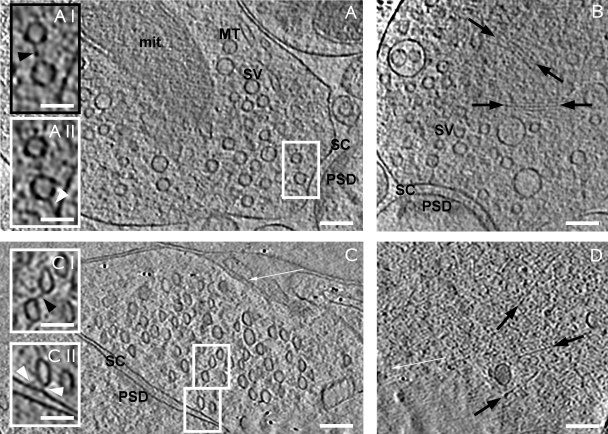
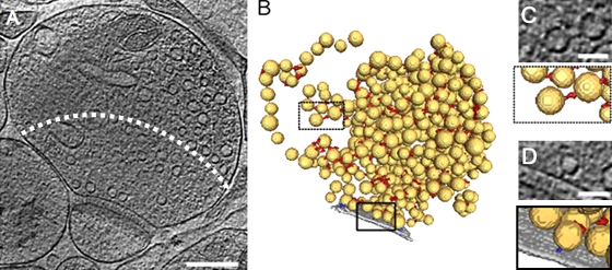
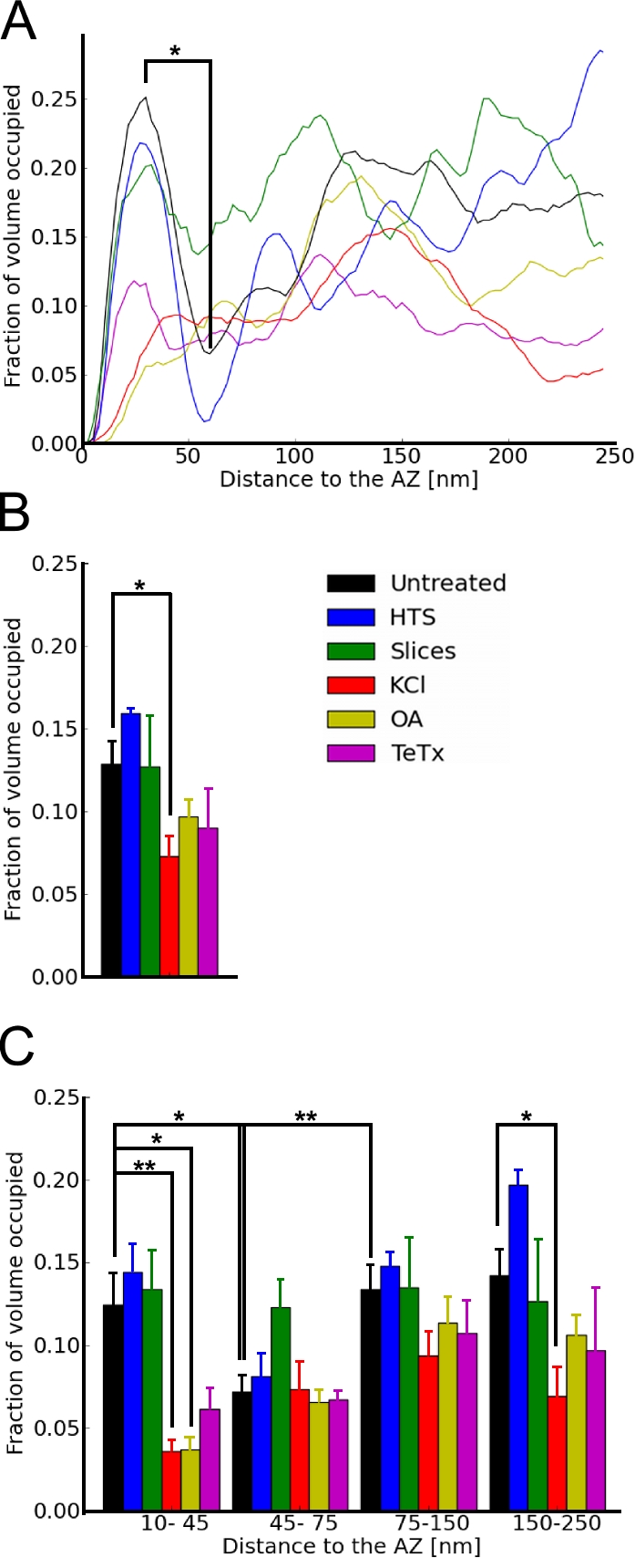
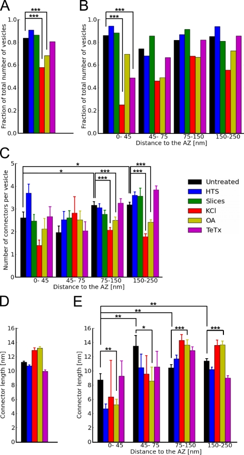
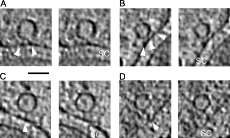
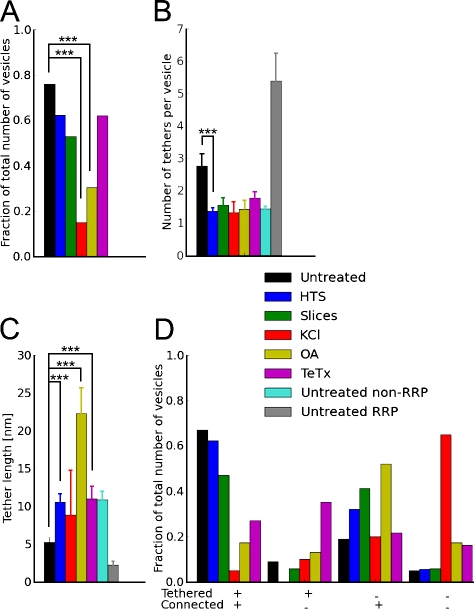
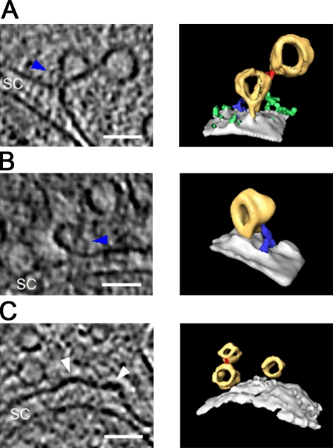
The presynaptic terminal contains a complex network of filaments whose precise organization and functions are not yet understood. The cryoelectron tomography experiments reported in this study indicate that these structures play a prominent role in synaptic vesicle release. Docked synaptic vesicles did not make membrane to membrane contact with the active zone but were instead linked to it by tethers of different length. Our observations are consistent with an exocytosis model in which vesicles are first anchored by long (>5 nm) tethers that give way to multiple short tethers once vesicles enter the readily releasable pool. The formation of short tethers was inhibited by tetanus toxin, indicating that it depends on soluble N-ethyl-maleimide sensitive fusion protein attachment protein receptor complex assembly. Vesicles were extensively interlinked via a set of connectors that underwent profound rearrangements upon synaptic stimulation and okadaic acid treatment, suggesting a role of these connectors in synaptic vesicle mobilization and neurotransmitter release.
Overview
The claims and statistics of this paper haven’t been checked yet.
Run the check — free, no account needed. It reads the open-access text and checks the sources it cites.
Integrity signals
Registry
Cites
Statistics
Figures
Verification
Claims the paper makes
The claims and statistics of this paper haven’t been checked yet.
Run the check — free, no account needed. It reads the open-access text and checks the sources it cites.
Claims other papers make about this one
Each is checked against this paper’s own text: does it say what the citing paper says it does?
The cited source doesn’t support the claim as written.
In the citing paper
“In cryo-EM studies of unfixed and unstained synapse preparations, however, no dense projections are detectable. The only structures visible are the plasma membrane, synaptic vesicles, and sparse filaments that either connect vesicles to each others (“connectors,” average length ~10 nm) or tether vesicles to the presynaptic plasma membrane (“tethers”—5–20 nm; Landis et al., 1988; Fernández-Busnadie”
From The Presynaptic Active Zone (2012) · cites this paper as Fernández-Busnadiego et al., 2010
Checked by a reasoning model — may be wrong.
Experimental — accuracy not yet measuredThe source backs part of the claim, not all of it — or backs it for a different population, condition or comparison.
In the citing paper
“Besides these proteins, actin was suggested to be an active zone component, but high-resolution electron microscopy (EM) shows that actin filaments are excluded from the active zone and the vesicle cluster (Fernández-Busnadiego et al., 2010).”
From The Presynaptic Active Zone (2012) · cites this paper as Fernández-Busnadiego et al., 2010
Experimental — accuracy not yet measured
Self-check
Percentages, sums and reported differences re-computed from the paper’s own numbers.
The self-check needs the paper’s full text.
Citations
Cites
A simple method for organotypic cultures of nervous tissue
Stoppini, L., Buchs, P., & Muller, D. (1991) · Journal of Neuroscience Methods · doi:10.1016/0165-0270(91)90128-m · Cited by 3,096
Südhof, T. C. (2004) · Annual Review of Neuroscience · doi:10.1146/annurev.neuro.26.041002.131412 · Cited by 2,491
Cryo-electron microscopy of vitrified specimens
Dubochet, J., Adrian, M., Chang, J.-J., Homo, J., Lepault, J., McDowall, A. W., & Schultz, P. (1988) · Quarterly Reviews of Biophysics · doi:10.1017/s0033583500004297 · Cited by 2,397
Soille, P. (1999) · Springer eBooks · doi:10.1007/978-3-662-03939-7 · Cited by 1,981
Okadaic acid: a new probe for the study of cellular regulation
Cohen, P., Holmes, C. F. B., & Tsukitani, Y. (1990) · Trends in Biochemical Sciences · doi:10.1016/0968-0004(90)90192-e · Cited by 1,373
Neurotoxins Affecting Neuroexocytosis
Schiavo, G., Matteoli, M., & Montecucco, C. (2000) · Physiological Reviews · doi:10.1152/physrev.2000.80.2.717 · Cited by 1,266
Definition of the readily releasable pool of vesicles at hippocampal synapses.
Rosenmund, C., & Stevens, C. (1996) · Neuron · doi:10.1016/s0896-6273(00)80146-4 · Cited by 1,084
Soille, P. (2004) · Springer eBooks · doi:10.1007/978-3-662-05088-0 · Cited by 1,065
Rizzoli, S. O., & Betz, W. J. (2005) · Nature Reviews Neuroscience · doi:10.1038/nrn1583 · Cited by 975
Mechanisms of membrane fusion: disparate players and common principles
Martens, S., & McMahon, H. T. (2008) · Nature Reviews Molecular Cell Biology · doi:10.1038/nrm2417 · Cited by 733
Cited by
Sorted by citations
Südhof, T. C. (2012) · Neuron · doi:10.1016/j.neuron.2012.06.012 · Cited by 1,062
Ultrastructure of Synapses in the Mammalian BrainPreprint
Harris, K. M., & Weinberg, R. J. (2012) · Cold Spring Harbor Perspectives in Biology · doi:10.1101/cshperspect.a005587 · Cited by 452
Ultrastructural analysis of adult mouse neocortex comparing aldehyde perfusion with cryo fixation
Korogod, N., Petersen, C. C., & Knott, G. W. (2015) · eLife · doi:10.7554/elife.05793 · Cited by 408
Cryo-electron tomography: The challenge of doing structural biology in situ
Lučić, V., Rigort, A., & Baumeister, W. P. (2013) · The Journal of Cell Biology · doi:10.1083/jcb.201304193 · Cited by 387
Self-citationThe Morphological and Molecular Nature of Synaptic Vesicle Priming at Presynaptic Active Zones
Imig, C., Min, S.-W., Krinner, S., Arancillo, M., Rosenmund, C., Südhof, T. C., Rhee, J., Brose, N., & Cooper, B. H. (2014) · Neuron · doi:10.1016/j.neuron.2014.10.009 · Cited by 389
Isotropic reconstruction for electron tomography with deep learning
Liu, Y.-T., Zhang, H., Wang, H., Tao, C.-L., Bi, G., & Zhou, Z. H. (2022) · Nature Communications · doi:10.1038/s41467-022-33957-8 · Cited by 397
Transcellular Nanoalignment of Synaptic Function
Biederer, T., Kaeser, P. S., & Blanpied, T. A. (2017) · Neuron · doi:10.1016/j.neuron.2017.10.006 · Cited by 352
Frank, T., Rutherford, M. A., Strenzke, N., Neef, A., Pangršič, T., Khimich, D., Fejtová, A., Gundelfinger, E. D., Liberman, M. C., Harke, B., et al. (2010) · Neuron · doi:10.1016/j.neuron.2010.10.027 · Cited by 298
Protein scaffolds in the coupling of synaptic exocytosis and endocytosis
Haucke, V., Neher, E., & Sigrist, S. J. (2011) · Nature reviews. Neuroscience · doi:10.1038/nrn2948 · Cited by 298
Perspectives on Kiss-and-Run: Role in Exocytosis, Endocytosis, and Neurotransmission
Alabi, A. A., & Tsien, R. W. (2011) · Annual Review of Physiology · doi:10.1146/annurev-physiol-020911-153305 · Cited by 261
α-Synuclein in synaptic function and dysfunction
Sharma, M., & Burré, J. (2022) · Trends in Neurosciences · doi:10.1016/j.tins.2022.11.007 · Cited by 270
Fernández‐Busnadiego, R., Saheki, Y., & Camilli, P. V. D. (2015) · Proceedings of the National Academy of Sciences · doi:10.1073/pnas.1503191112 · Cited by 236
Self-citationBassoon speeds vesicle reloading at a central excitatory synapse
Hallermann, S., Fejtova, A., Schmidt, H., Weyhersmüller, A., Silver, R. A., Gundelfinger, E. D., & Eilers, J. (2010) · Neuron · doi:10.1016/j.neuron.2010.10.026 · Cited by 223
Sensory Processing at Ribbon Synapses in the Retina and the Cochlea
Moser, T., Grabner, C. P., & Schmitz, F. (2019) · Physiological Reviews · doi:10.1152/physrev.00026.2018 · Cited by 215
Mechanism of neurotransmitter release coming into focus
Rizo, J. (2018) · Protein Science · doi:10.1002/pro.3445 · Cited by 210
Tao, C.-L., Liu, Y.-T., Sun, R., Zhang, B., Qi, L., Shivakoti, S., Tian, C.-L., Zhang, P., Lau, P.-M., Zhou, Z. H., et al. (2018) · The Journal of Neuroscience · doi:10.1523/jneurosci.1548-17.2017 · Cited by 207
Postsynaptic synaptotagmins mediate AMPA receptor exocytosis during LTP
Wu, D., Bacaj, T., Morishita, W., Goswami, D., Arendt, K. L., Xu, W., Chen, L., Malenka, R. C., & Südhof, T. C. (2017) · Nature · doi:10.1038/nature21720 · Cited by 195
GPCR mediated regulation of synaptic transmission
Betke, K. M., Wells, C. A., & Hamm, H. E. (2012) · Progress in Neurobiology · doi:10.1016/j.pneurobio.2012.01.009 · Cited by 172
Mahamid, J., Schampers, R., Persoon, H., Hyman, A. A., Baumeister, W. P., & Plitzko, J. M. (2015) · Journal of Structural Biology · doi:10.1016/j.jsb.2015.07.012 · Cited by 166
Self-citationHorstmann, H., Körber, C., Sätzler, K., Aydin, D., & Kuner, T. (2012) · PLoS ONE · doi:10.1371/journal.pone.0035172 · Cited by 161
Actin- and Myosin-Dependent Vesicle Loading of Presynaptic Docking Sites Prior to Exocytosis
Miki, T., Malagón, G., Pulido, C., Llano, I., Neher, E., & Marty, A. (2016) · Neuron · doi:10.1016/j.neuron.2016.07.033 · Cited by 164
Synucleins Have Multiple Effects on Presynaptic Architecture
Vargas, K. J., Schrod, N., Davis, T., Fernández‐Busnadiego, R., Taguchi, Y. V., Laugks, U., Lučić, V., & Chandra, S. S. (2017) · Cell Reports · doi:10.1016/j.celrep.2016.12.023 · Cited by 159
Self-citationHow to Make an Active Zone: Unexpected Universal Functional Redundancy between RIMs and RIM-BPs
Acuna, C., Liu, X., & Südhof, T. C. (2016) · Neuron · doi:10.1016/j.neuron.2016.07.042 · Cited by 158
Hemi-fused structure mediates and controls fusion and fission in live cells
Zhao, W., Hamid, E., Shin, W., Wen, P. J., Krystofiak, E., Villarreal, S. A., Chiang, H., Kachar, B., & Wu, L. (2016) · Nature · doi:10.1038/nature18598 · Cited by 150
Electron cryotomography of vitrified cells with a Volta phase plate
Fukuda, Y., Laugks, U., Lučić, V., Baumeister, W. P., & Danev, R. (2015) · Journal of Structural Biology · doi:10.1016/j.jsb.2015.03.004 · Cited by 140
Self-citationCryo‐EM—the first thirty years
Dubochet, J. (2011) · Journal of Microscopy · doi:10.1111/j.1365-2818.2011.03569.x · Cited by 137
The blockade of the neurotransmitter release apparatus by botulinum neurotoxins
Pantano, S., & Montecucco, C. (2013) · Cellular and Molecular Life Sciences · doi:10.1007/s00018-013-1380-7 · Cited by 137
Contact-induced clustering of syntaxin and munc18 docks secretory granules at the exocytosis site
Gandasi, N. R., & Barg, S. (2014) · Nature Communications · doi:10.1038/ncomms4914 · Cited by 130
Electron Microscopy of Biological Materials at the Nanometer Scale
Kourkoutis, L. F., Plitzko, J. M., & Baumeister, W. P. (2012) · Annual Review of Materials Research · doi:10.1146/annurev-matsci-070511-155004 · Cited by 130
Self-citationCryo–electron tomography reveals a critical role of RIM1α in synaptic vesicle tethering
Fernández‐Busnadiego, R., Asano, S. M., Oprişoreanu, A., Sakata, E., Doengi, M., Kochovski, Z., Zürner, M., Stein, V., Schoch, S., Baumeister, W. P., et al. (2013) · The Journal of Cell Biology · doi:10.1083/jcb.201206063 · Cited by 132
Self-citationRelating structure and function of inner hair cell ribbon synapses
Wichmann, C., & Moser, T. (2015) · Cell and Tissue Research · doi:10.1007/s00441-014-2102-7 · Cited by 123
Synapsin Condensates Recruit alpha-Synuclein
Hoffmann, C., Sansevrino, R., Morabito, G., Logan, C., Vabulas, R. M., Ulusoy, A., Ganzella, M., & Milovanović, D. (2021) · Journal of Molecular Biology · doi:10.1016/j.jmb.2021.166961 · Cited by 125
Computational methods for electron tomography
Fernández, J. (2012) · Micron · doi:10.1016/j.micron.2012.05.003 · Cited by 117
Stigloher, C., Zhan, H., Zhen, M., Richmond, J. E., & Bessereau, J. (2011) · Journal of Neuroscience · doi:10.1523/jneurosci.6164-10.2011 · Cited by 119
Synapsin regulation of vesicle organization and functional pools
Bykhovskaia, M. (2011) · Seminars in Cell and Developmental Biology · doi:10.1016/j.semcdb.2011.07.003 · Cited by 120
Synaptic Vesicle Clusters at Synapses: A Distinct Liquid Phase?
Milovanović, D., & Camilli, P. V. D. (2017) · Neuron · doi:10.1016/j.neuron.2017.02.013 · Cited by 120
Unconventional molecular regulation of synaptic vesicle replenishment in cochlear inner hair cells
Vogl, C., Cooper, B. H., Neef, J., Wojcik, S. M., Reim, K., Reisinger, E., Brose, N., Rhee, J., Moser, T., & Wichmann, C. (2015) · Journal of Cell Science · doi:10.1242/jcs.162099 · Cited by 103
Mesophasic organization of GABAA receptors in hippocampal inhibitory synapses
Liu, Y.-T., Tao, C.-L., Zhang, X., Xia, W., Shi, D., Qi, L., Xu, C., Sun, R., Li, X., Lau, P.-M., et al. (2020) · Nature Neuroscience · doi:10.1038/s41593-020-00729-w · Cited by 103
How synapsin I may cluster synaptic vesicles
Shupliakov, O., Haucke, V., & Pechstein, A. (2011) · Seminars in Cell and Developmental Biology · doi:10.1016/j.semcdb.2011.07.006 · Cited by 102
Schnee, M. E., Santos‐Sacchi, J., Castellano-Muñoz, M., Kong, J., & Ricci, A. J. (2011) · Neuron · doi:10.1016/j.neuron.2011.01.031 · Cited by 98
A Programmable DNA Origami Platform to Organize SNAREs for Membrane Fusion
Xu, W., Nathwani, B., Lin, C., Wang, J., Karatekin, E., Pincet, F., Shih, W. M., & Rothman, J. E. (2016) · Journal of the American Chemical Society · doi:10.1021/jacs.5b13107 · Cited by 94
Membrane-Tethered Monomeric Neurexin LNS-Domain Triggers Synapse Formation
Gökçe, Ö., & Südhof, T. C. (2013) · Journal of Neuroscience · doi:10.1523/jneurosci.1232-13.2013 · Cited by 96
Limbach, C., Laue, M. M., Wang, X., Hu, B., Thiede, N., Hultqvist, G., & Kilimann, M. W. (2011) · Proceedings of the National Academy of Sciences · doi:10.1073/pnas.1101707108 · Cited by 92
Template-free detection and classification of membrane-bound complexes in cryo-electron tomograms
Martínez-Sánchez, A., Kochovski, Z., Laugks, U., Borgloh, J. M. Z. A., Chakraborty, S., Pfeffer, S., Baumeister, W. P., & Lučić, V. (2020) · Nature Methods · doi:10.1038/s41592-019-0675-5 · Cited by 88
Self-citationHypothesis – buttressed rings assemble, clamp, and release SNAREpins for synaptic transmission
Rothman, J. E., Krishnakumar, S. S., Grushin, K. S., & Pincet, F. (2017) · FEBS Letters · doi:10.1002/1873-3468.12874 · Cited by 87
Short-distance vesicle transport via phase separation
Qiu, H., Wu, X., Ma, X., Li, S., Cai, Q., Ganzella, M., Ge, L., Zhang, H., & Zhang, M. (2024) · Cell · doi:10.1016/j.cell.2024.03.003 · Cited by 88
Fusion pores and their control of neurotransmitter and hormone release
Chang, C.-W., Chiang, C.-W., & Jackson, M. B. (2017) · The Journal of General Physiology · doi:10.1085/jgp.201611724 · Cited by 89
Neuromodulator Signaling Bidirectionally Controls Vesicle Numbers in Human Synapses
Patzke, C., Brockmann, M. M., Dai, J., Gan, K. J., Grauel, M. K., Fenske, P., Liu, Y., Acuna, C., Rosenmund, C., & Südhof, T. C. (2019) · Cell · doi:10.1016/j.cell.2019.09.011 · Cited by 85
Sustaining rapid vesicular release at active zones: potential roles for vesicle tethering
Hallermann, S., & Silver, R. A. (2012) · Trends in Neurosciences · doi:10.1016/j.tins.2012.10.001 · Cited by 88
Condensate biology of synaptic vesicle clusters
Sansevrino, R., Hoffmann, C., & Milovanović, D. (2023) · Trends in Neurosciences · doi:10.1016/j.tins.2023.01.001 · Cited by 83
Calcium-dependent docking of synaptic vesicles
Silva, M., Tran, V., & Marty, A. (2021) · Trends in Neurosciences · doi:10.1016/j.tins.2021.04.003 · Cited by 87
The actin cytoskeleton in presynaptic assembly
Nelson, J. C., Stavoe, A. K., & Colón‐Ramos, D. A. (2013) · Cell Adhesion & Migration · doi:10.4161/cam.24803 · Cited by 83
Electron tomographic analysis of synaptic ultrastructure
Burette, A. C., Lesperance, T., Crum, J., Martone, M., Volkmann, N., Ellisman, M. H., & Weinberg, R. J. (2012) · Journal of Comparative Neurology · doi:10.1002/cne.23067 · Cited by 85
Clarinet (CLA-1), a novel active zone protein required for synaptic vesicle clustering and release
Zhao, X., Manning, L., Nelson, J. C., Richmond, J. E., Colón‐Ramos, D. A., Shen, K., & Kurshan, P. T. (2017) · eLife · doi:10.7554/elife.29276 · Cited by 76
Synapsin condensation controls synaptic vesicle sequestering and dynamics
Hoffmann, C., Rentsch, J., Tsunoyama, T. A., Chhabra, A., Pérez, G. A., Chowdhury, R., Trnka, F., Коробейников, А., Shaib, A., Ganzella, M., et al. (2023) · Nature Communications · doi:10.1038/s41467-023-42372-6 · Cited by 78
Molecular Machines Regulating the Release Probability of Synaptic Vesicles at the Active Zone
Körber, C., & Kuner, T. (2016) · Frontiers in Synaptic Neuroscience · doi:10.3389/fnsyn.2016.00005 · Cited by 77
Cryo‐electron tomography—the cell biology that came in from the cold
Wagner, J., Schaffer, M., & Fernández‐Busnadiego, R. (2017) · FEBS Letters · doi:10.1002/1873-3468.12757 · Cited by 71
Self-citationSzule, J. A., Harlow, M. L., Jung, J. H., De‐Miguel, F. F., Marshall, R. M., & McMahan, U. J. (2012) · PLoS ONE · doi:10.1371/journal.pone.0033333 · Cited by 72
Cole, A. A., Chen, X., & Reese, T. S. (2016) · Journal of Neuroscience · doi:10.1523/jneurosci.2939-15.2016 · Cited by 69
Fife organizes synaptic vesicles and calcium channels for high-probability neurotransmitter release
Bruckner, J. J., Zhan, H., Gratz, S. J., Rao, M., Ukken, F. P., Zilberg, G., & O’Connor-Giles, K. M. (2016) · The Journal of Cell Biology · doi:10.1083/jcb.201601098 · Cited by 62
Imig, C., López-Murcia, F. J., Maus, L., García-Plaza, I. H., Mortensen, L. S., Schwark, M., Schwarze, V., Angibaud, J., Nägerl, U. V., Taschenberger, H., et al. (2020) · Neuron · doi:10.1016/j.neuron.2020.09.004 · Cited by 66
Synapsins and the Synaptic Vesicle Reserve Pool: Floats or Anchors?
Zhang, M., & Augustine, G. J. (2021) · Cells · doi:10.3390/cells10030658 · Cited by 66
Symmetrical arrangement of proteins under release-ready vesicles in presynaptic terminals
Radhakrishnan, A., Li, X., Grushin, K. S., Krishnakumar, S. S., Liu, J., & Rothman, J. E. (2021) · Proceedings of the National Academy of Sciences · doi:10.1073/pnas.2024029118 · Cited by 64
Trans-synaptic assemblies link synaptic vesicles and neuroreceptors
Martínez-Sánchez, A., Laugks, U., Kochovski, Z., Papantoniou, C., Zinzula, L., Baumeister, W. P., & Lučić, V. (2021) · Science Advances · doi:10.1126/sciadv.abe6204 · Cited by 64
Self-citationDiversity matters — extending sound intensity coding by inner hair cells via heterogeneous synapses
Moser, T., Karagulyan, N., Neef, J., & Tobón, L. M. J. (2023) · The EMBO Journal · doi:10.15252/embj.2023114587 · Cited by 58
Action potential-coupled Rho GTPase signaling drives presynaptic plasticity
Dube, S., Rácz, B., Brown, W. E., Gao, Y., Soderblom, E. J., Yasuda, R., & Soderling, S. H. (2021) · eLife · doi:10.7554/elife.63756 · Cited by 57
Cryo‐electron tomography: methodology, developments and biological applications
Vanhecke, D., Asano, S. M., Kochovski, Z., Fernández‐Busnadiego, R., Schrod, N., Baumeister, W. P., & Lučić, V. (2010) · Journal of Microscopy · doi:10.1111/j.1365-2818.2010.03478.x · Cited by 58
Self-citationMunc13- and SNAP25-dependent molecular bridges play a key role in synaptic vesicle priming
Papantoniou, C., Laugks, U., Betzin, J., Capitanio, C., Ferrero, J. J., Sánchez‐Prieto, J., Schoch, S., Brose, N., Baumeister, W. P., Cooper, B. H., et al. (2023) · Science Advances · doi:10.1126/sciadv.adf6222 · Cited by 55
Self-citationMultiple Modes of Endophilin-mediated Conversion of Lipid Vesicles into Coated Tubes
Mizuno, N., Jao, C. C., Langen, R., & Steven, A. C. (2010) · Journal of Biological Chemistry · doi:10.1074/jbc.m110.143776 · Cited by 54
A differential structure approach to membrane segmentation in electron tomography
Martínez-Sánchez, A., García, I., & Fernández, J. (2011) · Journal of Structural Biology · doi:10.1016/j.jsb.2011.05.010 · Cited by 52
Held, R. G., Liang, J., & Brünger, A. T. (2024) · Proceedings of the National Academy of Sciences · doi:10.1073/pnas.2403136121 · Cited by 56
Hoffmann, P. C., Giandomenico, S. L., Ganeva, I., Wozny, M. R., Sutcliffe, M., Lancaster, M. A., & Kukulski, W. (2021) · eLife · doi:10.7554/elife.70269 · Cited by 53
Heterogeneity of glutamatergic synapses: cellular mechanisms and network consequences
Wichmann, C., & Kuner, T. (2021) · Physiological Reviews · doi:10.1152/physrev.00039.2020 · Cited by 55
Yang, Y., Shin, J. Y., Oh, J.-M., Jung, C. H., Hwang, Y., Kim, S., Kim, J., Yoon, K., Ryu, J. Y., Shin, J., et al. (2010) · Proceedings of the National Academy of Sciences · doi:10.1073/pnas.1006899108 · Cited by 48
Isotropic Reconstruction of Electron Tomograms with Deep LearningPreprint
Liu, Y.-T., Zhang, H., Wang, H., Tao, C.-L., Bi, G., & Zhou, Z. H. (2021) · bioRxiv (Cold Spring Harbor Laboratory) · doi:10.1101/2021.07.17.452128 · Cited by 47
Transient docking of synaptic vesicles: Implications and mechanisms
Kusick, G. F., Ogunmowo, T. H., & Watanabe, S. (2022) · Current Opinion in Neurobiology · doi:10.1016/j.conb.2022.102535 · Cited by 40
Variable priming of a docked synaptic vesicle
Jung, J. H., Szule, J. A., Marshall, R. M., & McMahan, U. J. (2016) · Proceedings of the National Academy of Sciences · doi:10.1073/pnas.1523054113 · Cited by 44
ELKS active zone proteins as multitasking scaffolds for secretion
Held, R. G., & Kaeser, P. S. (2018) · Open Biology · doi:10.1098/rsob.170258 · Cited by 48
Vesicle sub‐pool organization at inner hair cell ribbon synapses
Chakrabarti, R., Michanski, S., & Wichmann, C. (2018) · EMBO Reports · doi:10.15252/embr.201744937 · Cited by 48
Ultrastructural organization of presynaptic terminals
Siksou, L., Triller, A., & Marty, S. (2011) · Current Opinion in Neurobiology · doi:10.1016/j.conb.2010.12.003 · Cited by 44
Harlow, M. L., Szule, J. A., Xu, J., Jung, J. H., Marshall, R. M., & McMahan, U. J. (2013) · PLoS ONE · doi:10.1371/journal.pone.0069410 · Cited by 46
Insights into the molecular organization of the neuron by cryo-electron tomography
Fernández‐Busnadiego, R., Schrod, N., Kochovski, Z., Asano, S. M., Vanhecke, D., Baumeister, W., & Lučić, V. (2011) · Microscopy · doi:10.1093/jmicro/dfr018 · Cited by 44
Self-citationJiao, W., Masich, S., Franzén, O., & Shupliakov, O. (2010) · Journal of Structural Biology · doi:10.1016/j.jsb.2010.07.007 · Cited by 42
Miki, T., Midorikawa, M., & Sakaba, T. (2020) · Proceedings of the National Academy of Sciences · doi:10.1073/pnas.2000265117 · Cited by 47
Nahmani, M., & Turrigiano, G. G. (2014) · Journal of Neuroscience · doi:10.1523/jneurosci.4600-13.2014 · Cited by 41
Protein Composition of Immunoprecipitated Synaptic Ribbons
Kantardzhieva, A., Peppi, M., Lane, W. S., & Sewell, W. F. (2011) · Journal of Proteome Research · doi:10.1021/pr2008972 · Cited by 41
Zucker, R. S., Kullmann, D. M., & Kaeser, P. S. (2014) · Elsevier eBooks · doi:10.1016/b978-0-12-397179-1.00015-4 · Cited by 44
Taking a Back Seat: Synaptic Vesicle Clustering in Presynaptic Terminals
Pechstein, A., & Shupliakov, O. (2010) · Frontiers in Synaptic Neuroscience · doi:10.3389/fnsyn.2010.00143 · Cited by 41
SNARE‐mediated membrane fusion arrests at pore expansion to regulate the volume of an organelle
D'Agostino, M., Risselada, H. J., Endter, L. J., Comte‐Miserez, V., & Mayer, A. (2018) · The EMBO Journal · doi:10.15252/embj.201899193 · Cited by 39
Voltage-gated calcium channels function as Ca2+-activated signaling receptors
Atlas, D. (2014) · Trends in Biochemical Sciences · doi:10.1016/j.tibs.2013.12.005 · Cited by 40
Pleomorphic linkers as ubiquitous structural organizers of vesicles in axons
Schrod, N., Vanhecke, D., Laugks, U., Stein, V., Fukuda, Y., Schaffer, M., Baumeister, W. P., & Lučić, V. (2018) · PLoS ONE · doi:10.1371/journal.pone.0197886 · Cited by 38
Self-citationPostsynaptic protein organization revealed by electron microscopy
Liu, Y.-T., Tao, C.-L., Lau, P.-M., Zhou, Z., & Bi, G. (2019) · Current Opinion in Structural Biology · doi:10.1016/j.sbi.2019.02.012 · Cited by 38
Disentangling the roles of RIM and Munc13 in synaptic vesicle localization and neurotransmission
Zarebidaki, F., Camacho, M., Brockmann, M. M., Trimbuch, T., Herman, M. A., & Rosenmund, C. (2020) · The Journal of Neuroscience · doi:10.1523/jneurosci.1922-20.2020 · Cited by 37
Optogenetics and electron tomography for structure-function analysis of cochlear ribbon synapses
Chakrabarti, R., Tobón, L. M. J., Slitin, L., Canales, M. R., Hoch, G., Slashcheva, M., Fritsch, E., Bodensiek, K., Özçete, Ö. D., Gültaş, M., et al. (2022) · eLife · doi:10.7554/elife.79494 · Cited by 37
A role for vesicular glutamate transporter 1 in synaptic vesicle clustering and mobility
Siksou, L., Silm, K., Biesemann, C., Nehring, R. B., Wojcik, S. M., Triller, A., Mestikawy, S. E., Marty, S., & Herzog, É. (2013) · European Journal of Neuroscience · doi:10.1111/ejn.12199 · Cited by 37
Lučić, V., Fernández‐Busnadiego, R., Laugks, U., & Baumeister, W. P. (2016) · Journal of Structural Biology · doi:10.1016/j.jsb.2016.10.004 · Cited by 34
Self-citationYim, Y. Y., Zurawski, Z., & Hamm, H. E. (2018) · Pharmacology & Therapeutics · doi:10.1016/j.pharmthera.2018.07.005 · Cited by 36
Molecular architecture of the presynaptic terminal
Zuber, B., & Lučić, V. (2019) · Current Opinion in Structural Biology · doi:10.1016/j.sbi.2019.01.008 · Cited by 33
Self-citationJung, J. H., Kirk, L. M., Bourne, J. N., & Harris, K. M. (2021) · Proceedings of the National Academy of Sciences · doi:10.1073/pnas.2018653118 · Cited by 32
Tandem SAM Domain Structure of Human Caskin1: A Presynaptic, Self-Assembling Scaffold for CASK
Stafford, R., Hinde, E., Knight, M. J., Pennella, M. A., Ear, J., Digman, M. A., Gratton, E., & Bowie, J. U. (2011) · Structure · doi:10.1016/j.str.2011.09.018 · Cited by 34
Metadata
- Authors
- Fernández-Busnadiego, R., Zuber, B., Maurer, U. E., Cyrklaff, M., Baumeister, W., & Lučić, V.
- Affiliations
- Department of Molecular Structural Biology, Max Planck Institute of Biochemistry, D-82152 Martinsried, Germany 1
- MRC Laboratory of Molecular Biology (GB)
- Max Planck Institute of Biochemistry (DE)
- Corresponding author
- Baumeister, Wolfgang
- Venue
- Journal of Cell Biology
- Publisher
- Rockefeller University Press
- Published
- 2010-01-11
- Volume
- 188
- Issue
- 1
- Pages
- 145-156
- ISSN
- 0021-9525 (ISSN-L)
- 1540-8140
- PMC
- PMC2812849
- OpenAlex
- W2132214171
- Cited by
- 267
- References
- 66
- Citations by year
- 2026: 12 · 2025: 16 · 2024: 21 · 2023: 18 · 2022: 15 · 2021: 14 · 2020: 16 · 2019: 13 · 2018: 16 · 2017: 18 · 2016: 13 · 2015: 17 · 2014: 17 · 2013: 14 · 2012: 16
- FWCI
- 8.54
- Type
- journal article
- Language
- en
- Open access
- hybrid
- doi.org
- Licence
- cc-by-nc-sa
- Locations
- The Journal of Cell Biology (publishedVersion, cc-by-nc-sa)
- PubMed Central (publishedVersion, cc-by-nc-sa)
- PubMed (publishedVersion)
- Bern Open Repository and Information System (University of Bern) (submittedVersion)
- Max Planck Institute for Plasma Physics (submittedVersion)
- Topics
- Life Sciences › Biochemistry, Genetics and Molecular Biology › Cell Biology › Cellular transport and secretion
- Life Sciences › Neuroscience › Cellular and Molecular Neuroscience › Neuroscience and Neuropharmacology Research
- Life Sciences › Biochemistry, Genetics and Molecular Biology › Structural Biology › Advanced Electron Microscopy Techniques and Applications
- Keywords
- synaptic vesicle release, docked vesicles, exocytosis, readily releasable pool, tetanus toxin, SNARE complex, okadaic acid, synaptic stimulation, neurotransmitter release, active zone, cryo-electron tomography
- MeSH terms
- Animals, Male, Synapses, Rats, Wistar, Tissue Culture Techniques, Rats, Electron Microscope Tomography
About these checks
Registry, retraction, Cites and statistics results are computed, not judged, with one exception: a printed reference that no index lists is read once by a model before it counts as a citation error. Claims are extracted by a model and kept only when their quote appears verbatim in the text; each cited source is then checked, with a model’s help, for whether it says what the claim relies on. Those verdicts can be wrong — read the claim’s quote and the cited source before relying on one.
Statistics and arithmetic are recomputed, not judged.
Confidence bands are shown only for fields calibrated on our annotated test set. None is calibrated yet, so no band is shown.
What counts as a claim ›How we measure confidence ›How we check this ›
Are you an author, or do you see a problem with this page? Request a review
