

The DOI matches its Crossref record; 45 of the 48 references it cites were found in a registry or an index.
A Proinflammatory Cytokine Inhibits P53 Tumor Suppressor Activity
Open accessAbstract
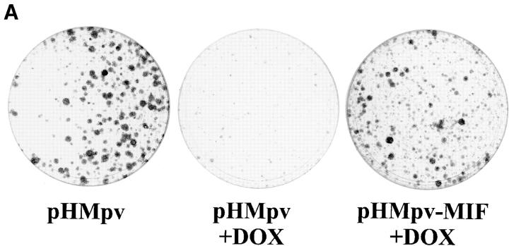
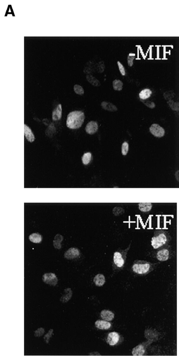
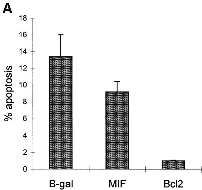
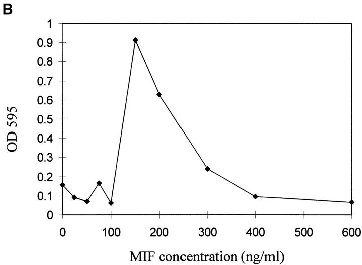
p53 has a key role in the negative regulation of cell proliferation, in the maintenance of genomic stability, and in the suppression of transformation and tumorigenesis. To identify novel regulators of p53, we undertook two functional screens to isolate genes which bypassed either p53-mediated growth arrest or apoptosis. In both screens, we isolated cDNAs encoding macrophage migration inhibitory factor (MIF), a cytokine that was shown previously to exert both local and systemic proinflammatory activities. Treatment with MIF overcame p53 activity in three different biological assays, and suppressed its activity as a transcriptional activator. The observation that a proinflammatory cytokine, MIF, is capable of functionally inactivating a tumor suppressor, p53, may provide a link between inflammation and tumorigenesis.
Overview
The claims and statistics of this paper haven’t been checked yet.
Run the check — free, no account needed. It reads the open-access text and checks the sources it cites.
Integrity signals
Registry
Cites
Statistics
Figures
Verification
Claims the paper makes
The claims and statistics of this paper haven’t been checked yet.
Run the check — free, no account needed. It reads the open-access text and checks the sources it cites.
Claims other papers make about this one
Each is checked against this paper’s own text: does it say what the citing paper says it does?
In the citing paper
“MIF is a potent cytokine that overcomes p53 function by suppressing its transcriptional activity33.”
From Inflammation and cancer (2002) · cites this paper as [33]
In the citing paper
“tetracycline regulated expression of wild-type p53 (GFP tagged) in the TGP53-4 mouse cell line (27) caused a significant decline in endogenous PGM activity (Fig. 5B).”
From Glycolytic Enzymes Can Modulate Cellular Life Span (2005) · cites this paper as [27]
Self-check
Percentages, sums and reported differences re-computed from the paper’s own numbers.
The self-check needs the paper’s full text.
Citations
Cites
The serial cultivation of human diploid cell strains
Hayflick, L., & Moorhead, P. S. (1961) · Experimental Cell Research · doi:10.1016/0014-4827(61)90192-6 · Cited by 8,508
WAF1, a potential mediator of p53 tumor suppression
El-Deiry, W. S., Tokino, T., Velculescu, V. E., Levy, D. B., Parsons, R., Trent, J. M., Lin, D., Mercer, W. E., Kinzler, K. W., & Vogelstein, B. (1993) · Cell · doi:10.1016/0092-8674(93)90500-p · Cited by 8,321
The p21 Cdk-interacting protein Cip1 is a potent inhibitor of G1 cyclin-dependent kinases.
Wade Harper, J. (1993) · Cell · doi:10.1016/0092-8674(93)90499-g · Cited by 5,651
Lane, D. P. (1992) · Nature · doi:10.1038/358015a0 · Cited by 5,138
NITRIC OXIDE AND MACROPHAGE FUNCTION
MacMicking, J. D., Xie, Q. W., & Nathan, C. (1997) · Annual Review of Immunology · doi:10.1146/annurev.immunol.15.1.323 · Cited by 4,245
Tumor suppressor p53 is a direct transcriptional activator of the human bax gene
Toshiyuki, M., & Reed, J. C. (1995) · Cell · doi:10.1016/0092-8674(95)90412-3 · Cited by 4,063
Participation of p53 protein in the cellular response to DNA damage.
Kastan, M. B., Onyekwere, O. C., Sidransky, D., Vogelstein, B., & Craig, R. W. (1991) · PubMed · Cited by 3,761
p21 is a universal inhibitor of cyclin kinases
Xiong, Y., Hannon, G., Zhang, H., Casso, D., Kobayashi, R., & Beach, D. (1993) · Nature · doi:10.1038/366701a0 · Cited by 3,691
Self-citationp53 is required for radiation-induced apoptosis in mouse thymocytes
Lowe, S. W., Schmitt, E. M., Smith, S. W., Osborne, B. A., & Jacks, T. (1993) · Nature · doi:10.1038/362847a0 · Cited by 2,864
Transcriptional Activation by Tetracyclines in Mammalian Cells
Gossen, M., Freundlieb, S., Bender, G., Müller, G., Hillen, W., & Bujard, H. (1995) · Science · doi:10.1126/science.7792603 · Cited by 2,488 · cited 1×
“the tetracycline inducible system (22), and pBabe-puro-tet-GFP-”
Research works printed but not linked: 1
- Christen, 1999, Chronic infection and inflammation lead to cancer
Cited by
Sorted by citations
Items 601–649 of 649
Genetic Diagnosis on Hepatic Metastasis from Colorectal Cancer
Zhang, S. (2016) · Springer eBooks · doi:10.1007/978-94-017-7755-1_6
Napolitano, L., Waku, M., & Innocenti, P. (2005) · European Journal of Inflammation · doi:10.1177/1721727x0500300101
Role of MIF in Experimental Autoimmune Encephalomyelitis and Multiple Sclerosis
Benedek, G., Meza‐Romero, R., & Vandenbark, A. A. (2017) · Springer eBooks · doi:10.1007/978-3-319-52354-5_6
Baker, T., Lee, P. J., & Sauler, M. (2017) · Springer eBooks · doi:10.1007/978-3-319-52354-5_8
Piva, M. R., & de Souza, L. B. (2009)
promote ovarian cancer growth via mobilization of protumor small peritoneal macrophages
Rei, M., Gonçalves‐Sousa, N., Lança, T., Thompson, R. G., Mensurado, S., Balkwill, F. R., Kulbe, H., Pennington, D. J., & Silva‐Santos, B. (2014)
de Freitas, T. E. (2017) · Digital Library of Theses and Dissertations (Universidade de São Paulo) · doi:10.11606/t.42.2017.tde-17112017-094057
The Tumor Microenvironment of Mif Deficient and Wild Type Mouse Injected of Colon Carcinoma
Tadesse, F. (2020) · Biomedical Journal of Scientific & Technical Research · doi:10.26717/bjstr.2020.25.004259
지질다당질로 유도된 백서 방광염에서 염증전구물질 MIF의 발현
Kim, J., 이광우, 김영호, & 김민의 (2007)
Mitochondrial metabolism and carcinogenesis
Aras, S., Purandare, N., & Grossman, L. I. (2021) · Elsevier eBooks · doi:10.1016/b978-0-12-822416-8.00001-4
Villalona-Calero, M. A. (2007) · Humana Press eBooks · doi:10.1007/978-1-59745-455-1_23
HSP90-stabilized proteins as therapeutic targets in cancer
Klemke, L. (2021) · eDiss (Georg-August-Universität Göttingen) · doi:10.53846/goediss-8679
Morand, E. F. (2009) · Faculty Opinions – Post-Publication Peer Review of the Biomedical Literature · doi:10.3410/f.1157882.619134
Satoskar, A. R., & Oghumu, S. (2012) · Faculty Opinions – Post-Publication Peer Review of the Biomedical Literature · doi:10.3410/f.717968371.793468086
Ying, L., Marino, J., Hussain, S. P., Khan, M., You, S., Hofseth, A. B., Trivers, G. E., Dixon, D. A., Harris, C. C., & Hofseth, L. J. (2023) · doi:10.1158/0008-5472.c.6494538.v1
Ying, L., Marino, J., Hussain, S. P., Khan, M. A. S., You, S., Hofseth, A. B., Trivers, G. E., Dixon, D. A., Harris, C. C., & Hofseth, L. J. (2023) · doi:10.1158/0008-5472.c.6494538
Data from Glycolytic Enzymes Can Modulate Cellular Life Span
(2023) · doi:10.1158/0008-5472.c.6493056.v1
Self-citationMiędzybrodzka, A., Kawczyk‐Krupka, A., Aebisher, D., Cieślar, G., & Czuba, Z. P. (2023) · European Journal of Clinical and Experimental Medicine · doi:10.15584/ejcem.2023.2.2
Gastric Cancer: H. pylori and Macrophage Migration Inhibitory Factor
Yoon, K. (2023) · Springer eBooks · doi:10.1007/978-981-97-0013-4_25
Thiele, M. (2024) · Elsevier eBooks · doi:10.1016/b978-0-443-14064-8.00004-7
Sinitsky, M. Y., Sinitskaya, А. V., Khutornaya, M. V., Асанов, М. А., Shishkova, D. K., Олеговна, П. А., & Ponasenko, A. V. (2024) · Российский физиологический журнал им И М Сеченова · doi:10.31857/s0869813924040087
The multifaceted role of macrophage migration inhibitory factor (MIF) in innate tumor immunity
Thiele, M. (2024) · Elsevier eBooks · doi:10.1016/b978-0-443-14064-8.90004-3
Gao, X., Cao, S., Ren, H., Wang, X., Chen, J., & Huang, H. (2025) · Discover Medicine · doi:10.1007/s44337-025-00361-2
The Role of Proinflammatory Biomarkers in Oral Cancer Pathophysiology
Yılmaz, B., & Ozpolat, B. (2025) · Advances in experimental medicine and biology · doi:10.1007/978-3-032-03176-1_17
Single-cell RNA sequencing revealed heterogeneity of the immune microenvironment in gastric cancerPreprint
Liu, G., Liu, Y., Ding, Q.-Q., Zhao, H.-C., Sun, J., & Yang, H. (2025) · Research Square · doi:10.21203/rs.3.rs-7504119/v1
Wang, Y. (2011) · Illinois Digital Environment for Access to Learning and Scholarship (University of Illinois at Urbana-Champaign)
Structure, function, and mechanism of human MIF and parasitic orthologs
Zierow, S., & Bernhagen, J. (2008) · RWTH Publications (RWTH Aachen)
Jia, X.-H., Geng, L.-Y., Jiang, P.-P., Xu, H., Nan, K.-J., Yao, Y., Jiang, L.-L., Sun, H., Qin, T.-J., & Guo, H. (2020) · Figshare · doi:10.6084/m9.figshare.13468451
Jia, X.-H., Geng, L.-Y., Jiang, P.-P., Xu, H., Nan, K.-J., Yao, Y., Jiang, L.-L., Sun, H., Qin, T.-J., & Guo, H. (2020) · Figshare · doi:10.6084/m9.figshare.13468447
Chronic pancreatitis and pancreatic cancer: It is in the genes!
Jain, K., & Midha, S. (2026) · Pancreatology · doi:10.1016/j.pan.2026.01.002
Bagratuni, T., Vlachos, C., Σάκκου, Μ., Kollias, I., Theologi, O., Koutsoukou, N. M., Liacos, C., Taouxi, K., Papadimou, A., Chrisostomidou, K., et al. (2026) · Research Square · doi:10.21203/rs.3.rs-8594385/v1
Impact of Targeting the Autophagy Related Gene Beclin 1 on the Immune Landscape of Melanoma
Arakelian, T. (2018) · theses.fr (ABES) · doi:10.70675/cd5f1ed6z9100z4dd3zb7fazb0537054f400
Analyse évolutive et fonctionnelle des Macrophage Migration Inhibitory Factors chez les eucaryotes
Michelet, C. (2018) · theses.fr (ABES) · doi:10.70675/8dfb07fcz29b8z47a4z9ca2z535bbf43cda6
Saccone, M. A. (2019) · theses.fr (ABES) · doi:10.70675/d8995276z6866z4372zbbc2z271914e8a438
Mutant p53 gains RNA-binding activity to reprogram translation and mitochondrial function
Zhou, W., Long, A., Douglas, C. J., Gallegos, D., Burke, J. M., MacMillan, D. W. C., Hacisuleyman, E., & Seath, C. P. (2026) · Cell Reports · doi:10.1016/j.celrep.2026.117856 · Cited by 1
Krohn, R. M., & Weber, C. (2008) · RWTH Publications (RWTH Aachen)
Metadata
- Authors
- Hudson, J. D., Shoaibi, M. A., Maestro, R., Carnero, A., Hannon, G. J., & Beach, D. H.
- Affiliations
- aUnit of Cancer Biology, Institute of Child Health, London WC1N 1EH, United Kingdom
- Centro di Riferimento Oncologico (IT)
- Cold Spring Harbor Laboratory (US)
- Venue
- The Journal of Experimental Medicine
- Publisher
- Rockefeller University Press
- Published
- 1999-11-15
- Volume
- 190
- Issue
- 10
- Pages
- 1375-1382
- ISSN
- 0022-1007 (ISSN-L)
- 1540-9538
- PMC
- PMC2195698
- OpenAlex
- W2110929110
- Cited by
- 649
- References
- 48
- Citations by year
- 2026: 8 · 2025: 12 · 2024: 14 · 2023: 10 · 2022: 12 · 2021: 18 · 2020: 25 · 2019: 17 · 2018: 14 · 2017: 14 · 2016: 27 · 2015: 24 · 2014: 25 · 2013: 25 · 2012: 38
- FWCI
- 9.23
- Type
- journal article
- Language
- en
- Open access
- bronze
- doi.org
- Locations
- The Journal of Experimental Medicine (publishedVersion)
- PubMed Central (publishedVersion, cc-by-nc-sa)
- PubMed (publishedVersion)
- Cold Spring Harbor Laboratory Institutional Repository (Cold Spring Harbor Laboratory) (acceptedVersion)
- DIGITAL.CSIC (Spanish National Research Council (CSIC)) (submittedVersion, other-oa)
- Funders
- University College London
- Associazione Italiana per la Ricerca sul Cancro
- Topics
- Life Sciences › Immunology and Microbiology › Immunology › Macrophage Migration Inhibitory Factor
- Health Sciences › Medicine › Oncology › Cancer-related Molecular Pathways
- Life Sciences › Biochemistry, Genetics and Molecular Biology › Molecular Biology › Mechanisms of cancer metastasis
- Keywords
- p53, macrophage migration inhibitory factor, inflammatory cytokines, tumor suppressor inactivation, inflammation and tumorigenesis, p53 transcriptional activation, apoptosis, cell proliferation, genome stability
- MeSH terms
- Animals, Cell Line, Chronic Disease, Fibroblasts, Inflammation, Macrophage Migration-Inhibitory Factors, Neoplasms, Nitric Oxide, Transcriptional Activation, Tumor Suppressor Protein p53, Apoptosis, Mice, Rats
About these checks
Registry, retraction, Cites and statistics results are computed, not judged, with one exception: a printed reference that no index lists is read once by a model before it counts as a citation error. Claims are extracted by a model and kept only when their quote appears verbatim in the text; each cited source is then checked, with a model’s help, for whether it says what the claim relies on. Those verdicts can be wrong — read the claim’s quote and the cited source before relying on one.
Statistics and arithmetic are recomputed, not judged.
Confidence bands are shown only for fields calibrated on our annotated test set. None is calibrated yet, so no band is shown.
What counts as a claim ›How we measure confidence ›How we check this ›
Are you an author, or do you see a problem with this page? Request a review
