

The DOI matches its Crossref record; 25 of the 25 references it cites were found in a registry or an index.
DC-STAMP is essential for cell–cell fusion in osteoclasts and foreign body giant cells
Open accessAbstract
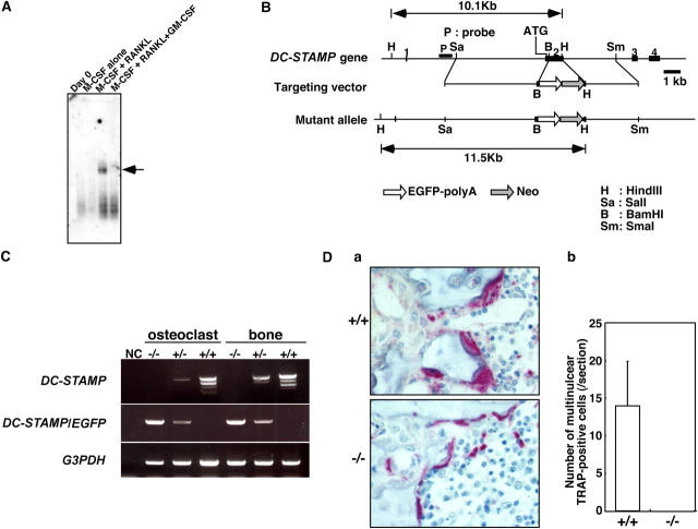
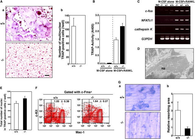
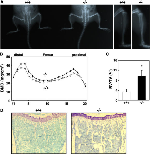
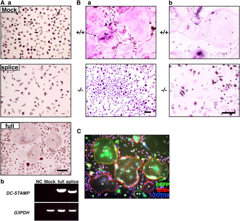
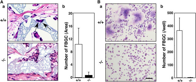
Osteoclasts are bone-resorbing cells that play a pivotal role in bone remodeling. Osteoclasts form large multinuclear giant cells by fusion of mononuclear osteoclasts. How cell fusion is mediated, however, is unclear. We identify the dendritic cell–specific transmembrane protein (DC-STAMP), a putative seven-transmembrane protein, by a DNA subtraction screen between multinuclear osteoclasts and mononuclear macrophages. DC-STAMP is highly expressed in osteoclasts but not in macrophages. DC-STAMP–deficient mice were generated, and osteoclast cell fusion was completely abrogated in homozygotes despite normal expression of osteoclast markers and cytoskeletal structure. As osteoclast multinucleation was restored by retroviral introduction of DC-STAMP, loss of cell fusion was directly attributable to a lack of DC-STAMP. Defects in osteoclast multinucleation reduce bone-resorbing activity, leading to osteopetrosis. Similar to osteoclasts, foreign body giant cell formation by macrophage cell fusion was also completely abrogated in DC-STAMP–deficient mice. We have thus identified an essential regulator of osteoclast and macrophage cell fusion, DC-STAMP, and an essential role of osteoclast multinucleation in bone homeostasis.
Overview
The claims and statistics of this paper haven’t been checked yet.
Run the check — free, no account needed. It reads the open-access text and checks the sources it cites.
Integrity signals
Registry
Cites
Statistics
Figures
Verification
Claims the paper makes
The claims and statistics of this paper haven’t been checked yet.
Run the check — free, no account needed. It reads the open-access text and checks the sources it cites.
Claims other papers make about this one
Each is checked against this paper’s own text: does it say what the citing paper says it does?
The cited source says what the claim attributes to it.
In the citing paper
“Macrophage fusion was completely abrogated in DC-STAMP deficient mice.[68]”
From Foreign body reaction to biomaterials (2008) · cites this paper as [68]
The cited source says what the claim attributes to it.
In the citing paper
“Dendritic cell-specific transmembrane protein (DC-STAMP) is necessary for fusion in the formation of FBGCs.”
From Foreign body reaction to biomaterials (2008) · cites this paper as [68]
In the citing paper
“NPBs induce osteoclasts to acquire a nonresorbing phenotype by inhibiting c-Src, which is essential for cytoskeletal construction,158 and by stimulating dendritic cell-specific transmembrane protein (DC-STAMP),159 which is necessary for cell fusion.”
From Bone remodeling: an operational process ensuring survival and bone mechanical competence (2022) · cites this paper as [159]
Self-check
Percentages, sums and reported differences re-computed from the paper’s own numbers.
The self-check needs the paper’s full text.
Citations
Cites
Takayanagi, H., Kim, S., Koga, T., Nishina, H., Isshiki, M., Yoshida, H., Saiura, A., Isobe, M., Yokochi, T., Inoue, J.-I., et al. (2002) · Developmental Cell · doi:10.1016/s1534-5807(02)00369-6 · Cited by 2,701
Yoshida, H., Hayashi, S.-I., Kunisada, T., Ogawa, M., Nishikawa, S., Okamura, H., Sudo, T., Shultz, L. D., & Nishikawa, S.-I. (1990) · Nature · doi:10.1038/345442a0 · Cited by 1,789
c-Fos: a Key Regulator of Osteoclast-Macrophage Lineage Determination and Bone Remodeling
Grigoriadis, A. E., Wang, Z., Cecchini, M. G., Hofstetter, W., Felix, R., Fleisch, H. A., & Wagner, E. F. (1994) · Science · doi:10.1126/science.7939685 · Cited by 1,264
ER–phagosome fusion defines an MHC class I cross-presentation compartment in dendritic cells
Guermonprez, P., Saveanu, L., Kleijmeer, M. J., Davoust, J. M., Endert, P. V., & Amigorena, S. (2003) · Nature · doi:10.1038/nature01911 · Cited by 753
Arai, F., Miyamoto, T., Ohneda, O., Inada, T., Sudo, T., Brasel, K., Miyata, T., Anderson, D. M., & Suda, T. (1999) · The Journal of Experimental Medicine · doi:10.1084/jem.190.12.1741 · Cited by 693
Self-citationNuclear Factor of Activated T-cells (NFAT) Rescues Osteoclastogenesis in Precursors Lacking c-Fos
Matsuo, K., Galson, D. L., Chen, Z., Peng, L., Laplace, C., Wang, K. Z. Q., Bachler, M. A., Amano, H., Aburatani, H., Ishikawa, H., et al. (2004) · Journal of Biological Chemistry · doi:10.1074/jbc.m313973200 · Cited by 570
Requirement of pp60c-src expression for osteoclasts to form ruffled borders and resorb bone in mice.
Boyce, B. F., Yoneda, T., Lowe, C., Soriano, P. M., & Mundy, G. R. (1992) · Journal of Clinical Investigation · doi:10.1172/jci116032 · Cited by 553
RANKL-induced DC-STAMP Is Essential for Osteoclastogenesis
Kukita, T., Wada, N., Kukita, A., Kakimoto, T., Sandra, F., Toh, K., Nagata, K., Iijima, T., Horiuchi, M., Matsusaki, H., et al. (2004) · The Journal of Experimental Medicine · doi:10.1084/jem.20040518 · Cited by 357
(1995) · Europe PMC (PubMed Central) · Cited by 294
Bifurcation of osteoclasts and dendritic cells from common progenitors
Miyamoto, T. K., Ohneda, O., Arai, F., Iwamoto, K., Okada, S., Takagi, K., Anderson, D. M., & Suda, T. (2001) · Blood · doi:10.1182/blood.v98.8.2544 · Cited by 280
Self-citation
Research works printed but not linked: 1
- 1995, Am. J. Pathol., 147, 1487
Cited by
Sorted by citations
Foreign body reaction to biomaterials
Anderson, J. M., Rodriguez, A., & Chang, D. T. (2008) · Seminars in Immunology · doi:10.1016/j.smim.2007.11.004 · Cited by 5,138
Alternative Activation of Macrophages: An Immunologic Functional Perspective
Martinez, F. O., Helming, L., & Gordon, S. (2009) · Annual Review of Immunology · doi:10.1146/annurev.immunol.021908.132532 · Cited by 2,745
Osteoimmunology: shared mechanisms and crosstalk between the immune and bone systems
Takayanagi, H. (2007) · Nature Reviews Immunology · doi:10.1038/nri2062 · Cited by 1,878
Functions of RANKL/RANK/OPG in bone modeling and remodeling
Boyce, B. F., & Xing, L. (2008) · Archives of Biochemistry and Biophysics · doi:10.1016/j.abb.2008.03.018 · Cited by 1,844
Franz, S., Rammelt, S., Scharnweber, D., & Simon, J. C. (2011) · Biomaterials · doi:10.1016/j.biomaterials.2011.05.078 · Cited by 1,391
Metadata
- Authors
- Yagi, M., Miyamoto, T., Sawatani, Y., Iwamoto, K., Hosogane, N., Fujita, N., Morita, K., Ninomiya, K., Suzuki, T., Miyamoto, K., Oike, Y., Takeya, M., Toyama, Y., & Suda, T.
- Affiliations
- Keio University (JP)
- Kumamoto University (JP)
- Kyushu University (JP)
- 1Department of Cell Differentiation, The Sakaguchi Laboratory
- ORCID
- Venue
- The Journal of Experimental Medicine
- Publisher
- Rockefeller University Press
- Published
- 2005-08-01
- Volume
- 202
- Issue
- 3
- Pages
- 345-351
- ISSN
- 0022-1007 (ISSN-L)
- 1540-9538
- PMC
- PMC2213087
- OpenAlex
- W2111239144
- Cited by
- 890
- References
- 24
- Citations by year
- 2026: 36 · 2025: 35 · 2024: 33 · 2023: 36 · 2022: 56 · 2021: 48 · 2020: 60 · 2019: 39 · 2018: 40 · 2017: 52 · 2016: 48 · 2015: 48 · 2014: 50 · 2013: 37 · 2012: 46
- FWCI
- 11.80
- Type
- journal article
- Language
- en
- Open access
- bronze
- doi.org
- Locations
- The Journal of Experimental Medicine (publishedVersion)
- Europe PMC (PubMed Central) (publishedVersion, cc-by-nc-sa)
- PubMed Central (publishedVersion, cc-by-nc-sa)
- PubMed (publishedVersion)
- Topics
- Life Sciences › Biochemistry, Genetics and Molecular Biology › Molecular Biology › Bone Metabolism and Diseases
- Life Sciences › Immunology and Microbiology › Immunology › Phagocytosis and Immune Regulation
- Health Sciences › Medicine › Rheumatology › Osteomyelitis and Bone Disorders Research
- Keywords
- DC-STAMP, cell fusion, osteoclasts, foreign body giant cells, multinucleation, osteoclastogenesis, bone resorption, osteopetrosis, macrophages, bone homeostasis, seven-transmembrane proteins
- MeSH terms
- Animals, Bone and Bones, Bone Resorption, Cell Fusion, Homeostasis, Macrophages, Membrane Proteins, Nerve Tissue Proteins, Osteoclasts, Giant Cells, Mice, Knockout, Mice
About these checks
Registry, retraction, Cites and statistics results are computed, not judged, with one exception: a printed reference that no index lists is read once by a model before it counts as a citation error. Claims are extracted by a model and kept only when their quote appears verbatim in the text; each cited source is then checked, with a model’s help, for whether it says what the claim relies on. Those verdicts can be wrong — read the claim’s quote and the cited source before relying on one.
Statistics and arithmetic are recomputed, not judged.
Confidence bands are shown only for fields calibrated on our annotated test set. None is calibrated yet, so no band is shown.
What counts as a claim ›How we measure confidence ›How we check this ›
Are you an author, or do you see a problem with this page? Request a review
