

The DOI matches its Crossref record; 76 of the 76 references it cites were found in a registry or an index.
Targeting iron homeostasis induces cellular differentiation and synergizes with differentiating agents in acute myeloid leukemia
Open accessAbstract
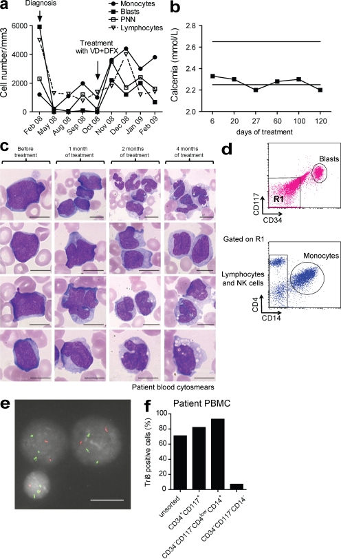
Differentiating agents have been proposed to overcome the impaired cellular differentiation in acute myeloid leukemia (AML). However, only the combinations of all-trans retinoic acid or arsenic trioxide with chemotherapy have been successful, and only in treating acute promyelocytic leukemia (also called AML3). We show that iron homeostasis is an effective target in the treatment of AML. Iron chelating therapy induces the differentiation of leukemia blasts and normal bone marrow precursors into monocytes/macrophages in a manner involving modulation of reactive oxygen species expression and the activation of mitogen-activated protein kinases (MAPKs). 30% of the genes most strongly induced by iron deprivation are also targeted by vitamin D3 (VD), a well known differentiating agent. Iron chelating agents induce expression and phosphorylation of the VD receptor (VDR), and iron deprivation and VD act synergistically. VD magnifies activation of MAPK JNK and the induction of VDR target genes. When used to treat one AML patient refractory to chemotherapy, the combination of iron-chelating agents and VD resulted in reversal of pancytopenia and in blast differentiation. We propose that iron availability modulates myeloid cell commitment and that targeting this cellular differentiation pathway together with conventional differentiating agents provides new therapeutic modalities for AML.
Overview
The claims and statistics of this paper haven’t been checked yet.
Run the check — free, no account needed. It reads the open-access text and checks the sources it cites.
Integrity signals
Registry
Cites
Statistics
Figures
Verification
Claims the paper makes
The claims and statistics of this paper haven’t been checked yet.
Run the check — free, no account needed. It reads the open-access text and checks the sources it cites.
Claims other papers make about this one
Each is checked against this paper’s own text: does it say what the citing paper says it does?
The cited source says what the claim attributes to it.
In the citing paper
“Through a comparable pathway, cellular superoxide generation can increase after exposure to iron chelators [48,49].”
From Asbestos and Iron (2023) · cites this paper as [49]
Self-check
Percentages, sums and reported differences re-computed from the paper’s own numbers.
The self-check needs the paper’s full text.
Citations
Cites
Analysis of Relative Gene Expression Data Using Real-Time Quantitative PCR and the 2−ΔΔCT Method
Livak, K. J., & Schmittgen, T. D. (2001) · Methods · doi:10.1006/meth.2001.1262 · Cited by 180,860
Chou, T.-C., & Talalay, P. (1984) · Advances in Enzyme Regulation · doi:10.1016/0065-2571(84)90007-4 · Cited by 7,178
Bennett, J. M., Catovsky, D., Mt, D., Flandrin, G., Galton, D. A. G., Gralnick, H. R., & Sultan, C. (1976) · British Journal of Haematology · doi:10.1111/j.1365-2141.1976.tb03563.x · Cited by 5,595
Mitogen-Activated Protein Kinase Pathways Mediated by ERK, JNK, and p38 Protein Kinases
Johnson, G. L., & Lapadat, R. (2002) · Science · doi:10.1126/science.1072682 · Cited by 4,295
Norman, A. W. (1979) · Cited by 3,769
Cellular response to oxidative stress: Signaling for suicide and survival*
Martindale, J. L., & Holbrook, N. J. (2002) · Journal of Cellular Physiology · doi:10.1002/jcp.10119 · Cited by 2,314
FoxOs Are Critical Mediators of Hematopoietic Stem Cell Resistance to Physiologic Oxidative Stress
Tothova, Z., Kollipara, R., Huntly, B. J., Lee, B. H., Castrillon, D. H., Cullen, D. E., McDowell, E. P., Lazo-Kallanian, S., Williams, I. R., Sears, C., et al. (2007) · Cell · doi:10.1016/j.cell.2007.01.003 · Cited by 1,562
Vitamin D signalling pathways in cancer: potential for anticancer therapeutics
Deeb, K. K., Trump, D. L., & Johnson, C. S. (2007) · Nature reviews. Cancer · doi:10.1038/nrc2196 · Cited by 1,419
The oxidative environment and protein damage
Davies, M. J. (2005) · Biochimica et Biophysica Acta (BBA) - Proteins and Proteomics · doi:10.1016/j.bbapap.2004.08.007 · Cited by 1,392
Löwenberg, B., Downing, J. R., & Burnett, A. K. (1999) · New England Journal of Medicine · doi:10.1056/nejm199909303411407 · Cited by 1,283
Cited by
Sorted by citations
IDH1(R132H) mutation increases murine haematopoietic progenitors and alters epigenetics
Sasaki, M., Knobbe, C. B., Munger, J. C., Lind, E. F., Brenner, D., Brüstle, A., Harris, I. S., Holmes, R., Wakeham, A., Haight, J., et al. (2012) · Nature · doi:10.1038/nature11323 · Cited by 513
Differentiation therapy revisited
de Thé, H. (2018) · Nature reviews. Cancer · doi:10.1038/nrc.2017.103 · Cited by 462
PML/RARA Oxidation and Arsenic Binding Initiate the Antileukemia Response of As2O3
Jeanne, M., Lallemand-Breitenbach, V., Ferhi, O., Koken, M. H. M., Bras, M. L., Duffort, S., Pérès, L., Berthier, C., Soilihi, H., Raught, B., et al. (2010) · Cancer Cell · doi:10.1016/j.ccr.2010.06.003 · Cited by 332
Inhibition of Endosteal Vascular Niche Remodeling Rescues Hematopoietic Stem Cell Loss in AML
Duarte, D., Hawkins, E. D., Akinduro, O., Ang, H. Y.-K., Filippo, K. D., Kong, I. Y., Haltalli, M. L. R., Ruivo, N., Straszkowski, L., Vervoort, S. J., et al. (2017) · Cell stem cell · doi:10.1016/j.stem.2017.11.006 · Cited by 328
Satoh‐Takayama, N., Lesjean‐Pottier, S., Vieira, P., Sawa, S., Eberl, G., Vosshenrich, C. A. J., & Santo, J. P. D. (2010) · The Journal of Experimental Medicine · doi:10.1084/jem.20092029 · Cited by 307
Metadata
- Authors
- Callens, C., Coulon, S., Naudin, J., Radford-Weiss, I., Boissel, N., Raffoux, E., Wang, P. H. M., Agarwal, S., Tamouza, H., Paubelle, E., Asnafi, V., Ribeil, J.-A., Dessen, P., Canioni, D., Chandesris, O., Rubio, M. T., Beaumont, C., Benhamou, M., Dombret, H., Macintyre, E., Monteiro, R. C., Moura, I. C., & Hermine, O.
- Affiliations
- Centre National de la Recherche Scientifique UMR 8147, Paris 75015, France 1
- Centre National de la Recherche Scientifique (FR)
- Faculté de Médecine, Université René Descartes Paris V, Institut Fédératif Necker, Paris 75015, France 2
- Assistance Publique – Hôpitaux de Paris (FR)
- Inserm (FR)
- Hôpital Necker-Enfants Malades (FR)
- Institut Gustave Roussy (FR)
- Corresponding author
- Hermine, Olivier
- ORCID
- Venue
- Journal of Experimental Medicine
- Publisher
- Rockefeller University Press
- Published
- 2010-04-05
- Volume
- 207
- Issue
- 4
- Pages
- 731-750
- ISSN
- 0022-1007 (ISSN-L)
- 1540-9538
- PMC
- PMC2856037
- OpenAlex
- W2137774959
- Cited by
- 197
- References
- 76
- Citations by year
- 2026: 2 · 2025: 8 · 2024: 5 · 2023: 14 · 2022: 11 · 2021: 15 · 2020: 13 · 2019: 9 · 2018: 13 · 2017: 12 · 2016: 11 · 2015: 13 · 2014: 13 · 2013: 9 · 2012: 20
- FWCI
- 11.44
- Type
- journal article
- Language
- en
- Open access
- bronze
- doi.org
- Locations
- The Journal of Experimental Medicine (publishedVersion)
- PubMed Central (publishedVersion, cc-by-nc-sa)
- PubMed (publishedVersion)
- HAL (submittedVersion)
- Funders
- Institut National de la Santé et de la Recherche Médicale
- Fondation pour la Recherche Médicale
- Fondation de France
- Topics
- Health Sciences › Medicine › Hematology › Acute Myeloid Leukemia Research
- Life Sciences › Biochemistry, Genetics and Molecular Biology › Molecular Biology › Retinoids in leukemia and cellular processes
- Health Sciences › Medicine › Hematology › Iron Metabolism and Disorders
- Keywords
- acute myeloid leukemia, iron homeostasis, iron chelation, iron starvation, cell differentiation, vitamin D3, vitamin D receptor, reactive oxygen species, monocyte-to-macrophage differentiation, pancytopenia, combination therapy, refractory leukemia
- MeSH terms
- Iron Deficiencies, Aged, Animals, Antibodies, Monoclonal, Blood Cell Count, Cell Differentiation, Cholecalciferol, Drug Synergism, Female, Granulocytes, Hematopoietic Stem Cells, Homeostasis, Humans, Hydroxycholecalciferols, Iron, Iron Chelating Agents, Male, Mice, Nude, Monocytes, Phosphorylation, Receptors, Transferrin, Leukemia, Myeloid, Acute, Antigens, CD, Gene Expression, Apoptosis, Reactive Oxygen Species, Receptors, Calcitriol, Lipopolysaccharide Receptors, Gene Expression Profiling, Mitogen-Activated Protein Kinases, Xenograft Model Antitumor Assays, CD11b Antigen, Cell Line, Tumor, JNK Mitogen-Activated Protein Kinases, Cell Proliferation, Mice, Kaplan-Meier Estimate
About these checks
Registry, retraction, Cites and statistics results are computed, not judged, with one exception: a printed reference that no index lists is read once by a model before it counts as a citation error. Claims are extracted by a model and kept only when their quote appears verbatim in the text; each cited source is then checked, with a model’s help, for whether it says what the claim relies on. Those verdicts can be wrong — read the claim’s quote and the cited source before relying on one.
Statistics and arithmetic are recomputed, not judged.
Confidence bands are shown only for fields calibrated on our annotated test set. None is calibrated yet, so no band is shown.
What counts as a claim ›How we measure confidence ›How we check this ›
Are you an author, or do you see a problem with this page? Request a review
