Cited by 247Inflammasome and immune disordersImmune Response and InflammationNF-κB Signaling Pathways

The DOI matches its Crossref record; 62 of the 62 references it cites were found in a registry or an index.

The linear ubiquitin assembly complex (LUBAC) is essential for NLRP3 inflammasome activation

Open access

Abstract

Linear ubiquitination is a newly discovered posttranslational modification that is currently restricted to a small number of known protein substrates. The linear ubiquitination assembly complex (LUBAC), consisting of HOIL-1L, HOIP, and Sharpin, has been reported to activate NF-κB–mediated transcription in response to receptor signaling by ligating linear ubiquitin chains to Nemo and Rip1. Despite recent advances, the detailed roles of LUBAC in immune cells remain elusive. We demonstrate a novel HOIL-1L function as an essential regulator of the activation of the NLRP3/ASC inflammasome in primary bone marrow–derived macrophages (BMDMs) independently of NF-κB activation. Mechanistically, HOIL-1L is required for assembly of the NLRP3/ASC inflammasome and the linear ubiquitination of ASC, which we identify as a novel LUBAC substrate. Consequently, we find that HOIL-1L−/− mice have reduced IL-1β secretion in response to in vivo NLRP3 stimulation and survive lethal challenge with LPS. Together, these data demonstrate that linear ubiquitination is required for NLRP3 inflammasome activation, defining the molecular events of NLRP3 inflammasome activation and expanding the role of LUBAC as an innate immune regulator. Furthermore, our observation is clinically relevant because patients lacking HOIL-1L expression suffer from pyogenic bacterial immunodeficiency, providing a potential new therapeutic target for enhancing inflammation in immunodeficient patients.

Overview

The claims and statistics of this paper haven’t been checked yet.

Run the check — free, no account needed. It reads the open-access text and checks the sources it cites.

A few free checks a day · papers already verified open instantly · results are public

Integrity signals

Registry

Crossref · 10.1084/jem.20132486

Cites

62 of 62 references verified0 citation errors · 0 not checkable

Statistics

Figures

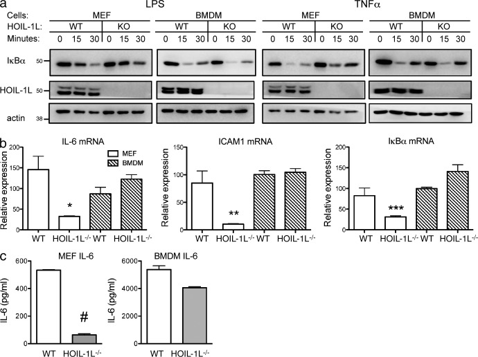
Figure 1. HOIL-1L has cell type–specific roles in NF-κB activation. (a) WT and HOIL-1L−/− MEFs and BMDMs were stimulated with 1 ng/ml TNF or 10 ng/ml LPS for 0, 15, or 30 min and probed for indicated protein levels by immunoblotting. Data are representative of three independent experiments. (b) The mRNA transcript abundance for selected NF-κB–regulated genes was quantified after 2 h of 10 ng/ml LPS stimulation by qRT-PCR (biological n = 2). Data are representative of two independent experiments. (c) IL-6 secretion was quantified by ELISA at 24 h after LPS treatment (biological n = 3). Data are representative of three independent experiments. Protein size markers are indicated in kD. All data are presented as the mean ± SEM by a Student’s t test. *, P = 0.013; **, P = 0.015; ***, P = 0.034; #, P = 10−6.
Figure 1. HOIL-1L has cell type–specific roles in NF-κB activation. (a) WT and HOIL-1L−/− MEFs and BMDMs were stimulated with 1 ng/ml TNF or 10 ng/ml LPS for 0, 15, or 30 min and probed for indicated protein levels by immunoblotting. Data are representative of three independent experiments. (b) The mRNA transcript abundance for selected NF-κB–regulated genes was quantified after 2 h of 10 ng/ml LPS stimulation by qRT-PCR (biological n = 2). Data are representative of two independent experiments. (c) IL-6 secretion was quantified by ELISA at 24 h after LPS treatment (biological n = 3). Data are representative of three independent experiments. Protein size markers are indicated in kD. All data are presented as the mean ± SEM by a Student’s t test. *, P = 0.013; **, P = 0.015; ***, P = 0.034; #, P = 10−6.CC BY-NC-SA · PubMed Central
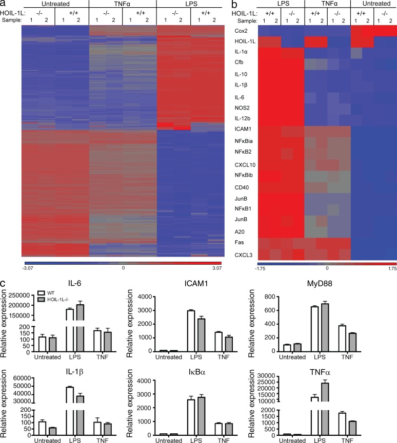
Figure 2. Global transcriptional profiles of WT and HOIL-1L−/− BMDMs in response to LPS and TNF. (a) Heat map of genes with fivefold or greater changes in expression by whole genome microarray comparison were grouped by hierarchal clustering for WT and HOIL-1L−/− BMDMs treated with 1 ng/ml TNF for 4 h, 10 ng/ml LPS for 4 h, or untreated for 4 h (biological n = 2, data for both individual replicates are presented as samples 1 and 2 for each condition). (b) Heat map of selected NF-κB genes compared by hierarchal clustering (biological n = 2, data for both individual replicates are presented as samples 1 and 2 for each condition). (c) Analysis of mRNA transcript abundance by qRT-PCR for selected genes as indicated in WT and HOIL-1L−/− BMDMs stimulated as in a (biological n = 2 and analytical n = 2 for each biological replicate). All data are presented as the mean ± SEM by a Student’s t test.
Figure 2. Global transcriptional profiles of WT and HOIL-1L−/− BMDMs in response to LPS and TNF. (a) Heat map of genes with fivefold or greater changes in expression by whole genome microarray comparison were grouped by hierarchal clustering for WT and HOIL-1L−/− BMDMs treated with 1 ng/ml TNF for 4 h, 10 ng/ml LPS for 4 h, or untreated for 4 h (biological n = 2, data for both individual replicates are presented as samples 1 and 2 for each condition). (b) Heat map of selected NF-κB genes compared by hierarchal clustering (biological n = 2, data for both individual replicates are presented as samples 1 and 2 for each condition). (c) Analysis of mRNA transcript abundance by qRT-PCR for selected genes as indicated in WT and HOIL-1L−/− BMDMs stimulated as in a (biological n = 2 and analytical n = 2 for each biological replicate). All data are presented as the mean ± SEM by a Student’s t test.CC BY-NC-SA · PubMed Central
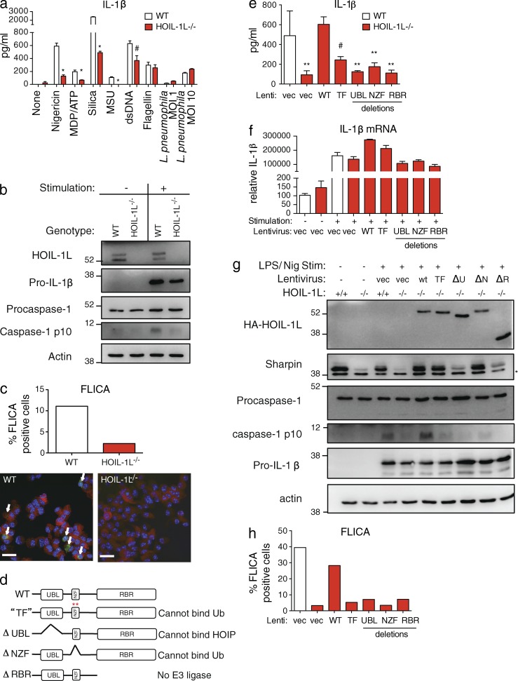
Figure 3. LUBAC is required for NLRP3 inflammasome activation in BMDMs. (a) IL-1β secretion as determined by ELISA of WT or HOIL-1L−/− BMDMs primed with LPS overnight and stimulated with nigericin, silica, MSU, MDP followed by ATP, dsDNA transfected by lipofectamine, or infected with adenovirus-Flagellin as indicated. Unprimed BMDMs were infected with L. pneumophila at indicated multiplicities of infection (MOIs). Data are presented as the mean ± SEM by a Student’s t test (biological n = 3 and are representative of two independent experiments; *, P < 0.00033; #, P = 0.011). (b) Protein expression and caspase-1 cleavage of WT or HOIL-1L−/− BMDMs untreated or primed with LPS and stimulated with nigericin was analyzed by immunoblotting. (c) BMDMs primed with LPS and stimulated with nigericin were treated with FAM-YVAD-FLICA fluorescent caspase-1 substrate during MDP treatment, fixed, and then stained for caspase-1. The percentage of FLICA-positive cells was determined for 250 total cells. Arrows point to FLICA-stained spots. Bars, 20 µm. (d–h) WT or HOIL-1L−/− BMDMs were complemented with lentiviral vectors expressing mutants described in d and analyzed for: (e) IL-1β secretion by ELISA (biological n = 3 and analytical n = 2; data presented as the mean ± SEM; **, P < 0.0075; #, P < 0.013 by a Student’s t test); (f) mRNA quantification by qRT-PCR; (g) protein expression by immunoblot; and (h) quantification of FLICA staining for active caspase-1. All data are representative of three independent experiments except for c and h, which depict combined data for all three experiments. Protein size markers are indicated in kD.
Figure 3. LUBAC is required for NLRP3 inflammasome activation in BMDMs. (a) IL-1β secretion as determined by ELISA of WT or HOIL-1L−/− BMDMs primed with LPS overnight and stimulated with nigericin, silica, MSU, MDP followed by ATP, dsDNA transfected by lipofectamine, or infected with adenovirus-Flagellin as indicated. Unprimed BMDMs were infected with L. pneumophila at indicated multiplicities of infection (MOIs). Data are presented as the mean ± SEM by a Student’s t test (biological n = 3 and are representative of two independent experiments; *, P < 0.00033; #, P = 0.011). (b) Protein expression and caspase-1 cleavage of WT or HOIL-1L−/− BMDMs untreated or primed with LPS and stimulated with nigericin was analyzed by immunoblotting. (c) BMDMs primed with LPS and stimulated with nigericin were treated with FAM-YVAD-FLICA fluorescent caspase-1 substrate during MDP treatment, fixed, and then stained for caspase-1. The percentage of FLICA-positive cells was determined for 250 total cells. Arrows point to FLICA-stained spots. Bars, 20 µm. (d–h) WT or HOIL-1L−/− BMDMs were complemented with lentiviral vectors expressing mutants described in d and analyzed for: (e) IL-1β secretion by ELISA (biological n = 3 and analytical n = 2; data presented as the mean ± SEM; **, P < 0.0075; #, P < 0.013 by a Student’s t test); (f) mRNA quantification by qRT-PCR; (g) protein expression by immunoblot; and (h) quantification of FLICA staining for active caspase-1. All data are representative of three independent experiments except for c and h, which depict combined data for all three experiments. Protein size markers are indicated in kD.CC BY-NC-SA · PubMed Central
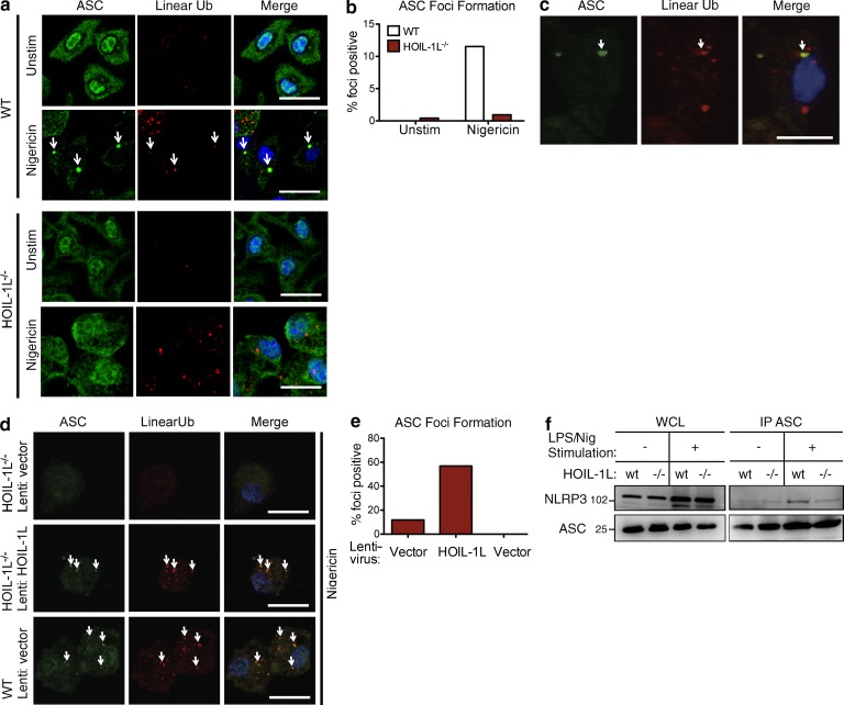
Figure 4. Assembly of the NLRP3/ASC inflammasome requires HOIL-1L. (a) Representative images of ASC foci formation (arrows) upon NLRP3 stimulation in WT or HOIL-1L−/− BMDMs with ASC staining in green, linear ubiquitin in red, and nuclei in blue. Bar, 20 µm. Images are from one of four independent experiments. (b) Quantification of ASC foci in 100 cells counted from images in a. (c) Z-stack of WT BMDMs in a. Bar, 10 µm. (d) Representative images of ASC foci formation (arrows) upon NLPR3 stimulation in HOIL-1L−/− and WT BMDMs infected with indicated lentiviruses and stained as described for a. Bar, 20 µm. Images are from one of two independent experiments. (e) Quantification of ASC foci from 30 cells combined from three experiments from images in d. (f) IP of ASC from untreated WT and HOIL-1L−/− BMDMs or after LPS priming and nigericin stimulation, data are representative of four independent experiments. Protein size markers are indicated in kD.
Figure 4. Assembly of the NLRP3/ASC inflammasome requires HOIL-1L. (a) Representative images of ASC foci formation (arrows) upon NLRP3 stimulation in WT or HOIL-1L−/− BMDMs with ASC staining in green, linear ubiquitin in red, and nuclei in blue. Bar, 20 µm. Images are from one of four independent experiments. (b) Quantification of ASC foci in 100 cells counted from images in a. (c) Z-stack of WT BMDMs in a. Bar, 10 µm. (d) Representative images of ASC foci formation (arrows) upon NLPR3 stimulation in HOIL-1L−/− and WT BMDMs infected with indicated lentiviruses and stained as described for a. Bar, 20 µm. Images are from one of two independent experiments. (e) Quantification of ASC foci from 30 cells combined from three experiments from images in d. (f) IP of ASC from untreated WT and HOIL-1L−/− BMDMs or after LPS priming and nigericin stimulation, data are representative of four independent experiments. Protein size markers are indicated in kD.CC BY-NC-SA · PubMed Central
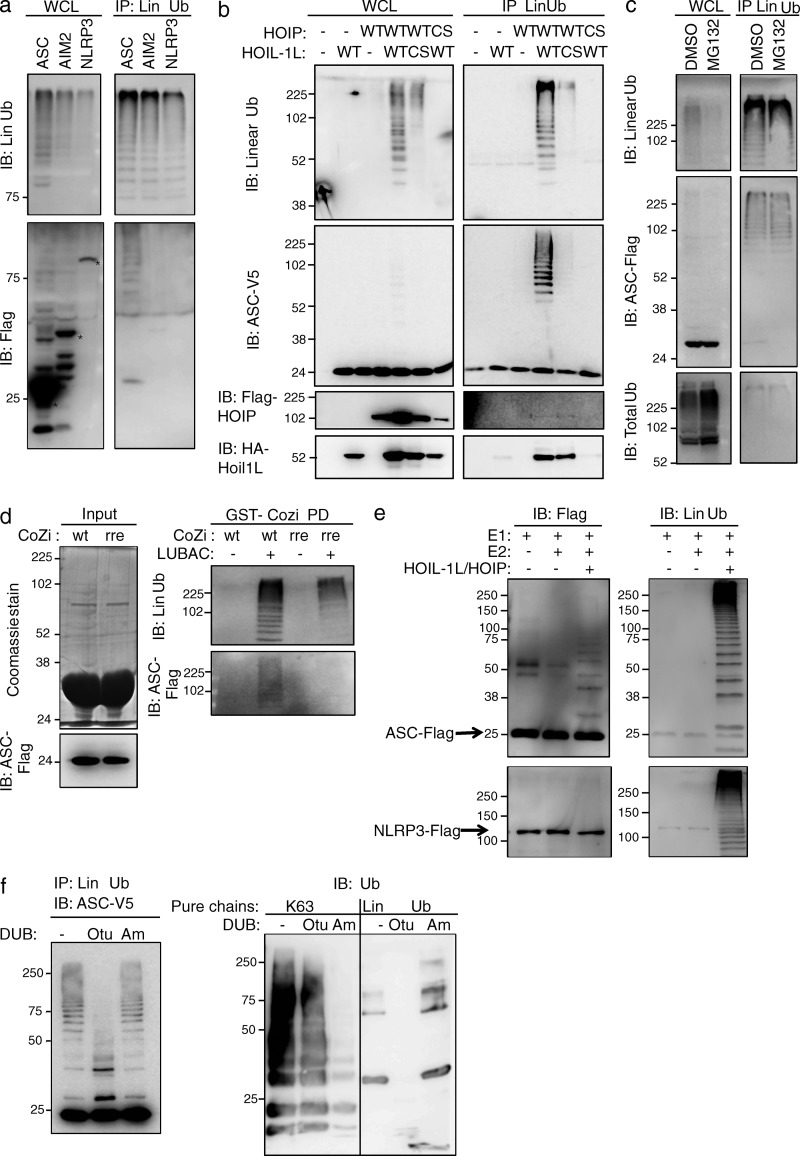
Figure 5. ASC is linear ubiquitinated by LUBAC in 293T cells and in vitro. (a) Flag-tagged ASC, AIM2, or NLRP3 were cotransfected with HOIL-1L-HA and myc-HOIP into 293T cells. Linear ubiquitinated proteins were immunoprecipitated. WCL (whole cell lysate) or immunoprecipitates (IP Lin Ub) were immunoblotted for Flag and linear ubiquitin. (b) 293T cells were transfected with a panel of WT HOIL-1L-HA and Flag-HOIP or RBR E3 ligase domain cysteine to serine point mutants (cs) and probed by linear ubiquitin IP for ASC-V5, HA, and Flag as indicated. (c) Transfected 293T cells were treated for 6 h with 20 µM MG132 or an equivalent volume of DMSO before lysates were collected and immunoprecipitated with linear ubiquitin antibody. Immunoprecipitates were probed for linear ubiquitin, Flag, and ubiquitin as indicated. (d) Bacterially purified GST-CoZi wt and rre proteins were incubated with lysates from 293T cells transfected with ASC-Flag, HOIL-1L-HA, and myc-HOIP or vector control plasmids (left, input). Proteins that were pulled down by WT or rre mutant GST-CoZi were probed for linear ubiquitin or Flag by immunoblotting (right, GST-Cozi PD). (e) ASC-Flag and NLRP3-Flag were purified from 293T lysates in a nondenaturing high salt buffer (500 mM NaCl) and combined as indicated with E1 (Ube1), E2 (UbcH5c), and LUBAC (HOIL-1L/HOIP) purified from bacteria in a linear ubiquitination reaction that was incubated for 18 h at 37°C. Reactions were stopped by the addition of loading dye and immunoblotted for Flag. Membranes were then stripped and reprobed for linear ubiquitin. (f) Deubiquitinase treatment of linear ubiquitin immunoprecipitates from 293T cell lysates overexpressing HOIL-1L-HA, myc-HOIP, and ASC-V5. Reactions were immunoblotted for linear ubiquitin and V5. Control purified ubiquitin chains were incubated in parallel in deubiquitinase reactions that were immunoblotted with a linkage nonspecific antibody against ubiquitin (Ub). All data are representative of a minimum of three independent experiments. * indicates unmodified size. Protein size markers are indicated in kD.
Figure 5. ASC is linear ubiquitinated by LUBAC in 293T cells and in vitro. (a) Flag-tagged ASC, AIM2, or NLRP3 were cotransfected with HOIL-1L-HA and myc-HOIP into 293T cells. Linear ubiquitinated proteins were immunoprecipitated. WCL (whole cell lysate) or immunoprecipitates (IP Lin Ub) were immunoblotted for Flag and linear ubiquitin. (b) 293T cells were transfected with a panel of WT HOIL-1L-HA and Flag-HOIP or RBR E3 ligase domain cysteine to serine point mutants (cs) and probed by linear ubiquitin IP for ASC-V5, HA, and Flag as indicated. (c) Transfected 293T cells were treated for 6 h with 20 µM MG132 or an equivalent volume of DMSO before lysates were collected and immunoprecipitated with linear ubiquitin antibody. Immunoprecipitates were probed for linear ubiquitin, Flag, and ubiquitin as indicated. (d) Bacterially purified GST-CoZi wt and rre proteins were incubated with lysates from 293T cells transfected with ASC-Flag, HOIL-1L-HA, and myc-HOIP or vector control plasmids (left, input). Proteins that were pulled down by WT or rre mutant GST-CoZi were probed for linear ubiquitin or Flag by immunoblotting (right, GST-Cozi PD). (e) ASC-Flag and NLRP3-Flag were purified from 293T lysates in a nondenaturing high salt buffer (500 mM NaCl) and combined as indicated with E1 (Ube1), E2 (UbcH5c), and LUBAC (HOIL-1L/HOIP) purified from bacteria in a linear ubiquitination reaction that was incubated for 18 h at 37°C. Reactions were stopped by the addition of loading dye and immunoblotted for Flag. Membranes were then stripped and reprobed for linear ubiquitin. (f) Deubiquitinase treatment of linear ubiquitin immunoprecipitates from 293T cell lysates overexpressing HOIL-1L-HA, myc-HOIP, and ASC-V5. Reactions were immunoblotted for linear ubiquitin and V5. Control purified ubiquitin chains were incubated in parallel in deubiquitinase reactions that were immunoblotted with a linkage nonspecific antibody against ubiquitin (Ub). All data are representative of a minimum of three independent experiments. * indicates unmodified size. Protein size markers are indicated in kD.CC BY-NC-SA · PubMed Central
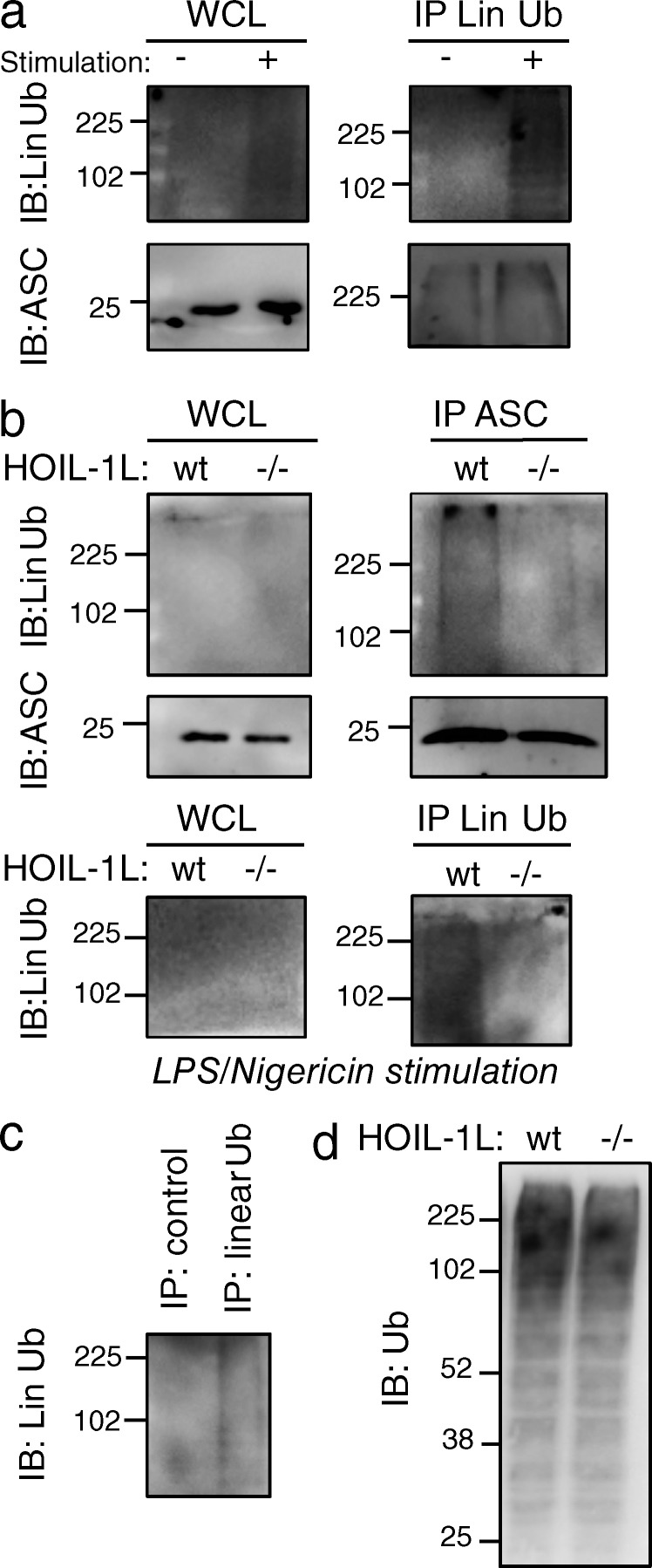
Figure 6. Endogenous mouse ASC is linear ubiquitinated in BMDMs. (a) WT BMDMs were untreated or primed with LPS overnight and stimulated with nigericin for 2 h before IP with a linear ubiquitin-specific antibody in 5M urea denaturing conditions. Immunoprecipitates (IP Lin Ub) and whole cell lysates (WCL) were immunoblotted for ASC and linear ubiquitin (Lin Ub). (b) WT and HOIL-1L−/− BMDMs were primed with LPS overnight and stimulated with nigericin for 2 h before IP with an ASC-specific antibody and extensive washing in high salt buffer (500 mM NaCl). Immunoprecipitates and WCLs were immunoblotted for ASC and linear ubiquitin. (c) WT BMDMs were primed with LPS overnight and stimulated with nigericin for 2 h before IP with a linear ubiquitin-specific antibody or an equivalent amount of control human antibody in 5M urea denaturing conditions. Immunoprecipitates and WCL were immunoblotted for linear ubiquitin. (d) WCL extracts from lysates in c were immunoblotted for ubiquitin with a linkage nonspecific antibody (Ub). All data are representative of two independent experiments. Protein size markers are indicated in kD.
Figure 6. Endogenous mouse ASC is linear ubiquitinated in BMDMs. (a) WT BMDMs were untreated or primed with LPS overnight and stimulated with nigericin for 2 h before IP with a linear ubiquitin-specific antibody in 5M urea denaturing conditions. Immunoprecipitates (IP Lin Ub) and whole cell lysates (WCL) were immunoblotted for ASC and linear ubiquitin (Lin Ub). (b) WT and HOIL-1L−/− BMDMs were primed with LPS overnight and stimulated with nigericin for 2 h before IP with an ASC-specific antibody and extensive washing in high salt buffer (500 mM NaCl). Immunoprecipitates and WCLs were immunoblotted for ASC and linear ubiquitin. (c) WT BMDMs were primed with LPS overnight and stimulated with nigericin for 2 h before IP with a linear ubiquitin-specific antibody or an equivalent amount of control human antibody in 5M urea denaturing conditions. Immunoprecipitates and WCL were immunoblotted for linear ubiquitin. (d) WCL extracts from lysates in c were immunoblotted for ubiquitin with a linkage nonspecific antibody (Ub). All data are representative of two independent experiments. Protein size markers are indicated in kD.CC BY-NC-SA · PubMed Central
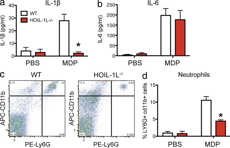
Figure 7. HOIL-1L is required for MDP-induced peritonitis in vivo. WT and HOIL-1L−/− mice were injected i.p. with PBS or MDP to induce peritonitis. 5 h later, IP wash fluid was analyzed for IL-1β (a) and IL-6 (b) levels by ELISA and infiltrating Ly6G/CD11b double-positive neutrophils (c and d) were quantified by flow cytometry (biological n = 8 from three independent experiments). All data are presented as the mean ± SEM. *, P < 0.00015 by a Student’s t test.
Figure 7. HOIL-1L is required for MDP-induced peritonitis in vivo. WT and HOIL-1L−/− mice were injected i.p. with PBS or MDP to induce peritonitis. 5 h later, IP wash fluid was analyzed for IL-1β (a) and IL-6 (b) levels by ELISA and infiltrating Ly6G/CD11b double-positive neutrophils (c and d) were quantified by flow cytometry (biological n = 8 from three independent experiments). All data are presented as the mean ± SEM. *, P < 0.00015 by a Student’s t test.CC BY-NC-SA · PubMed Central
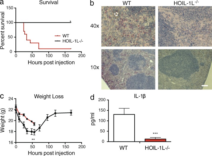
Figure 8. HOIL-1L is required for lethal inflammation upon LPS challenge in vivo. (a) Survival curve for WT or HOIL-1L−/− mice challenged with 30 mg/kg LPS (n = 10, combined from three independent experiments). (b) Apoptotic marker Annexin-V (red) and hematoxylin-eosin (H&E)–stained spleen sections from mice in a, sacrificed 24 h after LPS injection. Images are representative of three analyzed mice for each condition. Bar, 100 µm. (c) Weight curve of mice from a. (d) Serum IL-1β levels from WT and HOIL-1L−/− mice sacrificed 24 h after LPS injection were determined by ELISA (n = 4, combined from 2 independent experiments). Data are presented as the mean ± SEM. *, P < 0.0001 by a Log-rank Mantel-Cox test; **, P = 0.0015; ***, P < 0.008 by a Student’s t test.
Figure 8. HOIL-1L is required for lethal inflammation upon LPS challenge in vivo. (a) Survival curve for WT or HOIL-1L−/− mice challenged with 30 mg/kg LPS (n = 10, combined from three independent experiments). (b) Apoptotic marker Annexin-V (red) and hematoxylin-eosin (H&E)–stained spleen sections from mice in a, sacrificed 24 h after LPS injection. Images are representative of three analyzed mice for each condition. Bar, 100 µm. (c) Weight curve of mice from a. (d) Serum IL-1β levels from WT and HOIL-1L−/− mice sacrificed 24 h after LPS injection were determined by ELISA (n = 4, combined from 2 independent experiments). Data are presented as the mean ± SEM. *, P < 0.0001 by a Log-rank Mantel-Cox test; **, P = 0.0015; ***, P < 0.008 by a Student’s t test.CC BY-NC-SA · PubMed Central

Verification

Claims the paper makes

The claims and statistics of this paper haven’t been checked yet.

Run the check — free, no account needed. It reads the open-access text and checks the sources it cites.

A few free checks a day · papers already verified open instantly · results are public

Claims other papers make about this one

Each is checked against this paper’s own text: does it say what the citing paper says it does?

Self-check

Percentages, sums and reported differences re-computed from the paper’s own numbers.

The self-check needs the paper’s full text.

Citations

Cites

Show all 62 references

Cited by

Sorted by citations

See all 247

Metadata

Authors
Rodgers, M. A., Bowman, J. W., Fujita, H., Orazio, N., Shi, M., Liang, Q., Amatya, R., Kelly, T. J., Iwai, K., Ting, J., & Jung, J. U.
Affiliations
  • University of Southern California (US)
  • Kyoto University (JP)
  • Department of Molecular and Cellular Physiology, Graduate School of Medicine, Kyoto University, Sakyo-ku, Kyoto 606-8501, Japan 2
  • University of North Carolina at Chapel Hill (US)
Corresponding author
Jung, Jae U.
Venue
Journal of Experimental Medicine
Published
2014-06-23
Volume
211
Issue
7
Pages
1333-1347
ISSN
  • 0022-1007 (ISSN-L)
  • 1540-9538
OpenAlex
W2139206194
Cited by
247
References
62
Citations by year
2026: 10 · 2025: 10 · 2024: 14 · 2023: 20 · 2022: 17 · 2021: 23 · 2020: 18 · 2019: 22 · 2018: 22 · 2017: 29 · 2016: 31 · 2015: 27 · 2014: 4
FWCI
12.43
Type
journal article
Language
en
Open access
Licence
cc-by-nc-sa
Locations
  • The Journal of Experimental Medicine (publishedVersion)
  • Europe PMC (PubMed Central) (publishedVersion, cc-by-nc-sa)
  • PubMed Central (publishedVersion, cc-by-nc-sa)
  • PubMed (publishedVersion)
  • Carolina Digital Repository (University of North Carolina at Chapel Hill) (submittedVersion)
  • UNC Libraries
Funders
  • National Institutes of Health (1s10rr022508-01a1, 1f32ai096698-01)
  • Japan Society for the Promotion of Science (24112002)
  • National Heart, Lung, and Blood Institute (HL110609, R01HL110609)
  • National Cancer Institute (R01CA156330, CA180779, CA082057, P01CA180779, CA-31363, R01CA082057, CA115284, CA156330, R01CA031363, R01CA115284)
  • National Institute of Allergy and Infectious Diseases (AI073099, 1F32AI096698, R01AI073099, R37AI029564, F32-AI096698, U19AI083025, R56AI029564, AI083025, AI029564, R01AI029564)
  • National Institute of Diabetes and Digestive and Kidney Diseases (P30DK048522)
  • National Center for Research Resources (S10-RR022508)
Topics
  • Life Sciences › Biochemistry, Genetics and Molecular Biology › Molecular Biology › Inflammasome and immune disorders
  • Life Sciences › Immunology and Microbiology › Immunology › Immune Response and Inflammation
  • Life Sciences › Biochemistry, Genetics and Molecular Biology › Cancer Research › NF-κB Signaling Pathways
Keywords
LUBAC, linear ubiquitination, NLRP3 inflammasome, ASC, HOIL-1L, macrophages, IL-1β secretion, innate immunity, post-translational modification
MeSH terms
NLR Family, Pyrin Domain-Containing 3 Protein, Animals, Carrier Proteins, Humans, Immunity, Innate, Lipopolysaccharides, Macrophages, NF-kappa B, Common Variable Immunodeficiency, Mice, Knockout, GTPase-Activating Proteins, Ubiquitin-Protein Ligases, Multiprotein Complexes, Intracellular Signaling Peptides and Proteins, Mice, Interleukin-1beta, Ubiquitination, Inflammasomes

About these checks

Registry, retraction, Cites and statistics results are computed, not judged, with one exception: a printed reference that no index lists is read once by a model before it counts as a citation error. Claims are extracted by a model and kept only when their quote appears verbatim in the text; each cited source is then checked, with a model’s help, for whether it says what the claim relies on. Those verdicts can be wrong — read the claim’s quote and the cited source before relying on one.

Statistics and arithmetic are recomputed, not judged.

Confidence bands are shown only for fields calibrated on our annotated test set. None is calibrated yet, so no band is shown.

What counts as a claim ›How we measure confidence ›How we check this ›

Are you an author, or do you see a problem with this page? Request a review