

Acute mesenteric ischaemia
Open accessThe DOI matches its Crossref record; 30 of the 30 references it cites were found in a registry or an index; 24 of the 36 sources its claims cite were checked for support.
Abstract
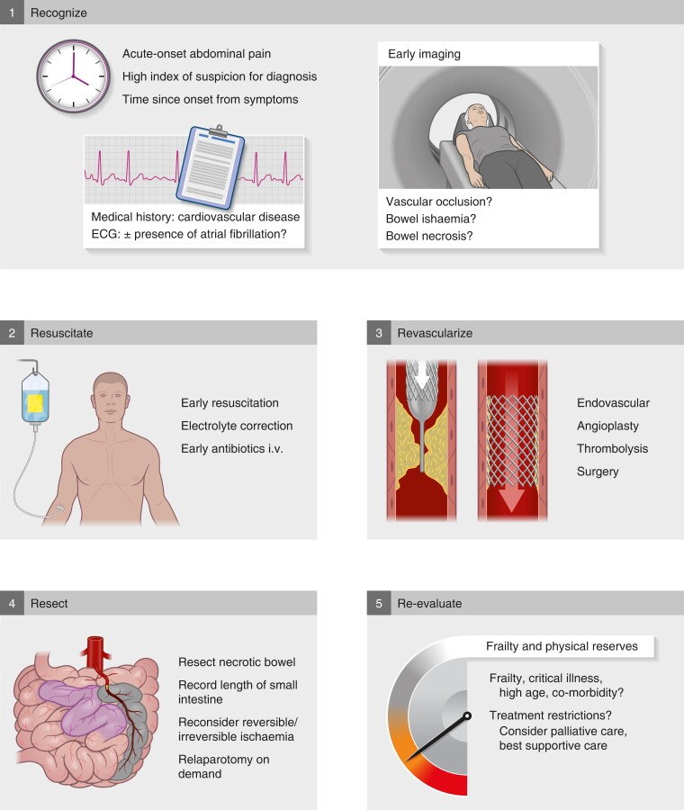
Acute mesenteric ischemia (AMI) is a relatively rare but highly lethal condition that requires emergency surgery. It is ranked as one of the most frequent causes of death among surgical diseases on a global level1. The estimated incidence is 6–12 events per 100 000 person-years2,3, and 1 patient with AMI in about every 2000 hospital admissions. The prevalence of AMI increases with age, with a median age of presentation of around 70 years, and a 10-fold higher risk in octogenarians with co-morbidities compared with the younger population2,3. Despite progress in care, the mortality rate is reported to be between 50 and 66 per cent, even in modern series. However, focus on clinical pathways and care bundles can increase awareness, speed up diagnosis, increase revascularization rates, and improve mortality rates compared with historical cohorts. Three surgical society guidelines4–6 exist for AMI, each deviating slightly from the others. The deviations likely depend on both the time of guideline development (from 2016 to 2022) and the various disciplines involved (emergency, gastrointestinal, and vascular surgeons). In addition, a separate set of guidelines7 has been provided by a radiology society, involving interventional radiology. Taken together, these recommendations for management highlight the emerging focus on AMI and the multidisciplinary approach to management that has evolved. Sudden onset of abdominal pain with symptoms out of proportion with clinical findings is typical of AMI. However, the presence of clinical symptoms may be more subtle in geriatric patients, and may depend on the underlying mechanism of mesenteric ischaemia (Fig. 1). Mechanisms of acute mesenteric ischaemia a Arterial occlusion from emboli or thrombus may occur on a background of atherosclerosis. Arterial occlusion is the most frequent cause of acute mesenteric ischaemia, in up to 70 per cent of patients. b Mesenteric occlusion is rare, but may occur in up to 20 per cent of patients. c Non-occlusive mesenteric ischaemia typically occurs in patients in the ICU or cardiac care unit, owing to hypotension from shock (for example, sepsis) or low cardiac output (for example, heart failure). The frequency of non-occlusive mesenteric ischaemia may be difficult to assess as the underlying condition may prevail over abdominal symptoms, and the correct diagnosis may not be made or suspected. About 70 per cent of cases of AMI are caused by arterial occlusion2. In patients with cardiovascular co-morbidity and atrial fibrillation (known or new-onset fibrillation) and abdominal pain, the suspicion of AMI due to arterial occlusion should be raised (Fig. 1a). A high index of clinical suspicion is important for an early diagnosis. As known for other emergency surgical conditions, diagnostic delay resulting from admission under the wrong specialty is also detrimental to outcomes for AMI, as time to intervention is crucial8,9. Mesenteric venous thrombosis is rare and accounts for 10–20 per cent of AMI cases (Fig. 1b)10. Mesenteric venous thrombosis may be caused by surgery to the upper gastrointestinal tract, and inflammatory conditions, including pancreatitis and thrombophilia. The stagnant venous flow can lead to venous gangrene of the bowel. Patients with non-occlusive mesenteric ischaemia (NOMI) typically come from the ICU or cardiac care unit11–13. NOMI is a result of hypoperfusion (from sepsis, shock, cardiac failure) and use of vasoconstrictors (Fig. 1c). Appropriate and timely imaging is the most crucial test for early diagnosis of AMI (Fig. 2). No currently available blood test is diagnostic of AMI. However, a negative D-dimer test rules out AMI. Plasma citrulline and intestinal fatty acid-binding protein have been proposed as potential biomarkers, but have not made it into clinical use owing to suboptimal accuracy11,14. Increased levels of inflammatory markers (white blood cell count, C-reactive protein) may indicate a systemic inflammatory response and systemic effects of bowel ischaemia, as well as translocation of gut bacteria. Five key steps in management of acute mesenteric ischaemia i.v., Intravenously. Arterial blood gas with lactate is also not diagnostic, and a high lactate level is unfortunately often a late sign of ischaemia. Hence, when clinical suspicion is raised, appropriate imaging should be initiated as an emergency procedure. Cross-sectional imaging with arterial contrast is warranted for rapid assessment. Resuscitation should commence immediately. CT of the abdomen with intravenous arterial contrast phase is mandatory for diagnosis4–6,15. Oral contrast is not needed for AMI assessment. The critical points to investigate include filling of the arteries and any signs of occlusions or arteriosclerosis, as well as assessment of the bowel for ischaemia. Typical signs of irreversible bowel ischaemia in the presence of AMI are intestinal dilatation and thickness, reduction, or absence of, visceral enhancement, pneumatosis intes
Overview
CoChat Summary
TL;DR
Acute mesenteric ischaemia (AMI) is a rare but highly lethal emergency surgical condition, with reported mortality of 50–70 per cent, whose outcomes depend on early clinical suspicion, prompt contrast-enhanced CT, and rapid revascularization. This narrative review summarizes presentation, diagnostic work-up, and treatment strategy across the three existing surgical society guidelines and one radiology guideline.
Key findings
- Arterial occlusion causes about 70 per cent of AMI cases; mesenteric venous thrombosis accounts for 10–20 per cent; non-occlusive mesenteric ischaemia (NOMI) arises from hypoperfusion and vasoconstrictors, typically in ICU or cardiac care patients.
- Estimated incidence is 6–12 events per 100 000 person-years, roughly 1 per 2000 hospital admissions, with median presentation around age 70 and a 10-fold higher risk in octogenarians with co-morbidities.
- No blood test is diagnostic; a negative D-dimer rules out AMI, and high lactate is often a late sign. CT with intravenous arterial contrast is mandatory, and bowel wall thinning and portomesenteric venous gas showed the highest specificity for transmural infarction (98 and 95 per cent).
- A Helsinki single-centre study reported 30-day mortality reduced to 25 per cent after implementing a multidisciplinary pathway and care bundle; a North American registry study found reduced risk of death with endovascular treatment without establishing causality. Anticoagulation for mesenteric venous thrombosis may lead to recanalization in about 80 per cent of patients.
Methods
- Narrative review synthesizing three surgical society guidelines (2016–2022) and one radiology society guideline, plus supporting literature on AMI presentation, diagnosis, and management.
- No systematic search strategy, inclusion criteria, or formal evidence appraisal is described in the text.
- Evidence cited is largely small retrospective single-centre studies, systematic reviews, and registry data; the text notes that endovascular versus open surgery has not been examined in RCTs.
Limitations
- The paper states no explicit limitations.
- Evident from the text: guideline recommendations deviate from one another (including conflicting advice on laparoscopy), and the evidence base for the open versus endovascular question rests on small retrospective single-centre studies with no RCTs.
- Evident from the text: there is a lack of data on patient-relevant outcomes other than survival, such as need for parenteral nutrition or presence of a stoma.
Model-written summary. Check it against the paper before relying on it.
Integrity signals
Registry
Cites
Statistics
Figures

Verification
Claims the paper makes
19 Valid · 0 Not valid · 3 Not checked · 8 Own results and interpretations
Every source the paper cites for this claim was checked and supports it.
In the paper
“The aim is to prevent bowel ischaemia, and the first line of treatment is anticoagulation and supportive treatment10.”
§ Acute mesenteric ischaemia > Treatment of mesenteric venous thrombosis
What the sources say
- Supportedcites [10] Acosta, 2021, Management of acute mesenteric venous thrombosis: a systematic review of contemporary studies, Scand J Surg source
The Non-Operative Therapy section says that patients diagnosed early without peritonitis are initially treated with bowel rest and full anticoagulation. This supports the cited first-line treatment statement; the review also makes clear that treatment depends on disease stage. (LLM, conf=0.90)
plain- Asserted: passes — The authors put it forward as true.
- Checkable: passes — It can be checked against evidence.
- Load-bearing: unclear — It is unclear whether the paper’s conclusions depend on it.
- Based: passes — The paper gives a reason why it holds.
- Supported
The cited sources support only part of this claim — read each source’s verdict below.
In the paper
“Patients with non-occlusive mesenteric ischaemia (NOMI) typically come from the ICU or cardiac care unit11–13.”
§ Acute mesenteric ischaemia > Presentation and clinical signs
What the sources say
- Partly supportedcites [11] Bourcier, 2022, A multicentric prospective observational study of diagnosis and prognosis features in ICU mesenteric ischemia: the DIAGOMI study, Ann Intensive Care sourceExperimental — accuracy not yet measured
The Introduction describes NOMI as the main form of mesenteric ischemia in the ICU setting, and the Discussion says it is frequently suspected there. The paper also discusses cardiac conditions associated with NOMI, but does not establish that patients typically come from a cardiac care unit. No page numbers were provided in our copy. (LLM, conf=0.84)
- Not checked — no full text of the sourcecites [12] Al-Diery, 2019, The pathogenesis of nonocclusive mesenteric ischemia: implications for research and clinical practice, J Intensive Care Med source
- Supportedcites [13] Yu, 2022, Non-occlusive mesenteric ischemia in critically ill patients, PLoS One source
The Introduction says ICU patients are frequently at risk of developing NOMI. The Results (Patient characteristics; page unavailable) report NOMI patients admitted to medical, surgical, and cardiac ICUs, supporting the cited sentence’s description of ICU and cardiac-care settings. (LLM, conf=0.85)
plainstates an association- Asserted: passes — The authors put it forward as true.
- Checkable: passes — It can be checked against evidence.
- Load-bearing: unclear — It is unclear whether the paper’s conclusions depend on it.
- Based: passes — The paper gives a reason why it holds.
- Partly supported
The cited sources support only part of this claim — read each source’s verdict below.
In the paper
“NOMI is a result of hypoperfusion (from sepsis, shock, cardiac failure) and use of vasoconstrictors (Fig. 1c).”
§ Acute mesenteric ischaemia > Presentation and clinical signs
What the sources say
- Partly supportedcites [11] Bourcier, 2022, A multicentric prospective observational study of diagnosis and prognosis features in ICU mesenteric ischemia: the DIAGOMI study, Ann Intensive Care sourceExperimental — accuracy not yet measured
The Introduction (page unavailable) says NOMI in critically ill patients is preceded by acute circulatory failure and low mesenteric flow. The Discussion (page unavailable) identifies septic and cardiogenic shock as conditions that may promote NOMI. The Results record norepinephrine use, but do not establish that vasoconstrictors cause NOMI; the claim also states a more definite causal relationship than the Discussion does. (LLM, conf=0.88)
- Not checked — no full text of the sourcecites [12] Al-Diery, 2019, The pathogenesis of nonocclusive mesenteric ischemia: implications for research and clinical practice, J Intensive Care Med source
- Partly supportedcites [13] Yu, 2022, Non-occlusive mesenteric ischemia in critically ill patients, PLoS One sourceExperimental — accuracy not yet measured
The Introduction (page unavailable) identifies septic and cardiogenic shock as risk factors for NOMI. The Discussion (page unavailable) says reports suggest an association between vasopressors and NOMI development and describes vasoconstriction reducing intestinal blood flow. The claim presents these factors as established causes and extends septic shock to sepsis generally; the paper does not make those stronger claims. (LLM, conf=0.84)
plainstates a cause- Asserted: passes — The authors put it forward as true.
- Checkable: passes — It can be checked against evidence.
- Load-bearing: unclear — It is unclear whether the paper’s conclusions depend on it.
- Based: passes — The paper gives a reason why it holds.
- Partly supported
The cited sources support only part of this claim — read each source’s verdict below.
In the paper
“As known for other emergency surgical conditions, diagnostic delay resulting from admission under the wrong specialty is also detrimental to outcomes for AMI, as time to intervention is crucial8,9.”
§ Acute mesenteric ischaemia > Presentation and clinical signs
What the sources say
- Not checked — no full text of the sourcecites [8] Magnus, 2022, Mortality and delays of management of acute mesenteric ischemia: the need of a dedicated program, Ann Vasc Surg source
- Partly supportedcites [9] Lemma, 2019, Choice of first emergency room affects the fate of patients with acute mesenteric ischaemia: the importance of referral patterns and triage, Eur J Vasc Endovasc Surg sourceExperimental — accuracy not yet measured
The Abstract (page unavailable) reports longer times to operation and higher 90-day mortality when an internist rather than a surgeon examined the patient first. The Results also report shorter times to diagnosis in the surgical-ER group. However, the Discussion says this study did not detect a correlation between time to operation and mortality, so it does not establish that the delay itself caused the worse outcomes. (LLM, conf=0.88)
boostedstates a cause- Asserted: passes — The authors put it forward as true.
- Checkable: passes — It can be checked against evidence.
- Load-bearing: unclear — It is unclear whether the paper’s conclusions depend on it.
- Based: passes — The paper gives a reason why it holds.
Every source the paper cites for this claim was checked and supports it.
In the paper
“The majority of patients require anticoagulant/antiplatelet therapy6.”
§ Acute mesenteric ischaemia > Short- and long-term outcomes
What the sources say
- Supportedcites [6] Bala, 2022, Acute mesenteric ischemia: updated guidelines of the World Society of Emergency Surgery, World J Emerg Surg source
The guidelines’ Pathophysiology and epidemiology section states that the majority of patients treated for AMI will require lifelong anticoagulant/antiplatelet therapy to prevent relapse. This directly supports the citing sentence. (LLM, conf=0.94)
plain- Asserted: passes — The authors put it forward as true.
- Checkable: passes — It can be checked against evidence.
- Load-bearing: unclear — It is unclear whether the paper’s conclusions depend on it.
- Based: passes — The paper gives a reason why it holds.
- Supported
4 more claims · 2 with their cited sources checked
Create a free account to see all 30 claims and every source verdict.
- It may state two separate claims as one. 15
- Probably not a substantial claim. 12
Citations
Cites
Editor's Choice – Management of the Diseases of Mesenteric Arteries and Veins
Björck, M., Koelemay, M., Acosta, S., Bastos Goncalves, F., Kölbel, T., Kolkman, J. J., Lees, T., Lefevre, J. H., Menyhei, G., Oderich, G., et al. (2017) · European Journal of Vascular and Endovascular Surgery · doi:10.1016/j.ejvs.2017.01.010 · Cited by 658
Global disease burden of conditions requiring emergency surgery
Stewart, B., Khanduri, P., McCord, C., Ohene-Yeboah, M., Uranues, S., Vega Rivera, F., & Mock, C. (2014) · Journal of British Surgery · doi:10.1002/bjs.9329 · Cited by 550
ESTES guidelines: acute mesenteric ischaemia
Tilsed, J. V. T., Casamassima, A., Kurihara, H., Mariani, D., Martinez, I., Pereira, J., Ponchietti, L., Shamiyeh, A., al-Ayoubi, F., Barco, L. A. B., et al. (2016) · European Journal of Trauma and Emergency Surgery · doi:10.1007/s00068-016-0634-0 · Cited by 382
Acute mesenteric ischemia: updated guidelines of the World Society of Emergency Surgery.
Bala, M., Catena, F., Kashuk, J., De Simone, B., Gomes, C. A., Weber, D., Sartelli, M., Coccolini, F., Kluger, Y., Abu-Zidan, F. M., et al. (2022) · World Journal of Emergency Surgery · doi:10.1186/s13017-022-00443-x · Cited by 380
Self-citationACR appropriateness criteria® imaging of mesenteric ischemia
Oliva, I. B., Davarpanah, A. H., Rybicki, F. J., Desjardins, B., Flamm, S. D., Francois, C. J., Gerhard-Herman, M. D., Kalva, S. P., Ashraf Mansour, M., Mohler, E. R., et al. (2013) · Abdominal Imaging · doi:10.1007/s00261-012-9975-2 · Cited by 200
Emergency general surgery in the geriatric patient
Desserud, K. F., Veen, T., & Søreide, K. (2016) · Journal of British Surgery · doi:10.1002/bjs.10044 · Cited by 176
Self-citationAl-Diery, H., Phillips, A., Evennett, N., Pandanaboyana, S., Gilham, M., & Windsor, J. A. (2019) · Journal of Intensive Care Medicine · doi:10.1177/0885066618788827 · Cited by 117
Incidence and outcomes of acute mesenteric ischaemia: a systematic review and meta-analysis
Tamme, K., Reintam Blaser, A., Laisaar, K.-T., Mändul, M., Kals, J., Forbes, A., Kiss, O., Acosta, S., Bjørck, M., & Starkopf, J. (2022) · BMJ Open · doi:10.1136/bmjopen-2022-062846 · Cited by 112
Emergency surgery in the elderly: the balance between function, frailty, fatality and futility
Søreide, K., & Desserud, K. F. (2015) · Scandinavian Journal of Trauma, Resuscitation and Emergency Medicine · doi:10.1186/s13049-015-0099-x · Cited by 103
Self-citationACR appropriateness criteria® imaging of mesenteric ischemia
Ginsburg, M., Obara, P., Lambert, D. L., Hanley, M., Steigner, M. L., Camacho, M. A., Chandra, A., Chang, K. J., Gage, K. L., Peterson, C. M., et al. (2018) · Journal of the American College of Radiology · doi:10.1016/j.jacr.2018.09.018 · Cited by 99
Oderich, G. S., Macedo, R., Stone, D. H., Woo, E. Y., Panneton, J. M., Resch, T., Dias, N. V., Sonesson, B., Schermerhorn, M. L., Lee, J. T., et al. (2018) · Journal of Vascular Surgery · doi:10.1016/j.jvs.2017.11.086 · Cited by 97
Accuracy of citrulline, I-FABP and d-lactate in the diagnosis of acute mesenteric ischemia
Nuzzo, A., Guedj, K., Curac, S., Hercend, C., Bendavid, C., Gault, N., Tran-Dinh, A., Ronot, M., Nicoletti, A., Bouhnik, Y., et al. (2021) · Scientific Reports · doi:10.1038/s41598-021-98012-w · Cited by 67
Management of Acute Mesenteric Venous Thrombosis: A Systematic Review of Contemporary Studies
Acosta, S., & Salim, S. (2021) · Scandinavian Journal of Surgery · doi:10.1177/1457496920969084 · Cited by 66
Ageno, W., Witt, D., Malato, A., Clark, N., Garcia, D., McCool, K., Siragusa, S., Dyke, S., Crowther, M., Dentali, F., et al. (2009) · Thrombosis and Haemostasis · doi:10.1160/th08-12-0842 · Cited by 61
Epidemiology of Acute Mesenteric Ischemia: A Population-Based Investigation
Kase, K., Reintam Blaser, A., Tamme, K., Mändul, M., Forbes, A., Talving, P., & Murruste, M. (2023) · World Journal of Surgery · doi:10.1007/s00268-022-06805-5 · Cited by 56
Salsano, G., Salsano, A., Sportelli, E., Petrocelli, F., Dahmane, M., Spinella, G., Pane, B., Mambrini, S., Palombo, D., & Santini, F. (2018) · CardioVascular and Interventional Radiology · doi:10.1007/s00270-017-1749-3 · Cited by 52
Tolonen, M., Lemma, A., Vikatmaa, P., Peltola, E., Mentula, P., Björkman, P., Leppäniemi, A., & Sallinen, V. (2021) · Journal of Trauma and Acute Care Surgery · doi:10.1097/ta.0000000000003305 · Cited by 46
Endovascular therapy for acute mesenteric ischemia: an NSQIP analysis
Branco, B. C., Montero-Baker, M. F., Aziz, H., Taylor, Z., & Mills, J. L. (2015) · The American Surgeon™ · doi:10.1177/000313481508101131 · Cited by 43
Zeng, Y., Yang, F., Hu, X., Zhu, F., Chen, W., & Lin, W. (2022) · European Radiology · doi:10.1007/s00330-022-09258-5 · Cited by 40
Bourcier, S., Ulmann, G., Jamme, M., Savary, G., Paul, M., Benghanem, S., Lavillegrand, J.-R., Schmidt, M., Luyt, C.-E., Maury, E., et al. (2022) · Annals of Intensive Care · doi:10.1186/s13613-022-01092-8 · Cited by 37
Björck, M., Orr, N., & Endean, E. D. (2015) · Journal of Vascular Surgery · doi:10.1016/j.jvs.2015.04.431 · Cited by 35
Lemma, A. N., Tolonen, M., Vikatmaa, P., Mentula, P., Vikatmaa, L., Kantonen, I., Leppäniemi, A., & Sallinen, V. (2019) · European Journal of Vascular and Endovascular Surgery · doi:10.1016/j.ejvs.2019.01.002 · Cited by 31
ACR Appropriateness Criteria® radiologic management of mesenteric ischemia: 2022 update
Lam, A., Kim, Y.-J., Fidelman, N., Higgins, M., Cash, B. D., Charalel, R. A., Guimaraes, M. S., Kwan, S. W., Patel, P. J., Plett, S., et al. (2022) · Journal of the American College of Radiology · doi:10.1016/j.jacr.2022.09.006 · Cited by 29
Open and endovascular management of acute mesenteric ischaemia: a systematic review
Murphy, B., Dejong, C. H. C., & Winter, D. C. (2019) · World Journal of Surgery · doi:10.1007/s00268-019-05149-x · Cited by 27
Non-occlusive mesenteric ischemia in critically ill patients
Yu, B., Ko, R.-E., Yoo, K., Gil, E., Choi, K.-J., & Park, C.-M. (2022) · PLOS ONE · doi:10.1371/journal.pone.0279196 · Cited by 27
Mortality and Delays of Management of Acute Mesenteric Ischemia: The Need of a Dedicated Program
Magnus, L., Lejay, A., Philouze, G., Chakfé, N., Collange, O., Thaveau, F., & Georg, Y. (2023) · Annals of Vascular Surgery · doi:10.1016/j.avsg.2022.12.070 · Cited by 21
Open abdomen in acute mesenteric ischemia
Acosta, S., & Kärkkäinen, J. M. (2019) · Anaesthesiology Intensive Therapy · doi:10.5114/ait.2019.86280 · Cited by 21
Local thrombolytic therapy in acute mesenteric ischemia
Yanar, F., Agcaoglu, O., Sarici, I. S., Sivrikoz, E., Ucar, A., Yanar, H., Aksoy, M., & Kurtoglu, M. (2013) · World Journal of Emergency Surgery · doi:10.1186/1749-7922-8-8 · Cited by 19
The virtual uncertainty of futility in emergency surgery
Javanmard-Emamghissi, H., & Moug, S. J. (2022) · British Journal of Surgery · doi:10.1093/bjs/znac313 · Cited by 11
Revisiting abdominal closure in mesenteric ischemia: is there an association with outcome?
Proaño-Zamudio, J. A., Argandykov, D., Renne, A. M., Gebran, A., Dorken‐Gallastegi, A., Paranjape, C. N., Kaafarani, H. M. A., King, D. R., Velmahos, G. C., & Hwabejire, J. O. (2022) · European Journal of Trauma and Emergency Surgery · doi:10.1007/s00068-022-02199-0 · Cited by 11
Cited by
Acute abdominal pain and emergency laparotomy: bundles of care to improve patient outcomes
Kokotovic, D., & Jensen, T. K. (2023) · British journal of surgery · doi:10.1093/bjs/znad224 · Cited by 29
Medical, Surgical and Experimental Approaches to Acute Mesenteric Ischemia and Reperfusion
Matković, Z., & Aleksic, Z. (2024) · Materia Socio Medica · doi:10.5455/msm.2024.36.77-81 · Cited by 14
Impact on the prognosis with the creation of a dedicated stroke to mesenteric ischaemia
Rudondy, V., Barral, P.-A., Markarian, T., Chopinet, S., Barraud, M., Gaudry, M., Bourenne, J., Nafati, C., Grigoresco, B., Lagier, D., et al. (2025) · World Journal of Emergency Surgery · doi:10.1186/s13017-025-00627-1 · Cited by 2
“Acute mesenteric ischaemia (AMI) is a very rare emergency condition, affecting approximately 0.09–0.2% of patients [1] and about 1.5% of those admitted to the Emergency Department with abdominal pain [2].”
InfluentialPredictive factors for mortality in acute mesenteric ischemia
Chen, Y., Wang, C., Tang, Y., Ma, M., Ma, Q., Zhao, Z., Liu, T., & Luo, F. (2026) · Frontiers in Surgery · doi:10.3389/fsurg.2026.1805697 · Cited by 2
Semenkova, T. N., Nikonov, A., Smetkin, A. A., Владимирович, К. В., & Кirov, М. Y. (2024) · Messenger of Anesthesiology and Resuscitation · doi:10.24884/2078-5658-2024-21-3-34-41 · Cited by 2
Metadata
- Authors
- Fuglseth, H., Søreide, K., & Vetrhus, M.
- Affiliations
- Stavanger University Hospital (NO)
- Corresponding author
- Søreide, Kjetil
- Venue
- British Journal of Surgery
- Publisher
- Oxford University Press (OUP)
- Published
- 2023-02-07
- Volume
- 110
- Issue
- 9
- Pages
- 1030-1034
- ISSN
- 0007-1323 (ISSN-L)
- 1365-2168
- PMC
- PMC10416693
- OpenAlex
- W4319333234
- Cited by
- 20
- References
- 30
- Citations by year
- 2026: 8 · 2025: 2 · 2024: 7 · 2023: 2
- FWCI
- 5.07
- Type
- journal article
- Language
- en
- Open access
- hybrid
- doi.org
- Licence
- Locations
- British journal of surgery (publishedVersion, cc-by)
- PubMed Central (publishedVersion, cc-by)
- PubMed (publishedVersion)
- NORA - Norwegian Open Research Archives (submittedVersion, cc-by)
- Topics
- Health Sciences › Medicine › Surgery › Abdominal vascular conditions and treatments
- Health Sciences › Medicine › Emergency Medicine › Appendicitis Diagnosis and Management
- Health Sciences › Medicine › Surgery › Omental and Epiploic Conditions
- Keywords
- acute mesenteric ischemia, nonocclusive mesenteric ischemia, arterial occlusion, mesenteric revascularization, emergency surgery, care pathways, mortality, elderly patients, sepsis, low cardiac output, diagnostic imaging, interventional radiology, surgical guidelines, multidisciplinary management, abdominal pain, atherosclerosis
- MeSH terms
- Acute Disease, Humans, Ischemia, Mesenteric Vascular Occlusion, Mesenteric Veins, Mesenteric Ischemia
About these checks
Registry, retraction, Cites and statistics results are computed, not judged, with one exception: a printed reference that no index lists is read once by a model before it counts as a citation error. Claims are extracted by a model and kept only when their quote appears verbatim in the text; each cited source is then checked, with a model’s help, for whether it says what the claim relies on. Those verdicts can be wrong — read the claim’s quote and the cited source before relying on one.
The claim verdicts this report counts are experimental: their accuracy is not yet measured.
A language model drew the claims from the paper on 2026-10-10, under version 3 of the claim definition (model deepseek-v4.1-flash, prompt extract-v3).
A second model labelled each claim on 2026-10-10 (model jev-1.13-20260917, definition revision gold-guideline-v0.2).
Cited sources are checked with a language model’s help (gpt-6-sol).
Statistics and arithmetic are recomputed, not judged.
Confidence bands are shown only for fields calibrated on our annotated test set. None is calibrated yet, so no band is shown.
What counts as a claim ›How we measure confidence ›How we check this ›
Are you an author, or do you see a problem with this page? Request a review
Data sources
Bibliographic data: Semantic Scholar, OpenAlex, Crossref.
Contains information from the Semantic Scholar Academic Graph Datasets, made available under the ODC Attribution License (ODC-BY 1.0).
Semantic Scholar’s data is described in: Kinney, R., et al. (2023). The Semantic Scholar Open Data Platform. arXiv:2301.10140.
