

The DOI matches its Crossref record; 105 of the 105 references it cites were found in a registry or an index.
Microglial large extracellular vesicles propagate early synaptic dysfunction in Alzheimer’s disease
Open accessAbstract
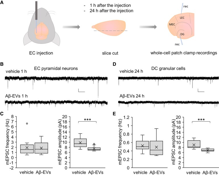
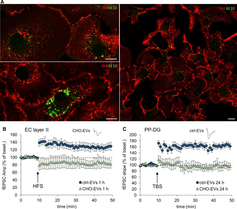
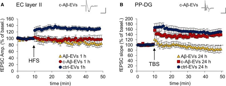
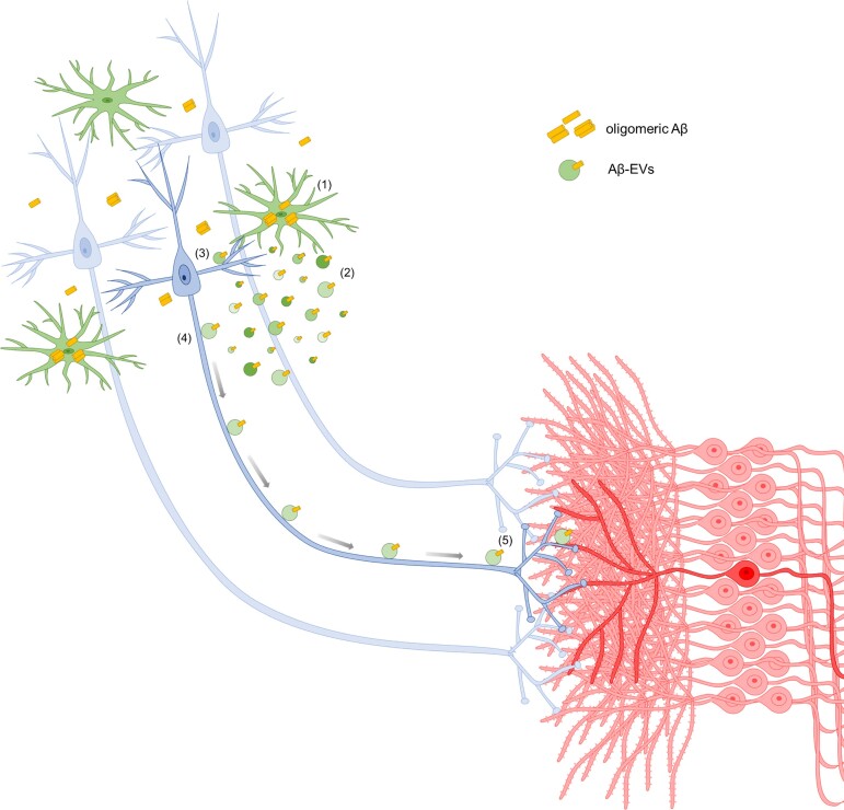
Synaptic dysfunction is an early mechanism in Alzheimer’s disease that involves progressively larger areas of the brain over time. However, how it starts and propagates is unknown. Here we show that amyloid-β released by microglia in association with large extracellular vesicles (Aβ-EVs) alters dendritic spine morphology in vitro, at the site of neuron interaction, and impairs synaptic plasticity both in vitro and in vivo in the entorhinal cortex–dentate gyrus circuitry. One hour after Aβ-EV injection into the mouse entorhinal cortex, long-term potentiation was impaired in the entorhinal cortex but not in the dentate gyrus, its main target region, while 24 h later it was also impaired in the dentate gyrus, revealing a spreading of long-term potentiation deficit between the two regions. Similar results were obtained upon injection of extracellular vesicles carrying Aβ naturally secreted by CHO7PA2 cells, while neither Aβ42 alone nor inflammatory extracellular vesicles devoid of Aβ were able to propagate long-term potentiation impairment. Using optical tweezers combined to time-lapse imaging to study Aβ-EV–neuron interaction, we show that Aβ-EVs move anterogradely at the axon surface and that their motion can be blocked through annexin-V coating. Importantly, when Aβ-EV motility was inhibited, no propagation of long-term potentiation deficit occurred along the entorhinal–hippocampal circuit, implicating large extracellular vesicle motion at the neuron surface in the spreading of long-term potentiation impairment. Our data indicate the involvement of large microglial extracellular vesicles in the rise and propagation of early synaptic dysfunction in Alzheimer’s disease and suggest a new mechanism controlling the diffusion of large extracellular vesicles and their pathogenic signals in the brain parenchyma, paving the way for novel therapeutic strategies to delay the disease.
Overview
The claims and statistics of this paper haven’t been checked yet.
Run the check — free, no account needed. It reads the open-access text and checks the sources it cites.
Integrity signals
Registry
Cites
Statistics
Figures
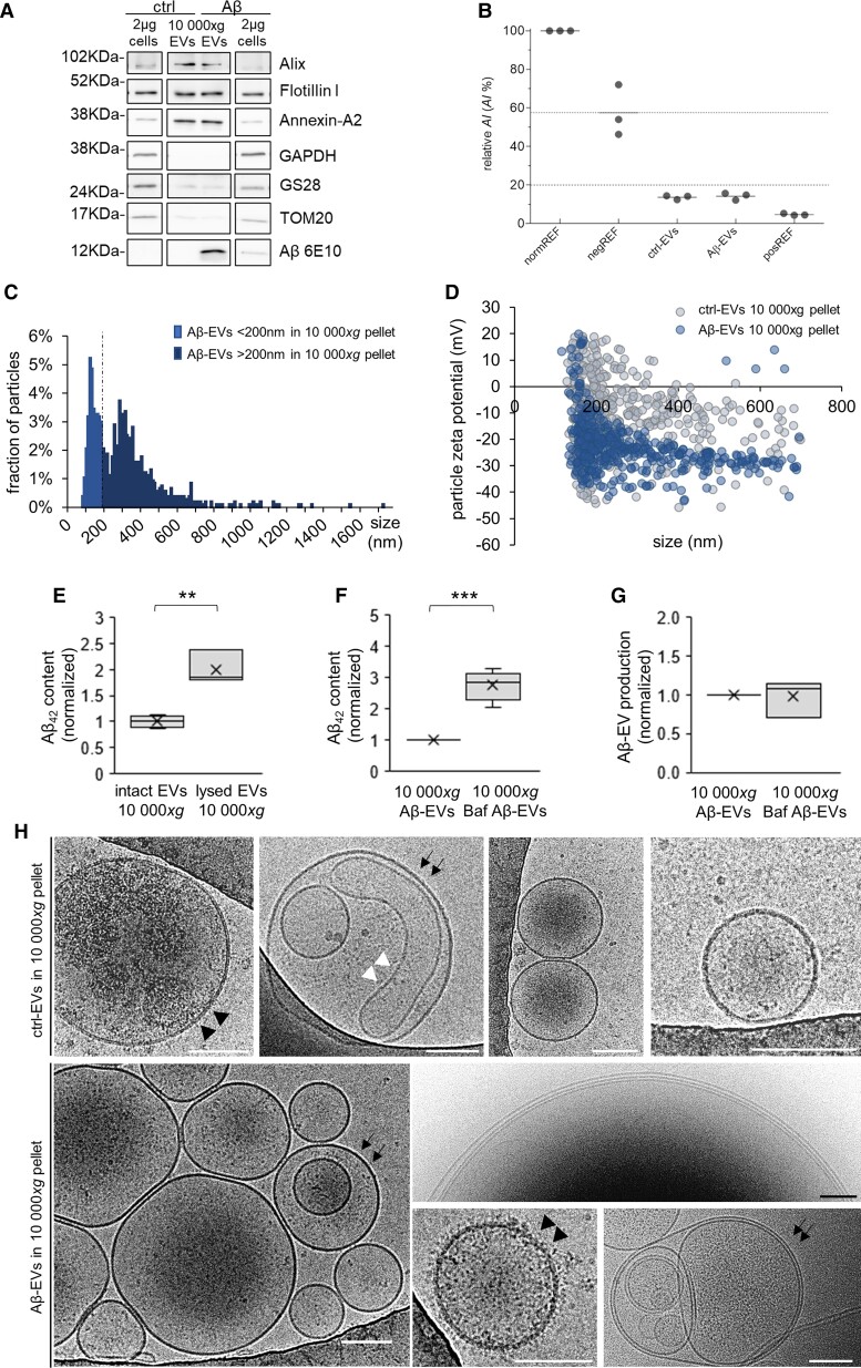
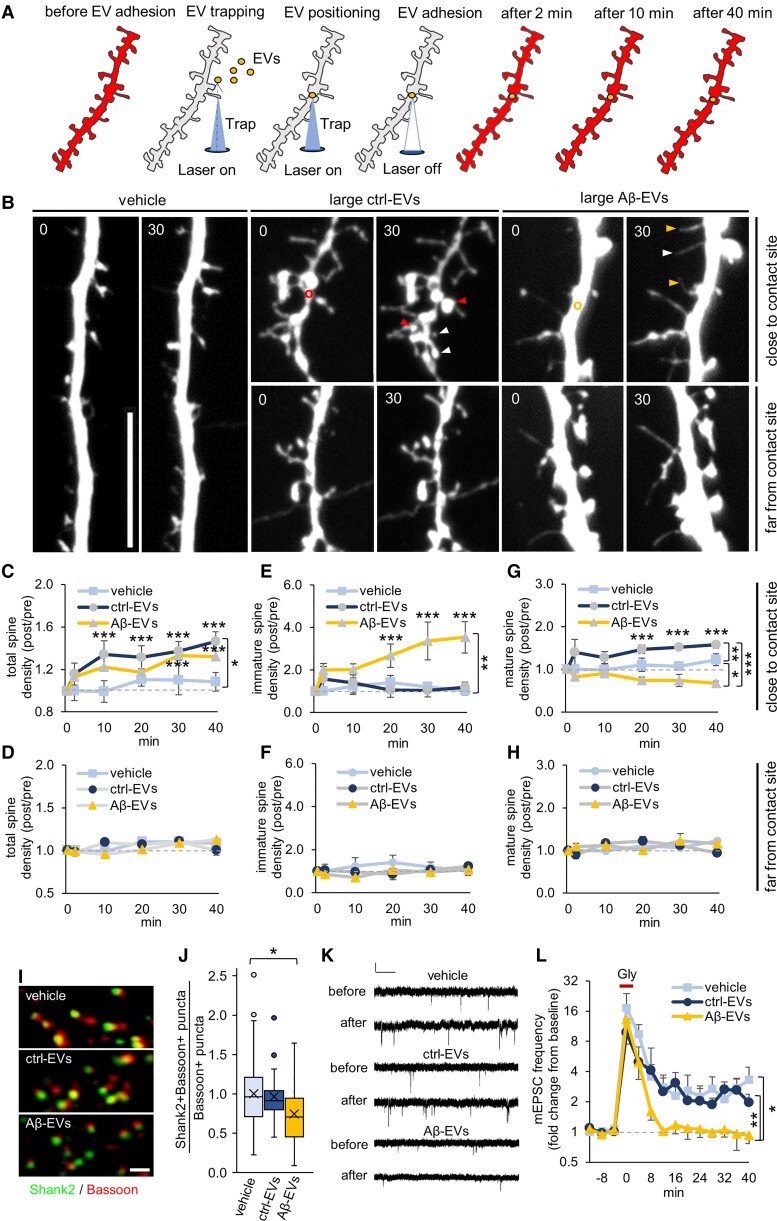
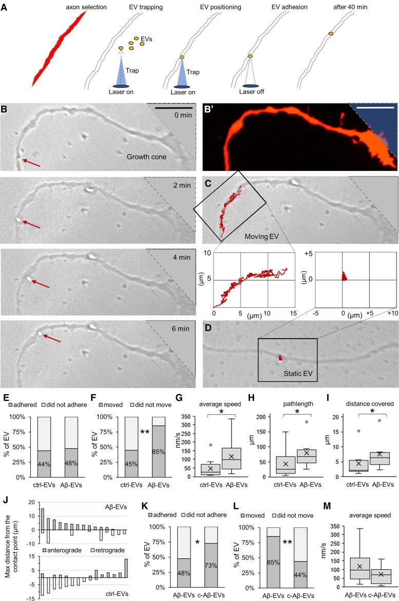
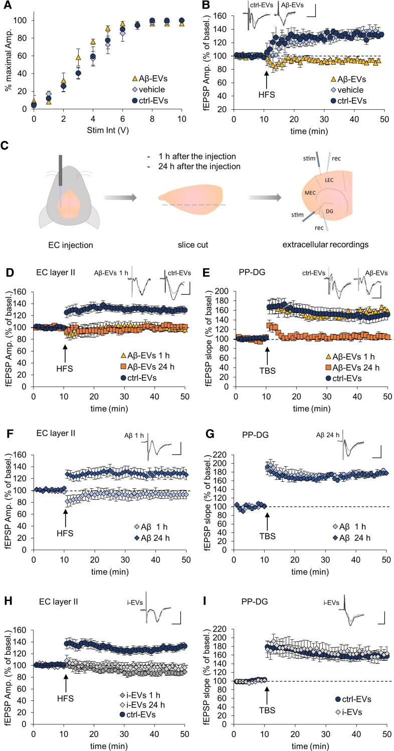
Verification
Claims the paper makes
The claims and statistics of this paper haven’t been checked yet.
Run the check — free, no account needed. It reads the open-access text and checks the sources it cites.
Claims other papers make about this one
Each is checked against this paper’s own text: does it say what the citing paper says it does?
In the citing paper
“Microglia could also lead to synaptic dysfunction in vivo and in vitro by releasing Aβ-containing EVs.”
From Microglia in neurodegenerative diseases: mechanism and potential therapeutic targets (2023) · cites this paper as [198]
Self-check
Percentages, sums and reported differences re-computed from the paper’s own numbers.
The self-check needs the paper’s full text.
Citations
Cites
Neuropathological stageing of Alzheimer-related changes.
Braak, H., & Braak, E. (1991) · Acta Neuropathologica · doi:10.1007/bf00308809 · Cited by 16,123
A synaptic model of memory: long-term potentiation in the hippocampus
Bliss, T. V. P., & Collingridge, G. L. (1993) · Nature · doi:10.1038/361031a0 · Cited by 11,566
Théry, C., Witwer, K. W., Aikawa, E., Alcaraz, M. J., Anderson, J. D., Andriantsitohaina, R., Antoniou, A., Arab, T., Archer, F., Atkin‐Smith, G. K., et al. (2018) · Journal of Extracellular Vesicles · doi:10.1080/20013078.2018.1535750 · Cited by 11,099
Self-citationAmyloid oligomers exacerbate tau pathology in a mouse model of tauopathy
Selenica, M.-L. B., Brownlow, M., Jimenez, J. P., Lee, D. C., Pena, G., Dickey, C. A., Gordon, M. N., & Morgan, D. (2013) · Neurodegenerative Diseases · doi:10.1159/000337230 · Cited by 10,359
Polazzi, E., Mengoni, I., Caprini, M., Peña‐Altamira, E., Kurtys, E., & Monti, B. (2012) · Neurosignals · doi:10.1159/000337115 · Cited by 9,023
Shedding light on the cell biology of extracellular vesicles
van Niel, G., D'Angelo, G., & Raposo, G. (2018) · Nature Reviews Molecular Cell Biology · doi:10.1038/nrm.2017.125 · Cited by 8,369
Neuroinflammation in Alzheimer's disease
Heneka, M. T., Carson, M. J., Khoury, J. E., Landreth, G. E., Brosseron, F., Feinstein, D. L., Jacobs, A. H., Wyss-Coray, T., Vitorica, J., Ransohoff, R. M., et al. (2015) · The Lancet Neurology · doi:10.1016/s1474-4422(15)70016-5 · Cited by 6,226
The amyloid hypothesis of Alzheimer's disease at 25 years
Selkoe, D. J., & Hardy, J. (2016) · EMBO Molecular Medicine · doi:10.15252/emmm.201606210 · Cited by 6,164
Alzheimer's Disease Is a Synaptic Failure
Selkoe, D. J. (2002) · Science · doi:10.1126/science.1074069 · Cited by 4,420
Terry, R. D., Masliah, E., Salmon, D. P., Butters, N., DeTeresa, R., Hill, R., Hansen, L. A., & Katzman, R. (1991) · Annals of Neurology · doi:10.1002/ana.410300410 · Cited by 4,397
Cited by
Sorted by citations
Microglia in neurodegenerative diseases: mechanism and potential therapeutic targets
Gao, C., Jiang, J., Tan, Y., & Chen, S. (2023) · Signal Transduction and Targeted Therapy · doi:10.1038/s41392-023-01588-0 · Cited by 1,616
Involvement of Astrocytes in the Formation, Maintenance, and Function of the Blood–Brain Barrier
Schiera, G., Liegro, C. M. D., Schirò, G., Sorbello, G., & Liegro, I. D. (2024) · Cells · doi:10.3390/cells13020150 · Cited by 128
Neuropathogenesis-on-chips for neurodegenerative diseases
Amartumur, S., Nguyen, H. M., Huynh, T., Kim, T. S., Woo, R., Oh, E., Kim, K. K., Lee, L. P., & Heo, C. (2024) · Nature Communications · doi:10.1038/s41467-024-46554-8 · Cited by 131
Extracellular vesicles set the stage for brain plasticity and recovery by multimodal signalling
Hermann, D. M., Peruzzotti‐Jametti, L., Giebel, B., & Pluchino, S. (2023) · Brain · doi:10.1093/brain/awad332 · Cited by 79
Research progress on the role of extracellular vesicles in neurodegenerative diseases
Li, Z., Wang, X., Wang, X., Yi, X., Wong, Y. K., Wu, J., Xie, F., Hu, D., Wang, Q., Wang, J., et al. (2023) · Translational Neurodegeneration · doi:10.1186/s40035-023-00375-9 · Cited by 67
Metadata
- Authors
- Gabrielli, M., Prada, I., Joshi, P., Falcicchia, C., D’Arrigo, G., Rutigliano, G., Battocchio, E., Zenatelli, R., Tozzi, F., Radeghieri, A., Arancio, O., Origlia, N., & Verderio, C.
- Affiliations
- CNR Institute of Neuroscience , Vedano al Lambro, MB 20854 , Italy
- Neuroscience Institute (IT)
- Scuola Superiore Sant'Anna (IT)
- University of Milano-Bicocca (IT)
- University of Brescia (IT)
- Scuola Normale Superiore (IT)
- Consorzio Interuniversitario per lo Sviluppo dei Sistemi a Grande Interfase (IT)
- Columbia University (US)
- Corresponding author
- Origlia, Nicola
- Verderio, Claudia
- ORCID
- Venue
- Brain
- Publisher
- Oxford University Press (OUP)
- Published
- 2022-03-03
- Volume
- 145
- Issue
- 8
- Pages
- 2849-2868
- ISSN
- 0006-8950 (ISSN-L)
- 1460-2156
- PMC
- PMC9420022
- OpenAlex
- W4214920994
- Cited by
- 106
- References
- 105
- Citations by year
- 2026: 22 · 2025: 29 · 2024: 27 · 2023: 22 · 2022: 6
- FWCI
- 12.99
- Type
- journal article
- Language
- en
- Open access
- hybrid
- doi.org
- Licence
- Locations
- Brain (publishedVersion, cc-by-nc)
- PubMed Central (publishedVersion, cc-by-nc)
- PubMed (publishedVersion)
- BOA (University of Milano-Bicocca) (submittedVersion, cc-by-nc)
- Institutional Research Information System (Università degli Studi di Brescia) (submittedVersion, cc-by-nc)
- Scuola Normale Superiore di Pisa (submittedVersion, cc-by-nc)
- Funders
- American National Institutes of Health (1R56AG056108-01)
- Alzheimer’s Association Research Fellowship (2018-AARF-588984)
- Italian Multiple Sclerosis Foundation
- Topics
- Life Sciences › Neuroscience › Cellular and Molecular Neuroscience › Alzheimer's disease research and treatments
- Life Sciences › Biochemistry, Genetics and Molecular Biology › Molecular Biology › Extracellular vesicles in disease
- Life Sciences › Neuroscience › Neurology › Neuroinflammation and Neurodegeneration Mechanisms
- Keywords
- Alzheimer's disease, microglia, large extracellular vesicles, amyloid-beta, synaptic dysfunction, long-term potentiation, dendritic spine morphology, synaptic plasticity, vesicle motility, annexin V, amyloid-beta 42, disease propagation
- MeSH terms
- Extracellular Vesicles, Alzheimer Disease, Animals, Hippocampus, Amyloid beta-Peptides, Microglia, Long-Term Potentiation, Mice
About these checks
Registry, retraction, Cites and statistics results are computed, not judged, with one exception: a printed reference that no index lists is read once by a model before it counts as a citation error. Claims are extracted by a model and kept only when their quote appears verbatim in the text; each cited source is then checked, with a model’s help, for whether it says what the claim relies on. Those verdicts can be wrong — read the claim’s quote and the cited source before relying on one.
Statistics and arithmetic are recomputed, not judged.
Confidence bands are shown only for fields calibrated on our annotated test set. None is calibrated yet, so no band is shown.
What counts as a claim ›How we measure confidence ›How we check this ›
Are you an author, or do you see a problem with this page? Request a review
