

Mitochondrial defects in acute multiple sclerosis lesions
Open accessThe DOI matches its Crossref record; 50 of the 50 references it cites were found in a registry or an index.
Abstract
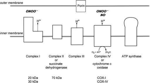
Multiple sclerosis is a chronic inflammatory disease, which leads to focal plaques of demyelination and tissue injury in the CNS. The structural and immunopathological patterns of demyelination suggest that different immune mechanisms may be involved in tissue damage. In a subtype of lesions, which are mainly found in patients with acute fulminant multiple sclerosis with Balo's type concentric sclerosis and in a subset of early relapsing remitting multiple sclerosis, the initial myelin changes closely resemble those seen in white matter stroke (WMS), suggesting a hypoxia-like tissue injury. Since mitochondrial injury may be involved in the pathogenesis of such lesions, we analysed a number of mitochondrial respiratory chain proteins in active lesions from acute multiple sclerosis and from WMS using immunohistochemistry. Functionally important defects of mitochondrial respiratory chain complex IV [cytochrome c oxidase (COX)] including its catalytic component (COX-I) are present in Pattern III but not in Pattern II multiple sclerosis lesions. The lack of immunohistochemically detected COX-I is apparent in oligodendrocytes, hypertrophied astrocytes and axons, but not in microglia. The profile of immunohistochemically detected mitochondrial respiratory chain complex subunits differs between multiple sclerosis and WMS. The findings suggest that hypoxia-like tissue injury in Pattern III multiple sclerosis lesions may be due to mitochondrial impairment.
Overview
The claims and statistics of this paper haven’t been checked yet.
Free: it reads the open-access text and checks the first 5 claims that cite a source we can check. Create a free account to check every claim.
Integrity signals
Registry
Cites
Statistics
Figures


Verification
Claims the paper makes
The claims and statistics of this paper haven’t been checked yet.
Free: it reads the open-access text and checks the first 5 claims that cite a source we can check. Create a free account to check every claim.
Claims other papers make about this one
Each is checked against this paper’s own text: does it say what the citing paper says it does?
The source backs part of the claim, not all of it — or backs it for a different population, condition or comparison.
In the citing paper
“Oxidative damage and mitochondrial dysfunction are key features of MS pathology [277,278,279,280].”
From Ageing, Cellular Senescence and Neurodegenerative Disease (2018) · cites this paper as [279]
Experimental — accuracy not yet measuredThe source backs part of the claim, not all of it — or backs it for a different population, condition or comparison.
In the citing paper
“Other features common to both cuprizone-induced and type 3 MS lesions are mitochondrial stress, selective loss of myelin-associated glycoprotein (MAG), lesions with indistinct borders that are not perivascular, pronounced glial activation, and lymphocyte infiltration [43, 46, 65, 69–71].”
From Oligodendrocyte death and myelin loss in the cuprizone model: an updated overview of the intrinsic and extrinsic causes of cuprizone demyelination (2022) · cites this paper as [69]
Experimental — accuracy not yet measured
Self-check
Percentages, sums and reported differences re-computed from the paper’s own numbers.
The self-check needs the paper’s full text.
Citations
Cites
Mitochondrial DNA mutations in human disease
Taylor, R. W., & Turnbull, D. M. (2005) · Nature Reviews Genetics · doi:10.1038/nrg1606 · Cited by 1,852 · cited 5×
“…c oxidase Abbreviations: CNPase=cyclic nucleotide phosphodiesterase; COX=cytochrome c oxidase; LFB=Luxol fast blue; MAG=myelin associated glycoprotein; MBP=myelin basic protein; mtDNA=mitochondrial DNA; NO=nitric oxide; NWM=normal white matter; PLP=proteolipid protein; WMS=white matter stroke”
BackgroundMethodInfluentialSelf-citationMultiple Sclerosis — The Plaque and Its Pathogenesis
Frohman, E. M., Racke, M. K., & Raine, C. S. (2006) · New England Journal of Medicine · doi:10.1056/nejmra052130 · Cited by 1,740 · cited 1×
“Multiple sclerosis is an inflammatory demyelinating disease of the central nervous system with axonal degeneration and astrogliosis (Frohman et al., 2006).”
BackgroundMitochondrial Respiratory-Chain Diseases
DiMauro, S., & Schon, E. A. (2003) · New England Journal of Medicine · doi:10.1056/nejmra022567 · Cited by 1,661 · cited 5×
“Keywords: multiple sclerosis; Pattern III lesion; mitochondria; cytochrome c oxidase Abbreviations: CNPase=cyclic nucleotide phosphodiesterase; COX=cytochrome c oxidase; LFB=Luxol fast blue; MAG=myelin associated glycoprotein; MBP=myelin basic protein; mtDNA=mitochondrial DNA; NO=nitric oxide;…”
BackgroundInfluentialvon Büdingen, H.-C., Palanichamy, A., Lehmann-Horn, K., Michel, B. A., & Zamvil, S. S. (2015) · European Neurology · doi:10.1159/000377675 · Cited by 1,463
Cleeter, M. W. J., Cooper, J. M., Darley-Usmar, V. M., Moncada, S., & Schapira, A. H. V. (1994) · FEBS Letters · doi:10.1016/0014-5793(94)00424-2 · Cited by 1,241 · cited 1×
“NO competitively inhibits the binding of oxygen to complex IV (Brown and Cooper, 1994; Cleeter et al., 1994).”
BackgroundHIF-1 Regulates Cytochrome Oxidase Subunits to Optimize Efficiency of Respiration in Hypoxic Cells
Fukuda, R., Zhang, H., Kim, J.-W., Shimoda, L., Dang, C. V., & Semenza, G. L. (2007) · Cell · doi:10.1016/j.cell.2007.01.047 · Cited by 1,225 · cited 5×
“Widespread defects of mitochondrial respiratory chain complexes inWMS lesions The transcription of mtDNA and nuclear DNA encoded subunits of the mitochondrial respiratory chain complexes are modulated by hypoxia (Vijayasarathy et al., 2003; Piruat and Lopez-Barneo, 2005; Fukuda et al., 2007).”
BackgroundInfluentialMicrodomains of Intracellular Ca2+: Molecular Determinants and Functional Consequences
Rizzuto, R., & Pozzan, T. (2006) · Physiological Reviews · doi:10.1152/physrev.00004.2005 · Cited by 1,206 · cited 1×
“Mitochondria also play an important role in apoptosis and calcium homeostasis (Rizzuto and Pozzan, 2006; Kalman et al., 2007).”
BackgroundRelapsing and remitting multiple sclerosis: Pathology of the newly forming lesion
Barnett, M. H., & Prineas, J. W. (2004) · Annals of Neurology · doi:10.1002/ana.20016 · Cited by 1,185 · cited 5×
“Mitochondrial defects in acute multiple sclerosis lesions Don Mahad,1 Iryna Ziabreva,1Hans Lassmann2 and DouglasTurnbull1 1The Mitochondrial Research Group, University of Newcastle uponTyne, UK and 2Centre for Brain Research, Medical University of Vienna, Austria Correspondence to: Prof. Dr Hans Lassmann, Center for B…”
BackgroundInfluentialBrown, G. C., & Cooper, C. E. (1994) · FEBS Letters · doi:10.1016/0014-5793(94)01290-3 · Cited by 1,055 · cited 2×
“…c oxidase Abbreviations: CNPase=cyclic nucleotide phosphodiesterase; COX=cytochrome c oxidase; LFB=Luxol fast blue; MAG=myelin associated glycoprotein; MBP=myelin basic protein; mtDNA=mitochondrial DNA; NO=nitric oxide; NWM=normal white matter; PLP=proteolipid protein; WMS=white matter stroke”
BackgroundSystemic infections and inflammation affect chronic neurodegeneration
Perry, V. H., Cunningham, C., & Holmes, C. (2007) · Nature Reviews Immunology · doi:10.1038/nri2015 · Cited by 1,027 · cited 1×
“These activated microglia, found in close proximity to apoptotic oligodendrocytes, may release a range of mediators (Barnett and Prineas, 2004; Perry et al., 2007).”
BackgroundFibrinogen Stimulates Macrophage Chemokine Secretion Through Toll-Like Receptor 4
Smiley, S. T., King, J. A., & Hancock, W. W. (2001) · The Journal of Immunology · doi:10.4049/jimmunol.167.5.2887 · Cited by 946 · cited 1×
“The microglia appear to be activated via Toll-like receptors in the early stages of Pattern III and not Pattern II lesions (Smiley et al., 2001; Marik et al., 2007).”
BackgroundClementi, E., Brown, G. C., Feelisch, M., & Moncada, S. (1998) · Proceedings of the National Academy of Sciences · doi:10.1073/pnas.95.13.7631 · Cited by 863 · cited 2×
“The loss of COX-I renders complex IV inactive as reported in patients with mitochondrial disease due to pathogenic mutations of COX-I gene and prevents utilization of oxygen (Clementi et al., 1998; Bruno et al., 1999; Kollberg et al., 2005).”
BackgroundMitochondrial dysfunction as a cause of axonal degeneration in multiple sclerosis patients
Dutta, R., McDonough, J., Yin, X., Peterson, J., Chang, A., Torres, T., Gudz, T., Macklin, W. B., Lewis, D. A., Fox, R. J., et al. (2006) · Annals of Neurology · doi:10.1002/ana.20736 · Cited by 841 · cited 2×
“, 2003) and decreased expression of mRNA for mitochondrial proteins (Dutta et al., 2006).”
BackgroundDoes nitric oxide modulate mitochondrial energy generation and apoptosis?
Moncada, S., & Erusalimsky, J. D. (2002) · Nature Reviews Molecular Cell Biology · doi:10.1038/nrm762 · Cited by 793 · cited 2×
“…c oxidase Abbreviations: CNPase=cyclic nucleotide phosphodiesterase; COX=cytochrome c oxidase; LFB=Luxol fast blue; MAG=myelin associated glycoprotein; MBP=myelin basic protein; mtDNA=mitochondrial DNA; NO=nitric oxide; NWM=normal white matter; PLP=proteolipid protein; WMS=white matter stroke”
BackgroundHeterogeneity of multiple sclerosis pathogenesis: implications for diagnosis and therapy
Lassmann, H., Brück, W., & Lucchinetti, C. (2001) · Trends in Molecular Medicine · doi:10.1016/s1471-4914(00)01909-2 · Cited by 627 · cited 1×
“The loss of myelin in multiple sclerosis may be the result of direct damage to myelin through immune mediated processes and/or dysfunction of oligodendrocytes (Lassmann et al., 2001; Barnett and Prineas, 2004).”
BackgroundSelf-citationThe role of nitric oxide in multiple sclerosis
Smith, K. J., & Lassmann, H. (2002) · The Lancet Neurology · doi:10.1016/s1474-4422(02)00102-3 · Cited by 531
Self-citationBolaños, J. P., Almeida, A., Stewart, V., Peuchen, S., Land, J. M., Clark, J. B., & Heales, S. J. R. (1997) · Journal of Neurochemistry · doi:10.1046/j.1471-4159.1997.68062227.x · Cited by 519 · cited 2×
“Reactive oxygen species and nitric oxide (NO), produced by these enzymes, may impair mitochondrial function (Bolanos et al., 1997) and thus may link 2008 The Author(s) This is an Open Access article distributed under the terms of the Creative Commons Attribution Non-Commercial License…”
BackgroundAlmeida, A., Moncada, S., & Bolaños, J. P. (2004) · Nature Cell Biology · doi:10.1038/ncb1080 · Cited by 480 · cited 1×
“This difference may be due to astrocytes being able to maintain ATP production through induction of glycolysis (Almeida et al., 2001, 2004).”
BackgroundAlmeida, A., Almeida, J., Bolaños, J. P., & Moncada, S. (2001) · Proceedings of the National Academy of Sciences · doi:10.1073/pnas.261560998 · Cited by 423 · cited 2×
“This difference may be due to astrocytes being able to maintain ATP production through induction of glycolysis (Almeida et al., 2001, 2004).”
BackgroundAxonal conduction and injury in multiple sclerosis: the role of sodium channels
Waxman, S. G. (2006) · Nature Reviews Neuroscience · doi:10.1038/nrn2023 · Cited by 397 · cited 2×
“In acutely demyelinated axons, the efflux of sodium (Na+) entering through the redistributed Na+ channels and ionic homeostasis are energy dependant processes (Waxman, 2006).”
BackgroundAboul-Enein, F., Rauschka, H., Kornek, B., Stadelmann, C., Stefferl, A., Brück, W., Lucchinetti, C., Schmidbauer, M., Jellinger, K., & Lassmann, H. (2003) · Journal of Neuropathology & Experimental Neurology · doi:10.1093/jnen/62.1.25 · Cited by 330 · cited 5×
“Mitochondrial defects in acute multiple sclerosis lesions Don Mahad,1 Iryna Ziabreva,1Hans Lassmann2 and DouglasTurnbull1 1The Mitochondrial Research Group, University of Newcastle uponTyne, UK and 2Centre for Brain Research, Medical University of Vienna, Austria Correspondence to: Prof. Dr Hans Lassmann, Center for B…”
BackgroundMethodResultInfluentialSelf-citationGeneral mechanisms of axonal damage and its prevention
Stys, P. K. (2005) · Journal of the Neurological Sciences · doi:10.1016/j.jns.2005.03.031 · Cited by 314 · cited 2×
“3g–i).”
BackgroundLesion genesis in a subset of patients with multiple sclerosis: a role for innate immunity?
Marik, C., Felts, P. A., Bauer, J., Lassmann, H., & Smith, K. J. (2007) · Brain · doi:10.1093/brain/awm236 · Cited by 292 · cited 4×
“HIF-1 is minimal or absent in LA stage of Pattern III lesions, where COX-IV is reduced (Marik et al., 2007).”
BackgroundInfluentialSelf-citationMitrovic, B., Ignarro, L. J., Montestruque, S., Smoll, A., & Merrill, J. E. (1994) · Neuroscience · doi:10.1016/0306-4522(94)90435-9 · Cited by 259 · cited 1×
“Oligodendrocytes are more susceptible to oxidative stress in vitro compared with astrocytes and microglia despite similar inhibition of mitochondrial activity (Mitrovic et al., 1994).”
BackgroundAxonal loss in multiple sclerosis: a pathological survey of the corticospinal and sensory tracts
DeLuca, G. C. (2004) · Brain · doi:10.1093/brain/awh118 · Cited by 255 · cited 1×
“Furthermore, the dominant loss of small calibre axons in multiple sclerosis lesions suggests energy deficiency as a major mechanism involved in axonal degeneration (DeLuca et al., 2004; Stys, 2005).”
BackgroundBlockers of sodium and calcium entry protect axons from nitric oxide‐mediated degeneration
Kapoor, R., Davies, M., Blaker, P. A., Hall, S. M., & Smith, K. J. (2003) · Annals of Neurology · doi:10.1002/ana.10443 · Cited by 252 · cited 2×
“The demyelinated axons can be protected from NO mediated degeneration, which involves an energy deficient state, using Na+ channel blockers or inhibitors of Na+/Ca2+ exchanger (Garthwaite et al., 2002; Kapoor et al., 2003).”
BackgroundCytochrome c Oxidase subunit I microdeletion in a patient with motor neuron disease
Comi, G. P., Bordoni, A., Salani, S., Franceschina, L., Sciacco, M., Prelle, A., Fortunato, F., Zeviani, M., Napoli, L., Bresolin, N., et al. (1998) · Annals of Neurology · doi:10.1002/ana.410430119 · Cited by 245 · cited 1×
“It is well established that mtDNA encoded subunit-I of complex IV (COX-I) is essential for the activity of complex IV or COX (Comi et al., 1998; Bruno et al., 1999; Kollberg et al., 2005).”
BackgroundItoyama, Y., Sterhberger, N. H., Webster, H. D., Quarles, R. H., Cohen, S. R., & Richardson, E. P. (1980) · Annals of Neurology · doi:10.1002/ana.410070212 · Cited by 241 · cited 2×
“A particular subtype of active multiple sclerosis lesions (Pattern III, Lucchinetti et al 2000), which are mainly found in patients with acute fulminant multiple sclerosis with Balo’s type concentric sclerosis and in a subset of early relapsing remitting multiple sclerosis, has been defined by the selective early loss…”
BackgroundAssembly of cytochrome‐c oxidase in cultured human cells
Nijtmans, L. G. J., Taanman, J., Muijsers, A. O., Speijer, D., & Van den Bogert, C. (1998) · European Journal of Biochemistry · doi:10.1046/j.1432-1327.1998.2540389.x · Cited by 241 · cited 1×
“Although COX-IV is not a catalytic component of complex IV, it is important for the assembly of subunits as well as for the stability of complex IV (Nijtmans et al., 1998; Taanman and Williams, 2001).”
BackgroundTissue preconditioning may explain concentric lesions in Baló's type of multiple sclerosis
Stadelmann, C., Ludwin, S., Tabira, T., Guseo, A., Lucchinetti, C. F., Leel-Össy, L., Ordinario, A. T., Brück, W., & Lassmann, H. (2005) · Brain · doi:10.1093/brain/awh457 · Cited by 234 · cited 4×
“A comparable loss of MAG and CNPase together with oligodendrocyte apoptosis is also seen in initial lesions in white matter stroke (WMS) and hypoxia inducible factor-1 , a transcription factor induced in hypoxic condition, is expressed in such multiple sclerosis and WMS lesions (Aboul-Enein et al., 2003; Stadelmann et…”
BackgroundInfluentialSelf-citationGraumann, U., Reynolds, R., Steck, A. J., & Schaeren‐Wiemers, N. (2003) · Brain Pathology · doi:10.1111/j.1750-3639.2003.tb00485.x · Cited by 233 · cited 2×
“Microarray studies from cortical and white matter tissue of patients with progressive multiple sclerosis showed up-regulation of genes involved in (hypoxic) preconditioning (Graumann et al., 2003) and decreased expression of mRNA for mitochondrial proteins (Dutta et al.”
BackgroundJackson, M. J., Schaefer, J. A., Johnson, M. A., Morris, A. A. M., Turnbull, D. M., & Bindoff, L. A. (1995) · Brain · doi:10.1093/brain/118.2.339 · Cited by 166 · cited 1×
“Furthermore, CSF lactate levels may be elevated in patients with primary mitochondrial disorders (Jackson et al., 1995).”
BackgroundSelf-citationBruno, C., Martinuzzi, A., Tang, Y., Andreu, A. L., Pallotti, F., Bonilla, E., Shanske, S., Fu, J., Sue, C. M., Angelini, C. I., et al. (1999) · The American Journal of Human Genetics · doi:10.1086/302546 · Cited by 147 · cited 2×
“It is well established that mtDNA encoded subunit-I of complex IV (COX-I) is essential for the activity of complex IV or COX (Comi et al., 1998; Bruno et al., 1999; Kollberg et al., 2005).”
BackgroundReversal of a mitochondrial DNA defect in human skeletal muscle
Clark, K. M., Bindoff, L. A., Lightowlers, R. N., Andrews, R. M., Griffiths, P. G., Johnson, M. A., Brierley, E. J., & Turnbull, D. M. (1997) · Nature Genetics · doi:10.1038/ng0797-222 · Cited by 141 · cited 3×
“Tissue from WMS cases (WMS1–WMS6, Table 1) containing lesions with active demyelination was provided by the University of Vienna (Aboul-Enein et al., 2003).”
MethodInfluentialSelf-citationNitric oxide toxicity in CNS white matter: an in vitro study using rat optic nerve
Garthwaite, G., Goodwin, D. A., Batchelor, A. M., Leeming, K., & Garthwaite, J. (2002) · Neuroscience · doi:10.1016/s0306-4522(01)00447-x · Cited by 112 · cited 2×
“The demyelinated axons can be protected from NO mediated degeneration, which involves an energy deficient state, using Na+ channel blockers or inhibitors of Na+/Ca2+ exchanger (Garthwaite et al., 2002; Kapoor et al., 2003).”
BackgroundZhang, J., Jin, B., Li, L., Block, E. R., & Patel, J. M. (2005) · American Journal of Physiology-Cell Physiology · doi:10.1152/ajpcell.00325.2004 · Cited by 102 · cited 1×
“NO, when overexpressed, may irreversibly impair the activity of mitochondrial respiratory chain complexes I and IV via perxoynitrite (Clementi et al., 1998; Smith and Lassmann, 2002; Zhang et al., 2005).”
BackgroundThe involvement of mitochondria in the pathogenesis of multiple sclerosis
Kálmán, B., Laitinen, K., & Komoly, S. (2007) · Journal of Neuroimmunology · doi:10.1016/j.jneuroim.2007.03.020 · Cited by 97 · cited 1×
“Mitochondria also play an important role in apoptosis and calcium homeostasis (Rizzuto and Pozzan, 2006; Kalman et al., 2007).”
BackgroundVijayasarathy, C., Damle, S., Prabu, S. K., Otto, C. M., & Avadhani, N. G. (2003) · European Journal of Biochemistry · doi:10.1046/j.1432-1033.2003.03447.x · Cited by 85 · cited 3×
“However, in vitro and in vivo studies have identified a decrease in transcription of mtDNA and nuclear DNA encoded subunits within 24 h of exposure to hypoxia (Vijayasarathy et al., 2003; Piruat and Lopez-Barneo, 2005; Fukuda et al., 2007).”
BackgroundMahad, D., Trebst, C., Kivisäkk, P., Staugaitis, S. M., Tucky, B. H., Wei, T., Lucchinetti, C. F., Lassmann, H., & Ransohoff, R. M. (2004) · Journal of Neuropathology & Experimental Neurology · doi:10.1093/jnen/63.3.262 · Cited by 79 · cited 1×
“The expression of chemokine receptors differs in Pattern II from Pattern III lesions (Mahad et al., 2004).”
BackgroundSelf-citationDobersen, M. J., Hammer, J. A., Noronha, A. B., MacIntosh, T. D., Trapp, B. D., Brady, R. O., & Quarles, R. H. (1985) · Neurochemical Research · doi:10.1007/bf00964654 · Cited by 72 · cited 3×
“…Encoding DNA Antibody type Source MBP (SMI 94) ^ mouse IgG1 Sternberger PLP ^ mouse IgG Serotec MAG (D7E10) ^ mouse IgG (Dobersen et al., 1985) CNPase ^ mouse IgG1 Sternberger HLA-DP, DQ and DR ^ mouse IgG1 Dako CD163 ^ mouse IgG1 Novocastra Glial fibrillary acidic…”
BackgroundInfluentialOxygen Tension Regulates Mitochondrial DNA-encoded Complex I Gene Expression
Piruat, J. I., & López‐Barneo, J. (2005) · Journal of Biological Chemistry · doi:10.1074/jbc.m507044200 · Cited by 59 · cited 3×
“However, in vitro and in vivo studies have identified a decrease in transcription of mtDNA and nuclear DNA encoded subunits within 24 h of exposure to hypoxia (Vijayasarathy et al., 2003; Piruat and Lopez-Barneo, 2005; Fukuda et al., 2007).”
BackgroundKollberg, G., Moslemi, A., Lindberg, C., Holme, E., & Oldfors, A. (2005) · Journal of Neuropathology & Experimental Neurology · doi:10.1093/jnen/64.2.123 · Cited by 50 · cited 2×
“It is well established that mtDNA encoded subunit-I of complex IV (COX-I) is essential for the activity of complex IV or COX (Comi et al., 1998; Bruno et al., 1999; Kollberg et al., 2005).”
BackgroundNijtmans, L. G. J., Spelbrink, J. N., Galen, M. J. M. V., Zwaan, M., Klement, P., & den Bogert, C. V. (1995) · Biochimica et Biophysica Acta (BBA) - Molecular Cell Research · doi:10.1016/0167-4889(94)00203-q · Cited by 44 · cited 2×
“A defect in COX-I leads to degradation of unassembled COX-IV, which may be the reason for a COX-IV defect in LA stage of Pattern III lesions (Nijtmans et al., 1995).”
BackgroundCytochrome oxidase immunohistochemistry: clues for genetic mechanisms
Rahman, S., Lake, B. D., Taanman, J. W., Hanna, M. G., Cooper, J. M., Schapira, A. H., & Leonard, J. V. (2000) · Brain · doi:10.1093/brain/123.3.591 · Cited by 43 · cited 2×
“subunits were included for the immunohistochemical detection (Rahman et al., 2000; Taylor and Turnbull, 2005).”
BackgroundMethodBaló's concentric sclerosis presenting as a stroke-like syndrome
Mowry, E. M., Woo, J. H., & Ances, B. M. (2007) · Nature Clinical Practice Neurology · doi:10.1038/ncpneuro0522 · Cited by 36 · cited 1×
“Indeed, magnetic resonance spectroscopic studies of Balo’s type lesions have found consistently increased lactate peaks (Lindquist et al., 2007; Mowry et al., 2007).”
BackgroundHistopathology and serial, multimodal magnetic resonance imaging in a multiple sclerosis variant
Lindquist, S., Bodammer, N., Kaufmann, J., König, F. B., Heinze, H. –., Brück, W., & Sailer, M. (2007) · Multiple Sclerosis Journal · doi:10.1177/1352458506071329 · Cited by 35 · cited 1×
“Indeed, magnetic resonance spectroscopic studies of Balo’s type lesions have found consistently increased lactate peaks (Lindquist et al., 2007; Mowry et al., 2007).”
BackgroundTaanman, J., & Williams, S. L. (2001) · Biochemical Society Transactions · doi:10.1042/bst0290446 · Cited by 32 · cited 5×
“…c oxidase Abbreviations: CNPase=cyclic nucleotide phosphodiesterase; COX=cytochrome c oxidase; LFB=Luxol fast blue; MAG=myelin associated glycoprotein; MBP=myelin basic protein; mtDNA=mitochondrial DNA; NO=nitric oxide; NWM=normal white matter; PLP=proteolipid protein; WMS=white matter stroke”
BackgroundInfluentialWei, J., Guo, H., & Kuo, P. C. (2002) · The Journal of Immunology · doi:10.4049/jimmunol.168.9.4721 · Cited by 23 · cited 2×
“Hence, we speculate that the mitochondrial defects in Pattern III lesions identified in this study are due to post-transcriptional modification of COX-I mRNA by NO and subsequent degradation of unassembled COX-IV (Nijtmans et al., 1995; Williams et al., 2001; Wei et al., 2002; Fukuda et al., 2007).”
BackgroundWilliams, S. L., Scholte, H. R., Gray, R. G. F., Leonard, J. V., Schapira, A. H. V., & Taanman, J. (2001) · Laboratory Investigation · doi:10.1038/labinvest.3780319 · Cited by 16 · cited 1×
“Hence, we speculate that the mitochondrial defects in Pattern III lesions identified in this study are due to post-transcriptional modification of COX-I mRNA by NO and subsequent degradation of unassembled COX-IV (Nijtmans et al., 1995; Williams et al., 2001; Wei et al., 2002; Fukuda et al., 2007).”
BackgroundDiagnostic criteria for primary progressive mutiple sclerosis: a position paper.
(2000) · Pure Amsterdam UMC · doi:10.1002/1531-8249(200006)47:6 · Cited by 2
Self-citation
Research works printed but not linked: 1
- Lucchinetti, 2000, Heterogeneity of multiple sclerosis lesions: implications for the pathogenesis of demyelination, Ann Neurol, 47, 707, 10.1002/1531-8249(200006)47:6<707::aid-ana3>3.0.co;2-q · DOI
Cited by
Compston, A., & Coles, A. (2008) · The Lancet · doi:10.1016/s0140-6736(08)61620-7 · Cited by 4,372
Pathological mechanisms in progressive multiple sclerosis
Mahad, D. H., Trapp, B. D., & Lassmann, H. (2015) · The Lancet Neurology · doi:10.1016/s1474-4422(14)70256-x · Cited by 1,137
Self-citationProgressive multiple sclerosis: pathology and pathogenesis
Lassmann, H., van Horssen, J., & Mahad, D. (2012) · Nature Reviews Neurology · doi:10.1038/nrneurol.2012.168 · Cited by 1,033
Self-citationLassmann, H. (2018) · Cold Spring Harbor Perspectives in Medicine · doi:10.1101/cshperspect.a028936 · Cited by 937
Self-citationOligodendrocytes: biology and pathology
Bradl, M., & Lassmann, H. (2010) · Acta Neuropathologica · doi:10.1007/s00401-009-0601-5 · Cited by 933
Self-citation
Metadata
- Authors
- Mahad, D., Ziabreva, I., Lassmann, H., & Turnbull, D.
- Affiliations
- Medical University of Vienna (AT)
- Corresponding author
- Lassmann, Hans
- Venue
- Brain
- Publisher
- Oxford University Press (OUP)
- Published
- 2008-05-30
- Volume
- 131
- Issue
- 7
- Pages
- 1722-1735
- ISSN
- 0006-8950 (ISSN-L)
- 1460-2156
- PMC
- PMC2442422
- OpenAlex
- W2149715738
- Cited by
- 406
- References
- 50
- Citations by year
- 2026: 8 · 2025: 8 · 2024: 22 · 2023: 18 · 2022: 33 · 2021: 25 · 2020: 30 · 2019: 26 · 2018: 13 · 2017: 23 · 2016: 25 · 2015: 28 · 2014: 28 · 2013: 30 · 2012: 24
- FWCI
- 15.42
- Type
- journal article
- Language
- en
- Open access
- hybrid
- doi.org
- Licence
- cc-by-nc
- Locations
- Brain (publishedVersion, cc-by-nc)
- Europe PMC (PubMed Central) (publishedVersion, cc-by-nc)
- PubMed Central (publishedVersion, cc-by-nc)
- PubMed (publishedVersion)
- Edinburgh Research Explorer (University of Edinburgh) (submittedVersion, cc-by-nc)
- Newcastle University ePrints (Newcastle Univesity)
- Funders
- Wellcome Trust (078415)
- Austrian Science Fund (P 19854)
- Topics
- Health Sciences › Medicine › Clinical Neurology › Multiple Sclerosis Research Studies
- Life Sciences › Biochemistry, Genetics and Molecular Biology › Molecular Biology › Mitochondrial Function and Pathology
- Health Sciences › Medicine › Clinical Neurology › Parkinson's Disease Mechanisms and Treatments
- Keywords
- multiple sclerosis, mitochondrial dysfunction, cytochrome c oxidase, coxI, mitochondrial respiratory chain, Balo's concentric sclerosis, demyelination, oligodendrocytes, astrocytes, microglia, relapsing-remitting multiple sclerosis
- MeSH terms
- Acute Disease, Adult, Aged, Aged, 80 and over, Astrocytes, Axons, Diffuse Cerebral Sclerosis of Schilder, Electron Transport Complex IV, Electron Transport, Female, Humans, Male, Middle Aged, Mitochondria, Multiple Sclerosis, Oligodendroglia, Mitochondrial Encephalomyopathies, Mitochondrial Proteins, Mitochondrial Diseases
About these checks
Registry, retraction, Cites and statistics results are computed, not judged, with one exception: a printed reference that no index lists is read once by a model before it counts as a citation error. Claims are extracted by a model and kept only when their quote appears verbatim in the text; each cited source is then checked, with a model’s help, for whether it says what the claim relies on. Those verdicts can be wrong — read the claim’s quote and the cited source before relying on one.
Statistics and arithmetic are recomputed, not judged.
Confidence bands are shown only for fields calibrated on our annotated test set. None is calibrated yet, so no band is shown.
What counts as a claim ›How we measure confidence ›How we check this ›
Are you an author, or do you see a problem with this page? Request a review
Data sources
Bibliographic data: Semantic Scholar, OpenAlex, Crossref.
Contains information from the Semantic Scholar Academic Graph Datasets, made available under the ODC Attribution License (ODC-BY 1.0).
Semantic Scholar’s data is described in: Kinney, R., et al. (2023). The Semantic Scholar Open Data Platform. arXiv:2301.10140.
