

Mitochondrial defects in acute multiple sclerosis lesions
Open accessThe DOI matches its Crossref record; 50 of the 50 references it cites were found in a registry or an index.
Abstract
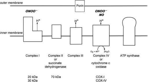
Multiple sclerosis is a chronic inflammatory disease, which leads to focal plaques of demyelination and tissue injury in the CNS. The structural and immunopathological patterns of demyelination suggest that different immune mechanisms may be involved in tissue damage. In a subtype of lesions, which are mainly found in patients with acute fulminant multiple sclerosis with Balo's type concentric sclerosis and in a subset of early relapsing remitting multiple sclerosis, the initial myelin changes closely resemble those seen in white matter stroke (WMS), suggesting a hypoxia-like tissue injury. Since mitochondrial injury may be involved in the pathogenesis of such lesions, we analysed a number of mitochondrial respiratory chain proteins in active lesions from acute multiple sclerosis and from WMS using immunohistochemistry. Functionally important defects of mitochondrial respiratory chain complex IV [cytochrome c oxidase (COX)] including its catalytic component (COX-I) are present in Pattern III but not in Pattern II multiple sclerosis lesions. The lack of immunohistochemically detected COX-I is apparent in oligodendrocytes, hypertrophied astrocytes and axons, but not in microglia. The profile of immunohistochemically detected mitochondrial respiratory chain complex subunits differs between multiple sclerosis and WMS. The findings suggest that hypoxia-like tissue injury in Pattern III multiple sclerosis lesions may be due to mitochondrial impairment.
Overview
The claims and statistics of this paper haven’t been checked yet.
Free: it reads the open-access text and checks the first 5 claims that cite a source we can check. Create a free account to check every claim.
Integrity signals
Registry
Cites
Statistics
Figures


Verification
Claims the paper makes
The claims and statistics of this paper haven’t been checked yet.
Free: it reads the open-access text and checks the first 5 claims that cite a source we can check. Create a free account to check every claim.
Claims other papers make about this one
Each is checked against this paper’s own text: does it say what the citing paper says it does?
The source backs part of the claim, not all of it — or backs it for a different population, condition or comparison.
In the citing paper
“Oxidative damage and mitochondrial dysfunction are key features of MS pathology [277,278,279,280].”
From Ageing, Cellular Senescence and Neurodegenerative Disease (2018) · cites this paper as [279]
Experimental — accuracy not yet measuredThe source backs part of the claim, not all of it — or backs it for a different population, condition or comparison.
In the citing paper
“Other features common to both cuprizone-induced and type 3 MS lesions are mitochondrial stress, selective loss of myelin-associated glycoprotein (MAG), lesions with indistinct borders that are not perivascular, pronounced glial activation, and lymphocyte infiltration [43, 46, 65, 69–71].”
From Oligodendrocyte death and myelin loss in the cuprizone model: an updated overview of the intrinsic and extrinsic causes of cuprizone demyelination (2022) · cites this paper as [69]
Experimental — accuracy not yet measured
Self-check
Percentages, sums and reported differences re-computed from the paper’s own numbers.
The self-check needs the paper’s full text.
Citations
Cites
Mitochondrial DNA mutations in human disease
Taylor, R. W., & Turnbull, D. M. (2005) · Nature Reviews Genetics · doi:10.1038/nrg1606 · Cited by 1,852 · cited 5×
“…c oxidase Abbreviations: CNPase=cyclic nucleotide phosphodiesterase; COX=cytochrome c oxidase; LFB=Luxol fast blue; MAG=myelin associated glycoprotein; MBP=myelin basic protein; mtDNA=mitochondrial DNA; NO=nitric oxide; NWM=normal white matter; PLP=proteolipid protein; WMS=white matter stroke”
BackgroundMethodInfluentialSelf-citationMultiple Sclerosis — The Plaque and Its Pathogenesis
Frohman, E. M., Racke, M. K., & Raine, C. S. (2006) · New England Journal of Medicine · doi:10.1056/nejmra052130 · Cited by 1,740 · cited 1×
“Multiple sclerosis is an inflammatory demyelinating disease of the central nervous system with axonal degeneration and astrogliosis (Frohman et al., 2006).”
BackgroundMitochondrial Respiratory-Chain Diseases
DiMauro, S., & Schon, E. A. (2003) · New England Journal of Medicine · doi:10.1056/nejmra022567 · Cited by 1,661 · cited 5×
“Keywords: multiple sclerosis; Pattern III lesion; mitochondria; cytochrome c oxidase Abbreviations: CNPase=cyclic nucleotide phosphodiesterase; COX=cytochrome c oxidase; LFB=Luxol fast blue; MAG=myelin associated glycoprotein; MBP=myelin basic protein; mtDNA=mitochondrial DNA; NO=nitric oxide;…”
BackgroundInfluentialvon Büdingen, H.-C., Palanichamy, A., Lehmann-Horn, K., Michel, B. A., & Zamvil, S. S. (2015) · European Neurology · doi:10.1159/000377675 · Cited by 1,463
Cleeter, M. W. J., Cooper, J. M., Darley-Usmar, V. M., Moncada, S., & Schapira, A. H. V. (1994) · FEBS Letters · doi:10.1016/0014-5793(94)00424-2 · Cited by 1,241 · cited 1×
“NO competitively inhibits the binding of oxygen to complex IV (Brown and Cooper, 1994; Cleeter et al., 1994).”
BackgroundHIF-1 Regulates Cytochrome Oxidase Subunits to Optimize Efficiency of Respiration in Hypoxic Cells
Fukuda, R., Zhang, H., Kim, J.-W., Shimoda, L., Dang, C. V., & Semenza, G. L. (2007) · Cell · doi:10.1016/j.cell.2007.01.047 · Cited by 1,225 · cited 5×
“Widespread defects of mitochondrial respiratory chain complexes inWMS lesions The transcription of mtDNA and nuclear DNA encoded subunits of the mitochondrial respiratory chain complexes are modulated by hypoxia (Vijayasarathy et al., 2003; Piruat and Lopez-Barneo, 2005; Fukuda et al., 2007).”
BackgroundInfluentialMicrodomains of Intracellular Ca2+: Molecular Determinants and Functional Consequences
Rizzuto, R., & Pozzan, T. (2006) · Physiological Reviews · doi:10.1152/physrev.00004.2005 · Cited by 1,206 · cited 1×
“Mitochondria also play an important role in apoptosis and calcium homeostasis (Rizzuto and Pozzan, 2006; Kalman et al., 2007).”
BackgroundRelapsing and remitting multiple sclerosis: Pathology of the newly forming lesion
Barnett, M. H., & Prineas, J. W. (2004) · Annals of Neurology · doi:10.1002/ana.20016 · Cited by 1,185 · cited 5×
“Mitochondrial defects in acute multiple sclerosis lesions Don Mahad,1 Iryna Ziabreva,1Hans Lassmann2 and DouglasTurnbull1 1The Mitochondrial Research Group, University of Newcastle uponTyne, UK and 2Centre for Brain Research, Medical University of Vienna, Austria Correspondence to: Prof. Dr Hans Lassmann, Center for B…”
BackgroundInfluentialBrown, G. C., & Cooper, C. E. (1994) · FEBS Letters · doi:10.1016/0014-5793(94)01290-3 · Cited by 1,055 · cited 2×
“…c oxidase Abbreviations: CNPase=cyclic nucleotide phosphodiesterase; COX=cytochrome c oxidase; LFB=Luxol fast blue; MAG=myelin associated glycoprotein; MBP=myelin basic protein; mtDNA=mitochondrial DNA; NO=nitric oxide; NWM=normal white matter; PLP=proteolipid protein; WMS=white matter stroke”
BackgroundSystemic infections and inflammation affect chronic neurodegeneration
Perry, V. H., Cunningham, C., & Holmes, C. (2007) · Nature Reviews Immunology · doi:10.1038/nri2015 · Cited by 1,027 · cited 1×
“These activated microglia, found in close proximity to apoptotic oligodendrocytes, may release a range of mediators (Barnett and Prineas, 2004; Perry et al., 2007).”
Background
Research works printed but not linked: 1
- Lucchinetti, 2000, Heterogeneity of multiple sclerosis lesions: implications for the pathogenesis of demyelination, Ann Neurol, 47, 707, 10.1002/1531-8249(200006)47:6<707::aid-ana3>3.0.co;2-q · DOI
Cited by
Compston, A., & Coles, A. (2008) · The Lancet · doi:10.1016/s0140-6736(08)61620-7 · Cited by 4,372
Pathological mechanisms in progressive multiple sclerosis
Mahad, D. H., Trapp, B. D., & Lassmann, H. (2015) · The Lancet Neurology · doi:10.1016/s1474-4422(14)70256-x · Cited by 1,137
Self-citationProgressive multiple sclerosis: pathology and pathogenesis
Lassmann, H., van Horssen, J., & Mahad, D. (2012) · Nature Reviews Neurology · doi:10.1038/nrneurol.2012.168 · Cited by 1,033
Self-citationLassmann, H. (2018) · Cold Spring Harbor Perspectives in Medicine · doi:10.1101/cshperspect.a028936 · Cited by 937
Self-citationOligodendrocytes: biology and pathology
Bradl, M., & Lassmann, H. (2010) · Acta Neuropathologica · doi:10.1007/s00401-009-0601-5 · Cited by 933
Self-citationOxidative damage in multiple sclerosis lesions
Haider, L., Fischer, M. T., Frischer, J. M., Bauer, J., Hoftberger, R., Botond, G., Esterbauer, H., Binder, C. J., Witztum, J. L., & Lassmann, H. (2011) · Brain · doi:10.1093/brain/awr128 · Cited by 753
“…may contribute to demye- lination and neurodegeneration in multiple sclerosis, it recently became clear that mitochondrial injury and subsequent energy failure is a major factor driving tissue injury (Lu et al., 2000; Dutta et al., 2006; Mahad et al., 2008; Trapp and Stys 2009; Witte et al., 2010).”
BackgroundInfluentialSelf-citationA reversible form of axon damage in experimental autoimmune encephalomyelitis and multiple sclerosis
Nikić, I., Merkler, D., Sorbara, C., Brinkoetter, M., Kreutzfeldt, M., Bareyre, F. M., Brück, W., Bishop, D., Misgeld, T., & Kerschensteiner, M. (2011) · Nature Medicine · doi:10.1038/nm.2324 · Cited by 713
“Our findings provide a mechanistic framework for recent histopathological studies that showed damage to neuronal mitochondria in acute and chronic stages of multiple sclerosi”
ResultBuendia, I., Michalska, P., Navarro, E., Gameiro, I., Egea, J., & León, R. (2016) · Pharmacology & Therapeutics · doi:10.1016/j.pharmthera.2015.11.003 · Cited by 678
Mechanisms of neurodegeneration and axonal dysfunction in multiple sclerosis
Friese, M. A., Schattling, B., & Fugger, L. (2014) · Nature Reviews Neurology · doi:10.1038/nrneurol.2014.37 · Cited by 628
Virtual hypoxia and chronic necrosis of demyelinated axons in multiple sclerosis
Trapp, B. D., & Stys, P. K. (2009) · The Lancet Neurology · doi:10.1016/s1474-4422(09)70043-2 · Cited by 621
Microglial cell dysregulation in brain aging and neurodegeneration
von Bernhardi, R., Eugenín-von Bernhardi, L., & Eugenín, J. (2015) · Frontiers in Aging Neuroscience · doi:10.3389/fnagi.2015.00124 · Cited by 556
Multiple sclerosis: experimental models and reality
Lassmann, H., & Bradl, M. (2017) · Acta Neuropathologica · doi:10.1007/s00401-016-1631-4 · Cited by 525
Self-citationIron and neurodegeneration in the multiple sclerosis brain
Hametner, S., Wimmer, I., Haider, L., Pfeifenbring, S., Brück, W., & Lassmann, H. (2013) · Annals of Neurology · doi:10.1002/ana.23974 · Cited by 519
Self-citationFischer, M. T., Sharma, R., Lim, J. L., Haider, L., Frischer, J. M., Drexhage, J., Mahad, D., Bradl, M., van Horssen, J., & Lassmann, H. (2012) · Brain · doi:10.1093/brain/aws012 · Cited by 507
Self-citationProgressive multiple sclerosis: from pathophysiology to therapeutic strategies
Faissner, S., Plemel, J. R., Gold, R., & Yong, V. W. (2019) · Nature Reviews Drug Discovery · doi:10.1038/s41573-019-0035-2 · Cited by 459
The Role of Environmental Exposures in Neurodegeneration and Neurodegenerative Diseases
Cannon, J. R., & Greenamyre, J. T. (2011) · Toxicological Sciences · doi:10.1093/toxsci/kfr239 · Cited by 459
“The high toxicity and neurological effects resulting from exposure to toxicants that inhibit cellular respiration illustrates the limited ability and selective sensitivity of neurons to energy depletion.”
BackgroundInfluentialMechanisms of neuronal dysfunction and degeneration in multiple sclerosis
Dutta, R., & Trapp, B. D. (2011) · Progress in Neurobiology · doi:10.1016/j.pneurobio.2010.09.005 · Cited by 457
“Excessive axoplasmic Ca2+ accumulation will cause a vicious cycle of impaired mitochondrial operation, reduced energy production and compromised axonal transport (Mahad et al., 2008a; Smith, 2007; Trapp and Stys, 2009; Waxman, 2006).”
Mitochondrial changes within axons in multiple sclerosis
Mahad, D. J., Ziabreva, I., Campbell, G., Lax, N., White, K., Hanson, P. S., Lassmann, H., & Turnbull, D. M. (2009) · Brain · doi:10.1093/brain/awp046 · Cited by 454
“We recently identified a defect in the main catalytic subunit of complex IV, COX-I, within acutely demyelinated axons in pattern III and Balo’s type MS lesions (Mahad et al., 2008).”
InfluentialSelf-citationAgeing, Cellular Senescence and Neurodegenerative Disease
Kritsilis, M., V. Rizou, S., Koutsoudaki, P. N., Evangelou, K., Gorgoulis, V. G., & Papadopoulos, D. (2018) · International Journal of Molecular Sciences · doi:10.3390/ijms19102937 · Cited by 448
Praet, J., Guglielmetti, C., Berneman, Z., Van der Linden, A., & Ponsaerts, P. (2014) · Neuroscience & Biobehavioral Reviews · doi:10.1016/j.neubiorev.2014.10.004 · Cited by 435
Exploring the origins of grey matter damage in multiple sclerosis
Calabrese, M., Magliozzi, R., Ciccarelli, O., Geurts, J. J. G., Reynolds, R., & Martin, R. (2015) · Nature Reviews Neuroscience · doi:10.1038/nrn3900 · Cited by 414
The topograpy of demyelination and neurodegeneration in the multiple sclerosis brain
Haider, L., Zrzavy, T., Hametner, S., Höftberger, R., Bagnato, F., Grabner, G., Trattnig, S., Pfeifenbring, S., Brück, W., & Lassmann, H. (2016) · Brain · doi:10.1093/brain/awv398 · Cited by 400
Self-citationPathology of Demyelinating Diseases
Popescu, B. F. G., & Lucchinetti, C. F. (2012) · Annual Review of Pathology: Mechanisms of Disease · doi:10.1146/annurev-pathol-011811-132443 · Cited by 397
The immunology of multiple sclerosis
Attfield, K. E., Jensen, L. T., Kaufmann, M., Friese, M. A., & Fugger, L. (2022) · Nature Reviews Immunology · doi:10.1038/s41577-022-00718-z · Cited by 380
Cell-type-specific profiling of brain mitochondria reveals functional and molecular diversity
Fecher, C., Trovò, L., Müller, S. A., Snaidero, N., Wettmarshausen, J., Heink, S., Ortiz, O., Wagner, I., Kühn, R., Hartmann, J., et al. (2019) · Nature Neuroscience · doi:10.1038/s41593-019-0479-z · Cited by 323
van Horssen, J., Schreibelt, G., Drexhage, J., Hazes, T., Dijkstra, C. D., van der Valk, P., & de Vries, H. E. (2008) · Free Radical Biology and Medicine · doi:10.1016/j.freeradbiomed.2008.09.023 · Cited by 318
Disease-specific molecular events in cortical multiple sclerosis lesions
Fischer, M. T., Wimmer, I., Höftberger, R., Gerlach, S., Haider, L., Zrzavy, T., Hametner, S., Mahad, D., Binder, C. J., Krumbholz, M., et al. (2013) · Brain · doi:10.1093/brain/awt110 · Cited by 311
“(Dutta et al., 2006; Mahad et al., 2008; Campbell et al., 2011; Lassmann et al., 2012).”
BackgroundSelf-citationPathology of multiple sclerosis and related inflammatory demyelinating diseases
(2014) · Handbook of Clinical Neurology · doi:10.1016/b978-0-444-52001-2.00002-9 · Cited by 303
Self-citationThe molecular basis of neurodegeneration in multiple sclerosis
Lassmann, H., & van Horssen, J. (2011) · FEBS Letters · doi:10.1016/j.febslet.2011.08.004 · Cited by 294
“In line with this concept recent data identified mitochondrial injury with subsequent energy deficiency as an important component of MS pathogenesis [78,101,113].”
BackgroundSelf-citationNeuroprotection and repair in multiple sclerosis
Franklin, R. J. M., ffrench-Constant, C., Edgar, J. M., & Smith, K. J. (2012) · Nature Reviews Neurology · doi:10.1038/nrneurol.2012.200 · Cited by 278
Mitochondrial dysfunction contributes to neurodegeneration in multiple sclerosis
Witte, M. E., Mahad, D. J., Lassmann, H., & van Horssen, J. (2014) · Trends in Molecular Medicine · doi:10.1016/j.molmed.2013.11.007 · Cited by 278
Self-citationSmouldering multiple sclerosis: the ‘real MS’
Giovannoni, G., Popescu, V., Wuerfel, J., Hellwig, K., Iacobaeus, E., Jensen, M. B., García-Domínguez, J. M., Sousa, L., De Rossi, N., Hupperts, R., et al. (2022) · Therapeutic Advances in Neurological Disorders · doi:10.1177/17562864211066751 · Cited by 271
Role of metabolism in neurodegenerative disorders
Procaccini, C., Santopaolo, M., Faicchia, D., Colamatteo, A., Formisano, L., de Candia, P., Galgani, M., De Rosa, V., & Matarese, G. (2016) · Metabolism · doi:10.1016/j.metabol.2016.05.018 · Cited by 258
The Ageing Brain: Molecular and Cellular Basis of Neurodegeneration
Azam, S., Haque, M. E., Balakrishnan, R., Kim, I.-S., & Choi, D.-K. (2021) · Frontiers in Cell and Developmental Biology · doi:10.3389/fcell.2021.683459 · Cited by 255
Iron in multiple sclerosis: roles in neurodegeneration and repair
Stephenson, E., Nathoo, N., Mahjoub, Y., Dunn, J. F., & Yong, V. W. (2014) · Nature Reviews Neurology · doi:10.1038/nrneurol.2014.118 · Cited by 248
Lactate at the crossroads of metabolism, inflammation, and autoimmunity
Pucino, V., Bombardieri, M., Pitzalis, C., & Mauro, C. (2017) · European Journal of Immunology · doi:10.1002/eji.201646477 · Cited by 242
“A mitochondrial dysfunction with defects of mitochondrial respiratory chain complex IV [cytochrome c oxidase (COX)] has been found in some active lesions of relapsing remitting MS patients (71).”
BackgroundRelapsing and progressive forms of multiple sclerosis: insights from pathology
Dutta, R., & Trapp, B. D. (2014) · Current Opinion in Neurology · doi:10.1097/wco.0000000000000094 · Cited by 242
Mitochondrial complex I activity in microglia sustains neuroinflammation
Peruzzotti-Jametti, L., Willis, C. M., Krzak, G., Hamel, R., Pirvan, L., Ionescu, R.-B., Reisz, J. A., Prag, H. A., Garcia-Segura, M. E., Wu, V., et al. (2024) · Nature · doi:10.1038/s41586-024-07167-9 · Cited by 239
“On the contrary, deficits in neuronal mitochondrial complexes and energy metabolism have been associated with persistent axonal damage, grey-matter atrophy and MS disease progression 10,11 .”
BackgroundOxidative stress and its impact on neurons and glia in multiple sclerosis lesions
Lassmann, H., & van Horssen, J. (2016) · Biochimica et Biophysica Acta (BBA) - Molecular Basis of Disease · doi:10.1016/j.bbadis.2015.09.018 · Cited by 238
Self-citationFormaldehyde in brain: an overlooked player in neurodegeneration?
Tulpule, K., & Dringen, R. (2013) · Journal of Neurochemistry · doi:10.1111/jnc.12356 · Cited by 237
“…in brain (Khokhlov et al. 1989 cited in Miao and He 2012; Tong et al. 2011, 2013a, b) show impaired mitochondrial function (Sullivan and Brown 2005; Mahad et al. 2008; Boumezbeur et al. 2010; Leuner et al. 2012) together with an increase in brain lactate content (Parnetti et al. 2000; Ross et al.…”
BackgroundZirngibl, M., Assinck, P., Sizov, A., Caprariello, A. V., & Plemel, J. R. (2022) · Molecular Neurodegeneration · doi:10.1186/s13024-022-00538-8 · Cited by 270
Misu, T., Höftberger, R., Fujihara, K., Wimmer, I., Takai, Y., Nishiyama, S., Nakashima, I., Konno, H., Bradl, M., Garzuly, F., et al. (2013) · Acta Neuropathologica · doi:10.1007/s00401-013-1116-7 · Cited by 232
“Since demyelination in a subset of MS lesions is associated with oxidative damage, mitochondrial injury [29] and a state of virtual hypoxia [1], it could also be that such lesions are induced”
BackgroundSelf-citationEnhanced number and activity of mitochondria in multiple sclerosis lesions
Witte, M. E., Bø, L., Rodenburg, R. J., Belien, J. A., Musters, R., Hazes, T., Wintjes, L. T., Smeitink, J. A., Geurts, J. J., De Vries, H. E., et al. (2009) · The Journal of Pathology · doi:10.1002/path.2582 · Cited by 225
“Recently, Mahad and colleagues reported a general reduction of mitochondria and a decrease in complex IV expression in pattern III MS lesions [32].”
BackgroundPathology and disease mechanisms in different stages of multiple sclerosis
Lassmann, H. (2013) · Journal of the Neurological Sciences · doi:10.1016/j.jns.2013.05.010 · Cited by 224
“Oxidative damage leads tomitochondrial injury, which is prominent in active MS lesions [41], and mitochondrial injury can explain many essential pathological features of MS lesions, such as demyelination, oligodendrocyte apoptosis, degeneration of thin caliber axons and structural and functional disturbances of astroc…”
Self-citationLiu, L., Belkadi, A., Darnall, L., Hu, T., Drescher, C., Cotleur, A. C., Padovani-Claudio, D., He, T., Choi, K., Lane, T. E., et al. (2010) · Nature Neuroscience · doi:10.1038/nn.2491 · Cited by 214
“and compromised mitochondrial function in pattern III lesions in particula”
Spaas, J., van Veggel, L., Schepers, M., Tiane, A., van Horssen, J., Wilson, D. M., Moya, P. R., Piccart, E., Hellings, N., Eijnde, B. O., et al. (2021) · Cellular and Molecular Life Sciences · doi:10.1007/s00018-021-03802-0 · Cited by 213
Review: Mitochondria and disease progression in multiple sclerosis
Mahad, D., Lassmann, H., & Turnbull, D. (2008) · Neuropathology and Applied Neurobiology · doi:10.1111/j.1365-2990.2008.00987.x · Cited by 193
Self-citationMechanisms of white matter damage in multiple sclerosis
Lassmann, H. (2014) · Glia · doi:10.1002/glia.22597 · Cited by 191
Self-citationInflammatory demyelinating diseases of the central nervous system
(2018) · Handbook of Clinical Neurology · doi:10.1016/b978-0-12-802395-2.00019-5 · Cited by 188
Self-citationDuarte, L. F., Farías, M. A., Álvarez, D. M., Bueno, S. M., Riedel, C. A., & González, P. A. (2019) · Frontiers in Cellular Neuroscience · doi:10.3389/fncel.2019.00046 · Cited by 183
Alves, F., Lane, D., Nguyen, T. P. M., Bush, A. I., & Ayton, S. (2025) · Signal Transduction and Targeted Therapy · doi:10.1038/s41392-024-02088-5 · Cited by 180
Multiple sclerosis: Prospects and promise
Hauser, S. L., Chan, J. R., & Oksenberg, J. R. (2013) · Annals of Neurology · doi:10.1002/ana.24009 · Cited by 179
A neurodegenerative perspective on mitochondrial optic neuropathies
Yu-Wai-Man, P., Votruba, M., Burté, F., La Morgia, C., Barboni, P., & Carelli, V. (2016) · Acta Neuropathologica · doi:10.1007/s00401-016-1625-2 · Cited by 176
Mechanisms underlying progression in multiple sclerosis
Absinta, M., Lassmann, H., & Trapp, B. D. (2020) · Current Opinion in Neurology · doi:10.1097/wco.0000000000000818 · Cited by 175
Self-citationMolecular mechanisms linking neuroinflammation and neurodegeneration in MS
Ellwardt, E., & Zipp, F. (2014) · Experimental Neurology · doi:10.1016/j.expneurol.2014.02.006 · Cited by 175
van Horssen, J., Singh, S., van der Pol, S., Kipp, M., Lim, J. L., Peferoen, L., Gerritsen, W., Kooi, E.-J., Witte, M. E., Geurts, J. J., et al. (2012) · Journal of Neuroinflammation · doi:10.1186/1742-2094-9-156 · Cited by 172
“Nonetheless, we cannot exclude the possibility that more subtle axonal alterations, such as changes in axonal ion transporters or mitochondrial dysfunction [30-33], lead to microglial activation and clustering.”
Brain atrophy in multiple sclerosis: mechanisms, clinical relevance and treatment options
Andravizou, A., Dardiotis, E., Artemiadis, A., Sokratous, M., Siokas, V., Tsouris, Z., Aloizou, A., Nikolaidis, I., Bakirtzis, C., Tsivgoulis, G. K., et al. (2019) · Autoimmunity Highlights · doi:10.1186/s13317-019-0117-5 · Cited by 170
Axonal and neuronal pathology in multiple sclerosis: What have we learnt from animal models
Lassmann, H. (2010) · Experimental Neurology · doi:10.1016/j.expneurol.2009.10.009 · Cited by 169
“In chronic established lesions mitochondrial number and function is increased in axons, suggesting a compensatory reaction of preserved axons to injury (Mahad et al., 2008b, 2009).”
InfluentialSelf-citationPathogenic implications of iron accumulation in multiple sclerosis
Williams, R., Buchheit, C. L., Berman, N. E. J., & LeVine, S. M. (2012) · Journal of Neurochemistry · doi:10.1111/j.1471-4159.2011.07536.x · Cited by 167
“This stress could allow mitochondria to become dysfunctional resulting in excess production of ROS (Mahad et al. 2008a; Mao and Reddy 2010).”
InfluentialStadelmann, C. (2011) · Current Opinion in Neurology · doi:10.1097/wco.0b013e328346056f · Cited by 166
Fatigue in multiple sclerosis – Insights into evaluation and management
Ayache, S. S., & Chalah, M. A. (2017) · Neurophysiologie Clinique/Clinical Neurophysiology · doi:10.1016/j.neucli.2017.02.004 · Cited by 161
Mitochondria and the Link Between Neuroinflammation and Neurodegeneration
Filippo, M. D., Chiasserini, D., Tozzi, A., Picconi, B., & Calabresi, P. (2010) · Journal of Alzheimer s Disease · doi:10.3233/jad-2010-100543 · Cited by 169
“Moreover, it has been recently shown that functionally important defects of mitochondrial respiratory chain complex IV including its catalytic component (COX-I) are present in a particular subtype of active MS lesions [15].”
Lee, D.-H., Gold, R., & Linker, R. A. (2012) · International Journal of Molecular Sciences · doi:10.3390/ijms130911783 · Cited by 160
“with this concept, mitochondrial injury with subsequent energy failure is declared as one of the main non-immune cell derived mechanisms which contributes to MS pathogenesis [82–84].”
BackgroundMultiple sclerosis: Lessons from molecular neuropathology
Lassmann, H. (2013) · Experimental Neurology · doi:10.1016/j.expneurol.2013.12.003 · Cited by 172
Self-citationThe role of immune cells, glia and neurons in white and gray matter pathology in multiple sclerosis
Mallucci, G., Peruzzotti‐Jametti, L., Bernstock, J. D., & Pluchino, S. (2015) · Progress in Neurobiology · doi:10.1016/j.pneurobio.2015.02.003 · Cited by 172
“Excessive axoplasmic Ca accumulation causes a vicious cycle of impaired mitochondrial function, reduced energy production and compromised axonal transport that further propagates damage (Mahad et al., 2008).”
InfluentialThe pathological spectrum of CNS inflammatory demyelinating diseases
Hu, W., & Lucchinetti, C. F. (2009) · Seminars in Immunopathology · doi:10.1007/s00281-009-0178-z · Cited by 165
“Mitochondrial defects, specifically functionally important defects of mitochondrial respiratory chain complex IV (cytochrome c oxidase (COX)), including its catalytic component (COX-I), were present in pattern III, but not in pattern II MS lesions [20], suggesting mitochondrial impairment may contribute to the hypoxia…”
BackgroundKlotz, L., Eschborn, M., Lindner, M., Liebmann, M., Herold, M. V. B., Janoschka, C., Garrido, B. T., Schulte‐Mecklenbeck, A., Groß, C. C., Breuer, J., et al. (2019) · Science Translational Medicine · doi:10.1126/scitranslmed.aao5563 · Cited by 152
Self-citationMorris, G., & Maes, M. H. J. (2013) · BMC Medicine · doi:10.1186/1741-7015-11-205 · Cited by 153
“In MS lesions, conformational changes are observed in proteins of the mitochondrial respiratory chain [255,256].”
Neuronal injury in chronic CNS inflammation
Zindler, E., & Zipp, F. (2010) · Best practice & research. Clinical anaesthesiology · doi:10.1016/j.bpa.2010.11.001 · Cited by 160
Fatigue in Multiple Sclerosis: Neural Correlates and the Role of Non-Invasive Brain Stimulation
Chalah, M. A., Riachi, N. J., Ahdab, R., Créange, A., Lefaucheur, J., & Ayache, S. S. (2015) · Frontiers in Cellular Neuroscience · doi:10.3389/fncel.2015.00460 · Cited by 159
The central role of mitochondria in axonal degeneration in multiple sclerosis
Campbell, G. R., Worrall, J. T., & Mahad, D. (2014) · Multiple Sclerosis Journal · doi:10.1177/1352458514544537 · Cited by 152
Self-citationColombo, E., Cordiglieri, C., Melli, G., Newcombe, J., Krumbholz, M., Parada, L. F., Medico, E., Hohlfeld, R., Meinl, E., & Farina, C. (2012) · Journal of Experimental Medicine · doi:10.1084/jem.20110698 · Cited by 146
Inflammation, Iron, Energy Failure, and Oxidative Stress in the Pathogenesis of Multiple Sclerosis
Haider, L. (2015) · Oxidative Medicine and Cellular Longevity · doi:10.1155/2015/725370 · Cited by 153
Multiple Sclerosis: Molecular Mechanisms and Therapeutic Opportunities
Miljković, D. M., & Spasojević, I. (2013) · Antioxidants and Redox Signaling · doi:10.1089/ars.2012.5068 · Cited by 165
“Without enough ATP to power Na + /K + ATPase, neurons suffer from Na + accumulation, which at higher concentrations leads to reverse action of the Na + /Ca 2 + exchanger (97), Ca 2 + import, and Ca 2 + -induced cell death (355).”
InfluentialPathologic heterogeneity persists in early active multiple sclerosis lesions
Metz, I., Weigand, S. D., Popescu, B. F. G., Frischer, J. M., Parisi, J. E., Guo, Y. X., Lassmann, H., Brück, W., & Lucchinetti, C. F. (2014) · Annals of Neurology · doi:10.1002/ana.24163 · Cited by 159
Self-citationFatigue in multiple sclerosis: Mechanisms and management
Vucic, S., Burke, D. J., & Kiernan, M. C. (2010) · Clinical Neurophysiology · doi:10.1016/j.clinph.2009.12.013 · Cited by 138
“(Dutta et al., 2006; Mahad et al., 2008; Waxman, 2006).”
Food, Nutrigenomics, and Neurodegeneration—Neuroprotection by What You Eat!
Virmani, A., Pinto, L., Binienda, Z., & Ali, S. (2013) · Molecular Neurobiology · doi:10.1007/s12035-013-8498-3 · Cited by 137
Physiopathology of the Permeability Transition Pore: Molecular Mechanisms in Human Pathology
Bonora, M., Patergnani, S., Ramaccini, D., Morciano, G., Pedriali, G., Kahsay, A., Bouhamida, E., Giorgi, C., Wieckowski, M., & Pinton, P. (2020) · Biomolecules · doi:10.3390/biom10070998 · Cited by 135
Microglial Phenotypes and Functions in Multiple SclerosisPreprint
O'Loughlin, E. K., Madore, C., Lassmann, H., & Butovsky, O. (2018) · Cold Spring Harbor Perspectives in Medicine · doi:10.1101/cshperspect.a028993 · Cited by 138
Self-citationNeuron-Oligodendrocyte Interactions in the Structure and Integrity of Axons
Duncan, G. J., Simkins, T., & Emery, B. (2021) · Frontiers in Cell and Developmental Biology · doi:10.3389/fcell.2021.653101 · Cited by 138
Sodium accumulation is associated with disability and a progressive course in multiple sclerosis
Paling, D., Solanky, B., Riemer, F., Tozer, D. J., Wheeler‐Kingshott, C. A. M. G., Kapoor, R., Golay, X., & Miller, D. H. (2013) · Brain · doi:10.1093/brain/awt149 · Cited by 133
“…studies suggest that neuroaxonal dysfunction and loss in multiple sclerosis may be caused by an inability of axons to maintain this electro- chemical gradient due to mitochondrial dysfunction (Mahad et al., 2008, 2009) and electrophysiological adaptation to demyelination (Craner et al., 2004a, b).”
BackgroundInfluentialHardy, T. A., & Miller, D. H. (2014) · The Lancet Neurology · doi:10.1016/s1474-4422(14)70052-3 · Cited by 138
Dulamea, A. O. (2017) · Advances in Experimental Medicine and Biology · doi:10.1007/978-3-319-47861-6_7 · Cited by 128
Redox metals homeostasis in multiple sclerosis and amyotrophic lateral sclerosis: a review
Sheykhansari, S., Kozielski, K., Bill, J., Sitti, M., Gemmati, D., Zamboni, P., & Singh, A. V. (2018) · Cell Death and Disease · doi:10.1038/s41419-018-0379-2 · Cited by 131
Oligodendrocyte Bioenergetics in Health and Disease
Rosko, L. M., Smith, V. N., Yamazaki, R., & Huang, J. K. (2018) · The Neuroscientist · doi:10.1177/1073858418793077 · Cited by 127
Neurodegeneration in autoimmune CNS inflammation
Herz, J., Zipp, F., & Siffrin, V. (2009) · Experimental Neurology · doi:10.1016/j.expneurol.2009.11.019 · Cited by 134
“Defects of mitochondrial respiration chain complex IV have been described in MS patients (Mahad et al., 2008, 2009), and have been associated with hypoxialike tissue injury (Lucchinetti et al., 2000) and reduced NAA (Lindquist et al., 2007).”
Lassmann, H. (2011) · Neuropathology and Applied Neurobiology · doi:10.1111/j.1365-2990.2011.01189.x · Cited by 134
Self-citationZhang, C. L., Ho, P. L., Kintner, D. B., Sun, D., & Chiu, S. Y. (2010) · Journal of Neuroscience · doi:10.1523/jneurosci.4551-09.2010 · Cited by 137
“Mitochondria (Andrews et al., 2005, 2006; Dutta et al., 2006; Einheber et al., 2006; Baloh et al., 2007; Mahad et al., 2008, 2009), Ca 2 (Sun and Chiu, 1999; Verbny et al., 2002; Zhang et al., 2004, 2006; Stys, 2005), and Na/K-ATPase (Waxman, 2008; Young et al., 2008) are three important players in…”
Clinical features of MS associated with Leber hereditary optic neuropathy mtDNA mutations
Pfeffer, G., Burke, A., Yu-Wai-Man, P., Compston, D. A. S., & Chinnery, P. F. (2013) · Neurology · doi:10.1212/01.wnl.0000437308.22603.43 · Cited by 122
“…have been described with coincident MS. Understanding the mechanism of this interaction is likely to provide novel insights into the 20 acute and chronic lesions, 21,22 it is conceivable that the 2 disorders interact at the biochemical level, through a defect of ATP ( triphosphate) synthesis.”
BackgroundHypoxia: molecular pathophysiological mechanisms in human diseases
Rocca, Y. D., Fonticoli, L., Rajan, T. S., Trubiani, O., Caputi, S., Diomede, F., Pizzicannella, J., & Marconi, G. D. (2022) · Journal of Physiology and Biochemistry · doi:10.1007/s13105-022-00912-6 · Cited by 135
Analysis of the mitochondrial proteome in multiple sclerosis cortex
Broadwater, L., Pandit, A., Clements, R. J., Azzam, S., Vadnal, J., Sulak, M. A., Yong, V. W., Freeman, E. J., Gregory, R. B., & McDonough, J. A. (2011) · Biochimica et Biophysica Acta (BBA) - Molecular Basis of Disease · doi:10.1016/j.bbadis.2011.01.012 · Cited by 126
Schuh, C., Wimmer, I., Hametner, S., Haider, L., van Dam, A., Liblau, R., Smith, K. J., Probert, L., Binder, C. J., Bauer, J., et al. (2014) · Acta Neuropathologica · doi:10.1007/s00401-014-1263-5 · Cited by 118
“Mitochondrial injury [11, 54] as well as accumulation of iron in the ageing brain and its liberation from intracellular stores in active MS lesions [35] was suggested to further amplify the oxidative injury.”
Self-citationMechanisms of neurodegeneration shared between multiple sclerosis and Alzheimer’s disease
Lassmann, H. (2011) · Journal of Neural Transmission · doi:10.1007/s00702-011-0607-8 · Cited by 112
“Mitochondrial injury in active MS lesions mainly affects complex IV of the respiratory chain and is seen in axons, oligodendrocyte as well as in astrocytes (Mahad et al. 2008).”
BackgroundSelf-citationSchirmer, L., Antel, J. P., Brück, W., & Stadelmann, C. (2010) · Brain Pathology · doi:10.1111/j.1750-3639.2010.00466.x · Cited by 119
Progressive multiple sclerosis
Bradl, M., & Lassmann, H. (2009) · Seminars in Immunopathology · doi:10.1007/s00281-009-0182-3 · Cited by 108
“Similar changes in mitochondrial proteins [47] and their messenger RNAs [48] have also been observed in chronic MS associated with active lesions [47].”
BackgroundSelf-citationInhibiting poly(ADP-ribose) polymerase: a potential therapy against oligodendrocyte death
Veto, S., Acs, P., Bauer, J., Lassmann, H., Berente, Z., Setalo, G., Borgulya, G., Sumegi, B., Komoly, S., Gallyas, F., et al. (2010) · Brain · doi:10.1093/brain/awp337 · Cited by 107
“Irrespective of the primary trigger for oligodendrocyte death in multiple sclerosis, mitochondrial dysfunction with subsequent apoptotic cell death is a cardinal feature in at least a subset of acute and chronic multiple sclerosis lesions (Aboul-Enein et al., 2003; Mahad et al., 2008) and this feature is shared betwee…”
InfluentialSelf-citationGray matter pathology in (chronic) MS: Modern views on an early observation
Geurts, J. J. G., Stys, P. K., Minagar, A., Amor, S., & Zivadinov, R. (2009) · Journal of the Neurological Sciences · doi:10.1016/j.jns.2009.01.018 · Cited by 119
“Furthermore, defects in mitochondrial function are known to exist in brains of MS patients [87,96].”
Targeting Keap1/Nrf2/ARE signaling pathway in multiple sclerosis
Michaličková, D., Hrnčíř, T., Canová, N. K., & Slanař, O. (2020) · European Journal of Pharmacology · doi:10.1016/j.ejphar.2020.172973 · Cited by 107
Huizinga, R., van der Star, B. J., Kipp, M., Jong, R., Gerritsen, W. H., Clarner, T., Puentes, F., Dijkstra, C. D., van der Valk, P., & Amor, S. (2011) · Glia · doi:10.1002/glia.22276 · Cited by 103
“, 1998), mitochondrial dysfunction (Mahad et al., 2008), and macrophage-derived reactive oxygen and nitrogen species (Nikic et al.”
Ziabreva, I., Campbell, G. R., Rist, J. M., Zambonin, J. L., Rorbach, J., Wydro, M. M., Lassmann, H., Franklin, R. J. M., & Mahad, D. (2010) · Glia · doi:10.1002/glia.21052 · Cited by 109
“Complex IV defects in acute MS lesions, based on the loss of the main catalytic subunit (COX-I), affected oligodendrocytes, astrocytes, and axons (Mahad et al., 2008).”
InfluentialSelf-citation
Metadata
- Authors
- Mahad, D., Ziabreva, I., Lassmann, H., & Turnbull, D.
- Affiliations
- Medical University of Vienna (AT)
- Corresponding author
- Lassmann, Hans
- Venue
- Brain
- Publisher
- Oxford University Press (OUP)
- Published
- 2008-05-30
- Volume
- 131
- Issue
- 7
- Pages
- 1722-1735
- ISSN
- 0006-8950 (ISSN-L)
- 1460-2156
- PMC
- PMC2442422
- OpenAlex
- W2149715738
- Cited by
- 406
- References
- 50
- Citations by year
- 2026: 8 · 2025: 8 · 2024: 22 · 2023: 18 · 2022: 33 · 2021: 25 · 2020: 30 · 2019: 26 · 2018: 13 · 2017: 23 · 2016: 25 · 2015: 28 · 2014: 28 · 2013: 30 · 2012: 24
- FWCI
- 15.42
- Type
- journal article
- Language
- en
- Open access
- hybrid
- doi.org
- Licence
- cc-by-nc
- Locations
- Brain (publishedVersion, cc-by-nc)
- Europe PMC (PubMed Central) (publishedVersion, cc-by-nc)
- PubMed Central (publishedVersion, cc-by-nc)
- PubMed (publishedVersion)
- Edinburgh Research Explorer (University of Edinburgh) (submittedVersion, cc-by-nc)
- Newcastle University ePrints (Newcastle Univesity)
- Funders
- Wellcome Trust (078415)
- Austrian Science Fund (P 19854)
- Topics
- Health Sciences › Medicine › Clinical Neurology › Multiple Sclerosis Research Studies
- Life Sciences › Biochemistry, Genetics and Molecular Biology › Molecular Biology › Mitochondrial Function and Pathology
- Health Sciences › Medicine › Clinical Neurology › Parkinson's Disease Mechanisms and Treatments
- Keywords
- multiple sclerosis, mitochondrial dysfunction, cytochrome c oxidase, coxI, mitochondrial respiratory chain, Balo's concentric sclerosis, demyelination, oligodendrocytes, astrocytes, microglia, relapsing-remitting multiple sclerosis
- MeSH terms
- Acute Disease, Adult, Aged, Aged, 80 and over, Astrocytes, Axons, Diffuse Cerebral Sclerosis of Schilder, Electron Transport Complex IV, Electron Transport, Female, Humans, Male, Middle Aged, Mitochondria, Multiple Sclerosis, Oligodendroglia, Mitochondrial Encephalomyopathies, Mitochondrial Proteins, Mitochondrial Diseases
About these checks
Registry, retraction, Cites and statistics results are computed, not judged, with one exception: a printed reference that no index lists is read once by a model before it counts as a citation error. Claims are extracted by a model and kept only when their quote appears verbatim in the text; each cited source is then checked, with a model’s help, for whether it says what the claim relies on. Those verdicts can be wrong — read the claim’s quote and the cited source before relying on one.
Statistics and arithmetic are recomputed, not judged.
Confidence bands are shown only for fields calibrated on our annotated test set. None is calibrated yet, so no band is shown.
What counts as a claim ›How we measure confidence ›How we check this ›
Are you an author, or do you see a problem with this page? Request a review
Data sources
Bibliographic data: Semantic Scholar, OpenAlex, Crossref.
Contains information from the Semantic Scholar Academic Graph Datasets, made available under the ODC Attribution License (ODC-BY 1.0).
Semantic Scholar’s data is described in: Kinney, R., et al. (2023). The Semantic Scholar Open Data Platform. arXiv:2301.10140.
