

The DOI matches its Crossref record; 57 of the 57 references it cites were found in a registry or an index.
Within-patient correspondence of amyloid-β and intrinsic network connectivity in Alzheimer’s disease
Open accessAbstract
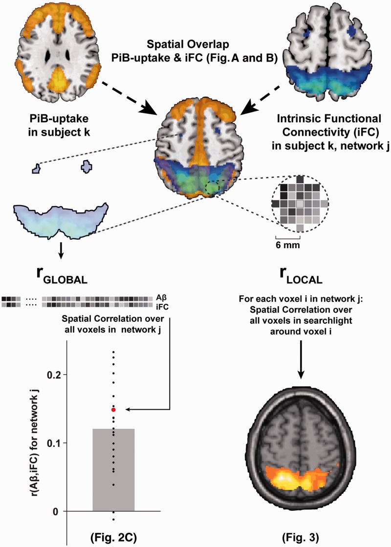
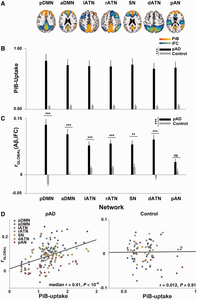
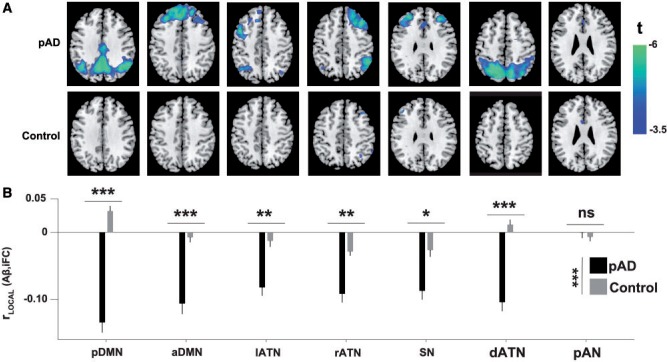
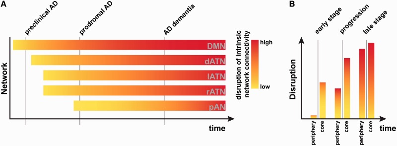
There is striking overlap between the spatial distribution of amyloid-β pathology in patients with Alzheimer's disease and the spatial distribution of high intrinsic functional connectivity in healthy persons. This overlap suggests a mechanistic link between amyloid-β and intrinsic connectivity, and indeed there is evidence in patients for the detrimental effects of amyloid-β plaque accumulation on intrinsic connectivity in areas of high connectivity in heteromodal hubs, and particularly in the default mode network. However, the observed spatial extent of amyloid-β exceeds these tightly circumscribed areas, suggesting that previous studies may have underestimated the negative impact of amyloid-β on intrinsic connectivity. We hypothesized that the known positive baseline correlation between patterns of amyloid-β and intrinsic connectivity may mask the larger extent of the negative effects of amyloid-β on connectivity. Crucially, a test of this hypothesis requires the within-patient comparison of intrinsic connectivity and amyloid-β distributions. Here we compared spatial patterns of amyloid-β-plaques (measured by Pittsburgh compound B positron emission tomography) and intrinsic functional connectivity (measured by resting-state functional magnetic resonance imaging) in patients with prodromal Alzheimer's disease via spatial correlations in intrinsic networks covering fronto-parietal heteromodal cortices. At the global network level, we found that amyloid-β and intrinsic connectivity patterns were positively correlated in the default mode and several fronto-parietal attention networks, confirming that amyloid-β aggregates in areas of high intrinsic connectivity on a within-network basis. Further, we saw an internetwork gradient of the magnitude of correlation that depended on network plaque-load. After accounting for this globally positive correlation, local amyloid-β-plaque concentration in regions of high connectivity co-varied negatively with intrinsic connectivity, indicating that amyloid-β pathology adversely reduces connectivity anywhere in an affected network as a function of local amyloid-β-plaque concentration. The local negative association between amyloid-β and intrinsic connectivity was much more pronounced than conventional group comparisons of intrinsic connectivity would suggest. Our findings indicate that the negative impact of amyloid-β on intrinsic connectivity in heteromodal networks is underestimated by conventional analyses. Moreover, our results provide first within-patient evidence for correspondent patterns of amyloid-β and intrinsic connectivity, with the distribution of amyloid-β pathology following functional connectivity gradients within and across intrinsic networks.
Overview
The claims and statistics of this paper haven’t been checked yet.
Run the check — free, no account needed. It reads the open-access text and checks the sources it cites.
Integrity signals
Registry
Cites
Statistics
Figures
Verification
Claims the paper makes
The claims and statistics of this paper haven’t been checked yet.
Run the check — free, no account needed. It reads the open-access text and checks the sources it cites.
Claims other papers make about this one
Each is checked against this paper’s own text: does it say what the citing paper says it does?
In the citing paper
“A certain overlap between the DMN and the distribution of Aβ fibrils in AD has also been noted in previous publications23–26.”
From Earliest accumulation of β-amyloid occurs within the default-mode network and concurrently affects brain connectivity (2017) · cites this paper as [25]
In the citing paper
“There is an increased Aβ-PET signal within the CEN and the SN in the progression of AD.244,245”
From Amyloid β-based therapy for Alzheimer’s disease: Challenges, successes and future (2023) · cites this paper as [244]
Self-check
Percentages, sums and reported differences re-computed from the paper’s own numbers.
The self-check needs the paper’s full text.
Citations
Cites
Neuropathological stageing of Alzheimer-related changes.
Braak, H., & Braak, E. (1991) · Acta Neuropathologica · doi:10.1007/bf00308809 · Cited by 16,123 · cited 1×
“The dissociation between heteromodal and primary networks is in line with findings that primary sensory and sensorimotor regions are relatively spared in earlier stages of Alzheimer’s disease (Braak and Braak, 1991).”
The Clinical Dementia Rating (CDR): Current version and scoring rules
Morris, J. C. (1993) · Neurology · doi:10.1212/wnl.43.11.2412-a · Cited by 9,882
Ashburner, J., & Friston, K. J. (2005) · NeuroImage · doi:10.1016/j.neuroimage.2005.02.018 · Cited by 7,507 · cited 1×
“…MRI image (using affine registration), and normalized to a template in the stereotactic space of the Montreal Neurological Institute (MNI) with the ‘segment’ function (SPM5), which uses an iterative combination of non-linear registration and cortical segmentation (Ashburner and Friston, 2005).”
Power, J. D., Barnes, K. A., Snyder, A. Z., Schlaggar, B. L., & Petersen, S. E. (2012) · NeuroImage · doi:10.1016/j.neuroimage.2011.10.018 · Cited by 7,429 · cited 2×
“Framewise displacement (Power et al., 2012) or the root-mean-”
Hypothetical model of dynamic biomarkers of the Alzheimer's pathological cascade
Jack, C. R., Knopman, D. S., Jagust, W. J., Shaw, L. M., Aisen, P. S., Weiner, M. W., Petersen, R. C., & Trojanowski, J. Q. (2010) · The Lancet Neurology · doi:10.1016/s1474-4422(09)70299-6 · Cited by 4,806
Alzheimer's Disease Is a Synaptic Failure
Selkoe, D. J. (2002) · Science · doi:10.1126/science.1074069 · Cited by 4,420 · cited 1×
“Aberrant clearance of amyloid-b precursor protein is thought to be a critical initial event in the disease’s pathogenesis, leading to amyloid-b peptide accumulation and plaque formation 20 to 30 years before cognitive symptoms arise (Selkoe, 2002; Jack et al., 2010; Bateman et al., 2012).”
Research criteria for the diagnosis of Alzheimer's disease: revising the NINCDS–ADRDA criteria
Dubois, B., Feldman, H. H., Jacova, C., DeKosky, S. T., Barberger-Gateau, P., Cummings, J., Delacourte, A., Galasko, D., Gauthier, S., Jicha, G., et al. (2007) · The Lancet Neurology · doi:10.1016/s1474-4422(07)70178-3 · Cited by 4,411 · cited 1×
“Prodromal Alzheimer’s disease has recently been defined by the coincidence of both mild cognitive impairment and the presence of at least one of five supportive biological signs for Alzheimer’s disease, such as medial temporal lobe atrophy or significant PiB uptake (Dubois et al., 2007).”
Moms, J. C., Heyman, A., Mohs, R. C., Hughes, J. P., van Belle, G., Fillenbaum, G., Mellits, E. D., & Clark, C. (1989) · Neurology · doi:10.1212/wnl.39.9.1159 · Cited by 4,130 · cited 1×
“…participant included medical history, neurological examination, informant interview (Morris, 1993), neuropsychological assessment by the neuropsychological assessment battery of the Consortium to Establish a Registry for Alzheimer’s disease (CERAD, Morris et al., 1989), structural MRI and PiB-PET.”
Gauthier, S. G., Reisberg, ., Zaudig, M., Petersen, R. C., Ritchie, K. A., Broich, K., Belleville, S., Brodaty, H., Bennett, D. A., Chertkow, H. M., et al. (2006) · The Lancet · doi:10.1016/s0140-6736(06)68542-5 · Cited by 4,094
Clinical and biomarker changes in dominantly inherited Alzheimer's disease
Bateman, R. J., Xiong, C., Benzinger, T. L. S., Fagan, A. M., Goate, A., Fox, N. C., Marcus, D. S., Cairns, N. J., Xie, X., Blazey, T. M., et al. (2012) · New England Journal of Medicine · doi:10.1056/nejmoa1202753 · Cited by 3,814 · cited 3×
“Given that significant PiB-uptake is measurable in the DMN even in early preclinical stages of Alzheimer’s disease (Jack et al., 2010; Bateman et al., 2012), where conventional measures of connectivity do not report robust connectivity reductions, the method presented here may help detect early, subtle reductions in c…”
Cited by
Sorted by citations
The Amyloid-β Pathway in Alzheimer’s Disease
Hampel, H., Hardy, J., Blennow, K., Chen, C., Perry, G., Kim, S. H., Villemagne, V. L., Aisen, P., Vendruscolo, M., Iwatsubo, T., et al. (2021) · Molecular Psychiatry · doi:10.1038/s41380-021-01249-0 · Cited by 1,952
The connectomics of brain disorders
Fornito, A., Zalesky, A., & Breakspear, M. (2015) · Nature Reviews Neuroscience · doi:10.1038/nrn3901 · Cited by 1,734
Amyloid β-based therapy for Alzheimer’s disease: Challenges, successes and future
Zhang, Y., Chen, H., Li, R., Sterling, K., & Song, W. (2023) · Signal Transduction and Targeted Therapy · doi:10.1038/s41392-023-01484-7 · Cited by 988
Palmqvist, S., Schöll, M., Strandberg, O., Mattsson, N., Stomrud, E., Zetterberg, H., Blennow, K., Landau, S., Jagust, W., & Hansson, O. (2017) · Nature Communications · doi:10.1038/s41467-017-01150-x · Cited by 911
The Evolution of Preclinical Alzheimer’s Disease: Implications for Prevention Trials
Sperling, R., Mormino, E., & Johnson, K. (2014) · Neuron · doi:10.1016/j.neuron.2014.10.038 · Cited by 725
Metadata
- Authors
- Myers, N., Pasquini, L., Göttler, J., Grimmer, T., Koch, K., Ortner, M., Neitzel, J., Mühlau, M., Förster, S., Kurz, A., Förstl, H., Zimmer, C., Wohlschläger, A. M., Riedl, V., Drzezga, A., & Sorg, C.
- Affiliations
- TUM Klinikum (DE)
- Technical University of Munich (DE)
- University of Cologne (DE)
- Corresponding author
- Myers, Nicholas
- ORCID
- Venue
- Brain
- Publisher
- Oxford University Press (OUP)
- Published
- 2014-04-26
- Volume
- 137
- Issue
- 7
- Pages
- 2052-2064
- ISSN
- 0006-8950 (ISSN-L)
- 1460-2156
- PMC
- PMC4065018
- OpenAlex
- W2031921419
- Cited by
- 132
- References
- 57
- Citations by year
- 2026: 3 · 2025: 3 · 2024: 9 · 2023: 13 · 2022: 9 · 2021: 15 · 2020: 5 · 2019: 10 · 2018: 13 · 2017: 20 · 2016: 11 · 2015: 14 · 2014: 7
- FWCI
- 9.67
- Type
- journal article
- Language
- en
- Open access
- hybrid
- doi.org
- Licence
- cc-by
- Locations
- Brain (publishedVersion, cc-by)
- PubMed Central (publishedVersion, cc-by)
- PubMed (publishedVersion)
- eScholarship (California Digital Library) (submittedVersion, cc-by)
- mediaTUM (Technical University of Munich) (submittedVersion)
- Funders
- Wellcome Trust
- Bundesministerium für Bildung und Forschung (01ER0803, 01EV0710)
- Topics
- Life Sciences › Neuroscience › Cognitive Neuroscience › Functional Brain Connectivity Studies
- Health Sciences › Medicine › Psychiatry and Mental health › Dementia and Cognitive Impairment Research
- Life Sciences › Neuroscience › Cellular and Molecular Neuroscience › Alzheimer's disease research and treatments
- Keywords
- amyloid plaques, intrinsic functional connectivity, Alzheimer's disease, prodromal Alzheimer's disease, default mode network, frontoparietal attention network, within-patient comparison, Pittsburgh compound B PET, resting-state fMRI
- MeSH terms
- Aged, Aged, 80 and over, Alzheimer Disease, Analysis of Variance, Aniline Compounds, Brain, Brain Mapping, Female, Humans, Magnetic Resonance Imaging, Male, Middle Aged, Neural Pathways, Neuropsychological Tests, Thiazoles, Amyloid beta-Peptides, Positron-Emission Tomography, Plaque, Amyloid
About these checks
Registry, retraction, Cites and statistics results are computed, not judged, with one exception: a printed reference that no index lists is read once by a model before it counts as a citation error. Claims are extracted by a model and kept only when their quote appears verbatim in the text; each cited source is then checked, with a model’s help, for whether it says what the claim relies on. Those verdicts can be wrong — read the claim’s quote and the cited source before relying on one.
Statistics and arithmetic are recomputed, not judged.
Confidence bands are shown only for fields calibrated on our annotated test set. None is calibrated yet, so no band is shown.
What counts as a claim ›How we measure confidence ›How we check this ›
Are you an author, or do you see a problem with this page? Request a review
