

The DOI matches its Crossref record; 56 of the 56 references it cites were found in a registry or an index.
Frontal white matter hyperintensities, clasmatodendrosis and gliovascular abnormalities in ageing and post-stroke dementia
Open accessAbstract
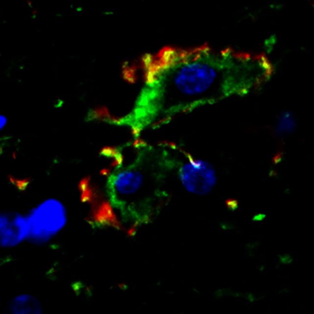
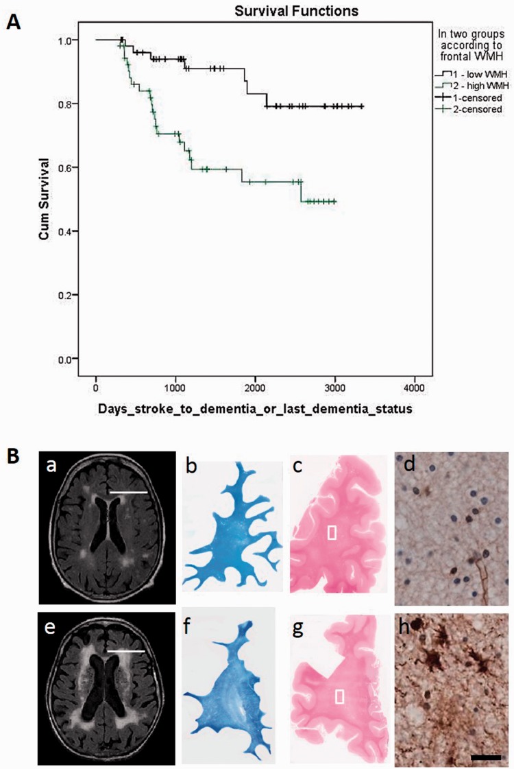
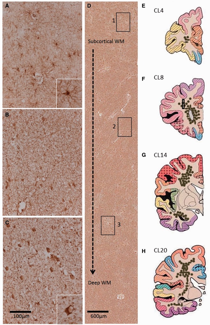
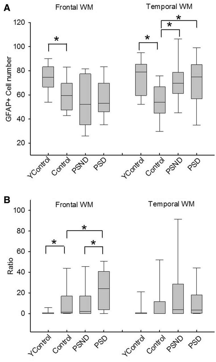
White matter hyperintensities as seen on brain T2-weighted magnetic resonance imaging are associated with varying degrees of cognitive dysfunction in stroke, cerebral small vessel disease and dementia. The pathophysiological mechanisms within the white matter accounting for cognitive dysfunction remain unclear. With the hypothesis that gliovascular interactions are impaired in subjects with high burdens of white matter hyperintensities, we performed clinicopathological studies in post-stroke survivors, who had exhibited greater frontal white matter hyperintensities volumes that predicted shorter time to dementia onset. Histopathological methods were used to identify substrates in the white matter that would distinguish post-stroke demented from post-stroke non-demented subjects. We focused on the reactive cell marker glial fibrillary acidic protein (GFAP) to study the incidence and location of clasmatodendrosis, a morphological attribute of irreversibly injured astrocytes. In contrast to normal appearing GFAP+ astrocytes, clasmatodendrocytes were swollen and had vacuolated cell bodies. Other markers such as aldehyde dehydrogenase 1 family, member L1 (ALDH1L1) showed cytoplasmic disintegration of the astrocytes. Total GFAP+ cells in both the frontal and temporal white matter were not greater in post-stroke demented versus post-stroke non-demented subjects. However, the percentage of clasmatodendrocytes was increased by >2-fold in subjects with post-stroke demented compared to post-stroke non-demented subjects (P = 0.026) and by 11-fold in older controls versus young controls (P < 0.023) in the frontal white matter. High ratios of clasmotodendrocytes to total astrocytes in the frontal white matter were consistent with lower Mini-Mental State Examination and the revised Cambridge Cognition Examination scores in post-stroke demented subjects. Double immunofluorescent staining showed aberrant co-localization of aquaporin 4 (AQP4) in retracted GFAP+ astrocytes with disrupted end-feet juxtaposed to microvessels. To explore whether this was associated with the disrupted gliovascular interactions or blood-brain barrier damage, we assessed the co-localization of GFAP and AQP4 immunoreactivities in post-mortem brains from adult baboons with cerebral hypoperfusive injury, induced by occlusion of three major vessels supplying blood to the brain. Analysis of the frontal white matter in perfused brains from the animals surviving 1-28 days after occlusion revealed that the highest intensity of fibrinogen immunoreactivity was at 14 days. At this survival time point, we also noted strikingly similar redistribution of AQP4 and GFAP+ astrocytes transformed into clasmatodendrocytes. Our findings suggest novel associations between irreversible astrocyte injury and disruption of gliovascular interactions at the blood-brain barrier in the frontal white matter and cognitive impairment in elderly post-stroke survivors. We propose that clasmatodendrosis is another pathological substrate, linked to white matter hyperintensities and frontal white matter changes, which may contribute to post-stroke or small vessel disease dementia.
Overview
The claims and statistics of this paper haven’t been checked yet.
Run the check — free, no account needed. It reads the open-access text and checks the sources it cites.
Integrity signals
Registry
Cites
Statistics
Figures
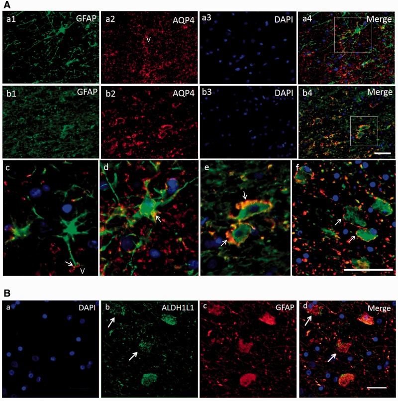
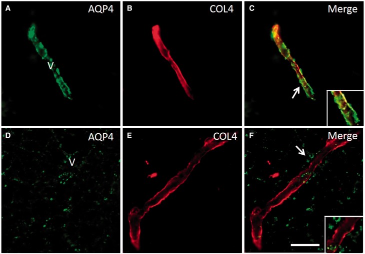
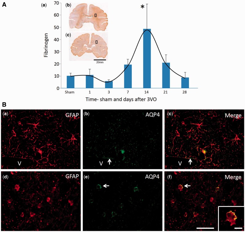
Verification
Claims the paper makes
The claims and statistics of this paper haven’t been checked yet.
Run the check — free, no account needed. It reads the open-access text and checks the sources it cites.
Claims other papers make about this one
Each is checked against this paper’s own text: does it say what the citing paper says it does?
The cited source says what the claim attributes to it.
In the citing paper
“These metabolic marker–related cell changes implicate alterations in cerebral perfusion and, therefore, potentially gliovascular changes in frontal white matter observed within PSD. 67”
From High vulnerability of medial prefrontal pyramidal neurons in post-stroke, vascular, Alzheimer's disease, and aging-related dementias (2026) · cites this paper as [67]
Self-check
Percentages, sums and reported differences re-computed from the paper’s own numbers.
The self-check needs the paper’s full text.
Citations
Cites
Astrocyte-endothelial interactions at the blood-brain barrier
Abbott, N. J., Rönnbäck, L., & Hansson, E. (2006) · Nature Reviews Neuroscience · doi:10.1038/nrn1824 · Cited by 5,595
Debette, S., & Markus, H. S. (2010) · BMJ · doi:10.1136/bmj.c3666 · Cited by 2,328
Pendlebury, S. T., & Rothwell, P. M. (2009) · The Lancet Neurology · doi:10.1016/s1474-4422(09)70236-4 · Cited by 1,853
Glycolytic oligodendrocytes maintain myelin and long-term axonal integrity
Fünfschilling, U., Supplie, L. M., Mahad, D., Boretius, S., Saab, A. S., Edgar, J., Brinkmann, B. G., Kassmann, C. M., Tzvetanova, I. D., Möbius, W., et al. (2012) · Nature · doi:10.1038/nature11007 · Cited by 1,523
de Leeuw, F.-E. (2001) · Journal of Neurology, Neurosurgery & Psychiatry · doi:10.1136/jnnp.70.1.9 · Cited by 1,291
Aquaporins in Brain: Distribution, Physiology, and Pathophysiology
Badaut, J., Lasbennes, F., Magistretti, P. J., & Regli, L. (2002) · Journal of Cerebral Blood Flow & Metabolism · doi:10.1097/00004647-200204000-00001 · Cited by 536
DeCarli, C., Murphy, D. G. M., Tranh, M., Grady, C. L., Haxby, J. V., Gillette, J. A., Salerno, J. A., Gonzales-Aviles, A., Honvitz, B., Rapoport, S. I., et al. (1995) · Neurology · doi:10.1212/wnl.45.11.2077 · Cited by 531
Pathogenic potential of IgG binding to water channel extracellular domain in neuromyelitis optica
Hinson, S. R., Pittock, S., Lucchinetti, C., Roemer, S., Fryer, J., Kryzer, T. J., & Lennon, V. (2007) · Neurology · doi:10.1212/01.wnl.0000289761.64862.ce · Cited by 482
The Pathology of an Autoimmune Astrocytopathy: Lessons Learned from Neuromyelitis Optica
Lucchinetti, C. F., Guo, Y. X., Popescu, B. F. G., Fujihara, K., Itoyama, Y., & Misu, T. (2013) · Brain Pathology · doi:10.1111/bpa.12099 · Cited by 422
Inzitari, D., Pracucci, G., Poggesi, A., Carlucci, G., Barkhof, F., Chabriat, H., Erkinjuntti, T., Fazekas, F., Ferro, J. M., Hennerici, M., et al. (2009) · BMJ · doi:10.1136/bmj.b2477 · Cited by 415
Self-citation
Cited by
Sorted by citations
Perivascular spaces in the brain: anatomy, physiology and pathology
Wardlaw, J. M., Benveniste, H., Nedergaard, M., Zlokovic, B. V., Mestre, H., Lee, H., Doubal, F. N., Brown, R., Ramirez, J., MacIntosh, B. J., et al. (2020) · Nature Reviews Neurology · doi:10.1038/s41582-020-0312-z · Cited by 870
Post-Stroke Cognitive Impairment and Dementia
Rost, N. S., Brodtmann, A. G., Pase, M. P., van Veluw, S. J., Biffi, A., Duering, M., Hinman, J. D., & Dichgans, M. (2022) · Circulation Research · doi:10.1161/circresaha.122.319951 · Cited by 864
van der Flier, W. M., Skoog, I. G., Schneider, J. A., Pantoni, L., Mok, V., Chen, C., & Scheltens, P. (2018) · Nature Reviews Disease Primers · doi:10.1038/nrdp.2018.3 · Cited by 606
Stroke injury, cognitive impairment and vascular dementia
Kalaria, R. N., Akinyemi, R., & Ihara, M. (2016) · Biochimica et Biophysica Acta (BBA) - Molecular Basis of Disease · doi:10.1016/j.bbadis.2016.01.015 · Cited by 647
Self-citationKalaria, R. N. (2016) · Acta Neuropathologica · doi:10.1007/s00401-016-1571-z · Cited by 441
Self-citationDuncombe, J., Kitamura, A., Hase, Y., Ihara, M., Kalaria, R. N., & Horsburgh, K. J. (2017) · Clinical Science · doi:10.1042/cs20160727 · Cited by 416
Self-citationThe role of the medial prefrontal cortex in cognition, ageing and dementia
Jobson, D. D., Hase, Y., Clarkson, A. N., & Kalaria, R. N. (2021) · Brain Communications · doi:10.1093/braincomms/fcab125 · Cited by 338
Self-citationAstrocytes: Role and Functions in Brain Pathologies
Siracusa, R., Fusco, R., & Cuzzocrea, S. (2019) · Frontiers in Pharmacology · doi:10.3389/fphar.2019.01114 · Cited by 361
The potential mechanism of postoperative cognitive dysfunction in older people
Lin, X., Chen, Y., Zhang, P., Chen, G., Zhou, Y., & Yu, X. (2019) · Experimental Gerontology · doi:10.1016/j.exger.2019.110791 · Cited by 357
Cuadrado‐Godia, E., Dwivedi, P., Sharma, S., Ois, Á., Roquer, J., Balcells, M., Laird, J. R., Turk, M., Suri, H. S., Nicolaides, A. N., et al. (2018) · Journal of Stroke · doi:10.5853/jos.2017.02922 · Cited by 297
Deciphering the astrocyte reaction in Alzheimer's disease
Perez-Nievas, B. G., & Serrano-Pozo, A. (2018) · Frontiers in Aging Neuroscience · doi:10.3389/fnagi.2018.00114 · Cited by 282
Wardlaw, J. M., Makin, S., Hernández, M. C. V., Armitage, P. A., Heye, A. K., Chappell, F. M., Maniega, S. M., Sakka, E., Shuler, K., Dennis, M. S., et al. (2016) · Alzheimer s & Dementia · doi:10.1016/j.jalz.2016.09.006 · Cited by 268
Post-Stroke Cognitive Impairment: Epidemiology, Risk Factors, and Management
Huang, Y.-Y., Chen, S.-D., Leng, X.-Y., Kuo, K., Wang, Z.-T., Cui, M., Tan, L., Wang, K., Dong, Q., & Yu, J.-T. (2022) · Journal of Alzheimer s Disease · doi:10.3233/jad-215644 · Cited by 252
White matter degeneration in vascular and other ageing‐related dementias
Hase, Y., Horsburgh, K. J., Ihara, M., & Kalaria, R. N. (2017) · Journal of Neurochemistry · doi:10.1111/jnc.14271 · Cited by 214
Self-citationExtracellular matrix inflammation in vascular cognitive impairment and dementia
Rosenberg, G. A. (2017) · Clinical Science · doi:10.1042/cs20160604 · Cited by 214
Astrocytes in Alzheimer's disease: pathological significance and molecular pathways
Preman, P., Alfonso-Triguero, M., Alberdi, E., Verkhratsky, A., & Arranz, A. M. (2021) · Cells · doi:10.3390/cells10030540 · Cited by 176
Animal Models of Chronic Cerebral Hypoperfusion: From Mouse to Primate
Washida, K., Hattori, Y., & Ihara, M. (2019) · International Journal of Molecular Sciences · doi:10.3390/ijms20246176 · Cited by 166
Self-citationRamirez, J., Berezuk, C., McNeely, A. A., Gao, F., McLaurin, J., & Black, S. E. (2016) · Cellular and Molecular Neurobiology · doi:10.1007/s10571-016-0343-6 · Cited by 164
Cells of the Blood–Brain Barrier: An Overview of the Neurovascular Unit in Health and Disease
McConnell, H., & Mishra, A. (2022) · Methods in molecular biology · doi:10.1007/978-1-0716-2289-6_1 · Cited by 174
Dysfunction of Cerebrovascular Endothelial Cells: Prelude to Vascular Dementia
Wang, F., Cao, Y., Ma, L., Pei, H., Rausch, W., & Li, H. (2018) · Frontiers in Aging Neuroscience · doi:10.3389/fnagi.2018.00376 · Cited by 163
Iadecola, C., Smith, E. E., Anrather, J., Gu, C., Mishra, A., Misra, S., Pérez‐Pinzón, M. A., Shih, A. Y., Sorond, F. A., van Veluw, S. J., et al. (2023) · Stroke · doi:10.1161/str.0000000000000431 · Cited by 161
Ding, R., Hase, Y., Ameen‐Ali, K. E., Ndung'u, M., Stevenson, W., Barsby, J., Gourlay, R., Akinyemi, T. O., Akinyemi, R., Uemura, M. T., et al. (2020) · Brain Pathology · doi:10.1111/bpa.12888 · Cited by 145
Self-citationHabes, M., Sotiras, A., Erus, G., Toledo, J. B., Janowitz, D., Wolk, D. A., Shou, H., Bryan, R. N., Doshi, J. J., Völzke, H., et al. (2018) · Neurology · doi:10.1212/wnl.0000000000006116 · Cited by 147
Post-mortem assessment in vascular dementia: advances and aspirations
McAleese, K. E., Alafuzoff, I. I., Charidimou, A., Reuck, J. D., Grinberg, L. T., Hainsworth, A. H., Hortobágyi, T., Ince, P. G., Jellinger, K. A., Gao, J., et al. (2016) · BMC Medicine · doi:10.1186/s12916-016-0676-5 · Cited by 144
Self-citationJoutel, A., & Chabriat, H. (2017) · Clinical Science · doi:10.1042/cs20160380 · Cited by 141
Separating vascular and neuronal effects of age on fMRI BOLD signals
Tsvetanov, K. A., Henson, R. N., & Rowe, J. B. (2020) · Philosophical Transactions of the Royal Society B Biological Sciences · doi:10.1098/rstb.2019.0631 · Cited by 136
Age-associated changes in the blood-brain barrier: comparative studies in human and mouse.
Goodall, E. F., Wang, C., Simpson, J. E., Baker, D. J., Drew, D. R., Heath, P. R., Saffrey, M. J., Romero, I. A., & Wharton, S. B. (2018) · Neuropathology and Applied Neurobiology · doi:10.1111/nan.12408 · Cited by 120
Yang, Q. Z., Wei, X., Deng, B., Chang, Z., Jin, D., Huang, Y., Zhang, J. H., Yenari, M. A., Jin, K., & Wang, Q. (2022) · Neurobiology of Disease · doi:10.1016/j.nbd.2022.105750 · Cited by 138
The pathogenesis of cerebral small vessel disease and vascular cognitive impairment
Markus, H. S., & Joutel, A. (2025) · Physiological Reviews · doi:10.1152/physrev.00028.2024 · Cited by 140
Waller, R., Baxter, L., Fillingham, D. J., Coelho, S., Pozo, J. M., Mozumder, M., Frangi, A. F., Ince, P. G., Simpson, J. E., & Highley, J. R. (2019) · PLoS ONE · doi:10.1371/journal.pone.0210888 · Cited by 117
Translational models for vascular cognitive impairment: a review including larger species
Hainsworth, A. H., Allan, S. M., Boltze, J., Cunningham, C. J., Farris, C. W., Head, E., Ihara, M., Isaacs, J. D., Kalaria, R. N., Oberstein, S. A. J. L., et al. (2017) · BMC Medicine · doi:10.1186/s12916-017-0793-9 · Cited by 111
Self-citationVascular dementia: A microglia's perspective
Yang, Y. R., Zhao, X., Zhu, Z., & Zhang, L. (2022) · Ageing Research Reviews · doi:10.1016/j.arr.2022.101734 · Cited by 103
McDonald, M. W., Black, S. E., Copland, D. A., Corbett, D., Dijkhuizen, R. M., Farr, T. D., Jeffers, M. S., Kalaria, R. N., Karayanidis, F., Leff, A. P., et al. (2019) · International Journal of Stroke · doi:10.1177/1747493019873600 · Cited by 95
Self-citationLana, D., Ugolini, F., & Giovannini, M. G. (2020) · Frontiers in Cellular Neuroscience · doi:10.3389/fncel.2020.585833 · Cited by 92
Mills, W. A., Woo, A. M., Jiang, S., Martin, J., Surendran, D., Bergstresser, M., Kimbrough, I. F., Eyo, U. B., Sofroniew, M. V., & Sontheimer, H. W. (2022) · Nature Communications · doi:10.1038/s41467-022-29475-2 · Cited by 95
Duncombe, J., Lennen, R. J., Jansen, M. A., Marshall, I., Wardlaw, J. M., & Horsburgh, K. J. (2016) · Neuropathology and Applied Neurobiology · doi:10.1111/nan.12375 · Cited by 85
Self-citationMcConnell, H., Li, Z., Woltjer, R. L., & Mishra, A. (2019) · Neurochemistry International · doi:10.1016/j.neuint.2019.04.005 · Cited by 74
Update on the Neurobiology of Vascular Cognitive Impairment: From Lab to Clinic
Vinciguerra, L., Lanza, G., Puglisi, V., Fisicaro, F., Pennisi, M., Bella, R., & Cantone, M. (2020) · International Journal of Molecular Sciences · doi:10.3390/ijms21082977 · Cited by 69
Lana, D., Ugolini, F., Nosi, D., Wenk, G. L., & Giovannini, M. G. (2021) · Frontiers in Aging Neuroscience · doi:10.3389/fnagi.2021.651973 · Cited by 73
Hase, Y., Craggs, L., Hase, M., Stevenson, W., Slade, J. Y., Lopez, D., Mehta, R., Chen, A., Liang, D., Oakley, A. E., et al. (2017) · Journal of Neuroinflammation · doi:10.1186/s12974-017-0850-5 · Cited by 68
Self-citationEffects of advanced age upon astrocyte-specific responses to acute traumatic brain injury in mice
Early, A. N., Gorman, A. A., Eldik, L. J. V., Bachstetter, A. D., & Morganti, J. M. (2020) · Journal of Neuroinflammation · doi:10.1186/s12974-020-01800-w · Cited by 64
Ferroptosis of Microglia in Aging Human White Matter Injury
Adeniyi, P. A., Gong, X., MacGregor, E., Degener-O’Brien, K., McClendon, E., Garcia, M., Romero, O., Russell, J., Srivastava, T., Miller, J. A., et al. (2023) · Annals of Neurology · doi:10.1002/ana.26770 · Cited by 62
Small vessels, dementia and chronic diseases – molecular mechanisms and pathophysiology
Horsburgh, K. J., Wardlaw, J. M., Agtmael, T. V., Allan, S. M., Ashford, M. L. J., Bath, P. M., Brown, R., Berwick, J., Cader, M. Z., Carare, R. O., et al. (2018) · Clinical Science · doi:10.1042/cs20171620 · Cited by 65
Self-citationCerebral small vessel disease and post-stroke cognitive impairment
Teng, Z., Dong, Y., Zhang, D., An, J., & Lv, P. (2016) · International Journal of Neuroscience · doi:10.1080/00207454.2016.1261291 · Cited by 63
Association between different acute stroke therapies and development of post stroke seizures
Naylor, J., Thevathasan, A., Churilov, L. P., Guo, R., Xiong, Y., Koome, M. E., Chen, Z., Chen, Z., Liu, X., Kwan, P., et al. (2018) · BMC Neurology · doi:10.1186/s12883-018-1064-x · Cited by 63
Homocysteine in Neurology: A Possible Contributing Factor to Small Vessel Disease
Moretti, R., Giuffrè, M., Caruso, P., Gazzin, S., & Tiribelli, C. (2021) · International Journal of Molecular Sciences · doi:10.3390/ijms22042051 · Cited by 64
Giovannini, M. G., Lana, D., Traini, C., & Vannucchi, M. G. (2021) · Journal of Clinical Medicine · doi:10.3390/jcm10112358 · Cited by 64
Forsberg, K., Zhang, Y., Reiners, J. C., Ander, M., Niedermayer, A., Fang, L., Neugebauer, H., Kassubek, J., Katona, I., Weis, J., et al. (2018) · Acta Neuropathologica Communications · doi:10.1186/s40478-018-0632-z · Cited by 56
Small Vessel Disease-Related Dementia: An Invalid Neurovascular Coupling?
Moretti, R., & Caruso, P. (2020) · International Journal of Molecular Sciences · doi:10.3390/ijms21031095 · Cited by 62
Lu, Q., Xiong, J., Yuan, Y., Ruan, Z., Zhang, Y., Chai, B., Li, L., Cai, S., Xiao, J., Wu, Y., et al. (2021) · International Journal of Biological Sciences · doi:10.7150/ijbs.64187 · Cited by 56
Vascular Contributions to Cognitive Impairment and Dementia
Madigan, J. B., Wilcock, D. M., & Hainsworth, A. H. (2016) · Stroke · doi:10.1161/strokeaha.116.012066 · Cited by 56
Severe white matter astrocytopathy in CADASIL
Hase, Y., Chen, A., Bates, L. L., Craggs, L., Yamamoto, Y., Gemmell, E., Oakley, A. E., Korolchuk, V. I., & Kalaria, R. N. (2018) · Brain Pathology · doi:10.1111/bpa.12621 · Cited by 58
Self-citationThe Pattern of AQP4 Expression in the Ageing Human Brain and in Cerebral Amyloid Angiopathy
Owasil, R., O’Neill, R., Keable, A., Nimmo, J., MacGregor Sharp, M., Kelly, L., Saito, S., Simpson, J. E., Weller, R. O., Smith, C., et al. (2020) · International Journal of Molecular Sciences · doi:10.3390/ijms21041225 · Cited by 48
Heterogeneity of white matter astrocytes in the human brain
Bugiani, M., Plug, B. C., Man, J. H. K., Breur, M., & van der Knaap, M. S. (2021) · Acta Neuropathologica · doi:10.1007/s00401-021-02391-3 · Cited by 55
Manso, Y., Holland, P. R., Kitamura, A., Szymkowiak, S., Duncombe, J., Hennessy, E., Searcy, J. L., Marangoni, M., Randall, A. D., Brown, J. T., et al. (2017) · Glia · doi:10.1002/glia.23190 · Cited by 51
Self-citationNeurovascular Ageing and Age-Related Diseases
Kalaria, R. N., & Hase, Y. (2019) · Sub-cellular biochemistry/Subcellular biochemistry · doi:10.1007/978-981-13-3681-2_17 · Cited by 55
Self-citationVascular Cognitive Impairment through the Looking Glass of Transcranial Magnetic Stimulation
Lanza, G., Bramanti, P., Cantone, M., Pennisi, M., Pennisi, G., & Bella, R. (2017) · Behavioural Neurology · doi:10.1155/2017/1421326 · Cited by 50
White Matter Disease and Outcomes of Mechanical Thrombectomy for Acute Ischemic Stroke
Mistry, E. A., Mistry, A. M., Mehta, T., Arora, N., Starosciak, A. K., Rosa, F. D. L. R. L., Siegler, J. E., Kasner, S. E., Chitale, R. V., Fusco, M. R., et al. (2020) · American Journal of Neuroradiology · doi:10.3174/ajnr.a6478 · Cited by 50
Cai, M., Zhang, W., Weng, Z., Stetler, R. A., Jiang, X., Shi, Y., Gao, Y., & Chen, J. (2017) · Aging and Disease · doi:10.14336/ad.2017.0520 · Cited by 49
Post-stroke Dementia: Epidemiology, Mechanisms and Management
Hu, G., & Chen, Y.-M. (2017) · International journal of gerontology · doi:10.1016/j.ijge.2017.07.004 · Cited by 46
Abdelkarim, D., Zhao, Y., Turner, M. P., Sivakolundu, D. K., Lu, H., & Bart Rypma, A. (2019) · Neuroscience & Biobehavioral Reviews · doi:10.1016/j.neubiorev.2019.09.005 · Cited by 47
Heterogeneous blood‐brain barrier dysfunction in cerebral small vessel diseases
Ying, Y., Li, Y., Yao, T., Shao, X., Tang, W., Montagne, A., Chabriat, H., Wang, D. J. J., Wang, C., Yang, Q., et al. (2024) · Alzheimer s & Dementia · doi:10.1002/alz.13874 · Cited by 42
Ding, R., Hase, Y., Burke, M., Foster, V., Stevenson, W., Polvikoski, T. M., & Kalaria, R. N. (2021) · Acta Neuropathologica Communications · doi:10.1186/s40478-021-01230-6 · Cited by 39
Self-citationHealthcare services relaxing natural selection may contribute to increase of dementia incidence
You, W., Henneberg, R. J., & Henneberg, M. (2022) · Scientific Reports · doi:10.1038/s41598-022-12678-4 · Cited by 37
Astrocytes in Neuropathologies Affecting the Frontal Cortex
Peteri, U., Niukkanen, M., & Castrén, M. L. (2019) · Frontiers in Cellular Neuroscience · doi:10.3389/fncel.2019.00044 · Cited by 38
Large household reduces dementia mortality: A cross-sectional data analysis of 183 populations
You, W., & Henneberg, M. (2022) · PLoS ONE · doi:10.1371/journal.pone.0263309 · Cited by 33
Hase, Y., Polvikoski, T. M., Ihara, M., Hase, M., Zafar, R., Stevenson, W., Allan, L. M., Ennaceur, A., Horsburgh, K. J., Gallart‐Palau, X., et al. (2019) · Neuropathology and Applied Neurobiology · doi:10.1111/nan.12550 · Cited by 33
Self-citationThe phenomenon of clasmatodendrosis
Balaban, D. T., Miyawaki, E. K., Bhattacharyya, S., & Torre, M. (2021) · Heliyon · doi:10.1016/j.heliyon.2021.e07605 · Cited by 30
Nikkar, R., Esmaeili‐bandboni, A., Badrikoohi, M., & Babaei, P. (2022) · Metabolic Brain Disease · doi:10.1007/s11011-022-00940-7 · Cited by 31
Atrial Fibrillation is Independently Associated with Cognitive Impairment after Ischemic Stroke
Chander, R. J., Lim, L., Handa, S., Hiu, S. K. W., Choong, A., Lin, X., Singh, R., Oh, D. C. T., & Kandiah, N. (2017) · Journal of Alzheimer s Disease · doi:10.3233/jad-170313 · Cited by 29
White Matter Hyperintensities: Complex Predictor of Complex Outcomes
Karvelas, N., & Elahi, F. M. (2023) · Journal of the American Heart Association · doi:10.1161/jaha.123.030351 · Cited by 29
Tachibana, M., Mohri, I., Hirata, I., Kuwada, A., Kimura-Ohba, S., Kagitani-Shimono, K., Fushimi, H., Inoue, T., Shiomi, M., Kakuta, Y., et al. (2019) · Brain and Development · doi:10.1016/j.braindev.2018.07.008 · Cited by 28
Kim, H. J., & Fraser, S. (2022) · Journal of Neurology · doi:10.1007/s00415-021-10944-5 · Cited by 30
Reactive astrocytes associated with prion disease impair the blood brain barrier
Kushwaha, R., Li, Y., Makarava, N., Pandit, N. P., Molesworth, K., Birukov, K. G., & Baskakov, I. V. (2023) · Neurobiology of Disease · doi:10.1016/j.nbd.2023.106264 · Cited by 30
The impact of cerebral vasomotor reactivity on cerebrovascular diseases and cognitive impairment
Sforza, M., Bianchini, E., Alivernini, D., Salvetti, M., Pontieri, F. E., & Sette, G. (2022) · Journal of Neural Transmission · doi:10.1007/s00702-022-02546-w · Cited by 27
Dettori, I., Gaviano, L., Ugolini, F., Lana, D., Bulli, I., Magni, G., Rossi, F., Giovannini, M. G., & Pedata, F. (2021) · Frontiers in Pharmacology · doi:10.3389/fphar.2020.588757 · Cited by 26
McDonald, M. W., Black, S. E., Copland, D. A., Corbett, D., Dijkhuizen, R. M., Farr, T. D., Jeffers, M. S., Kalaria, R. N., Karayanidis, F., Leff, A. P., et al. (2019) · Neurorehabilitation and neural repair · doi:10.1177/1545968319886444 · Cited by 29
Self-citationHase, Y., Ameen‐Ali, K. E., Waller, R., Simpson, J. E., Stafford, C., Mahesh, A., Ryan, L., Pickering, L., Bodman, C., Hase, M., et al. (2022) · Brain Pathology · doi:10.1111/bpa.13101 · Cited by 27
Self-citationKim, J.-E., & Kang, T. (2021) · Antioxidants · doi:10.3390/antiox10050655 · Cited by 25
Dufour, B. D., Bartley, T., McBride, E., Allen, E., McLennan, Y. A., Hagerman, R. J., & Martínez‐Cerdeño, V. (2023) · Annals of Neurology · doi:10.1002/ana.26851 · Cited by 22
Evidence of beta amyloid independent small vessel disease in familial Alzheimer's disease
Littau, J. L., Velilla, L., Hase, Y., Villalba‐Moreno, N. D., Hagel, C., Drexler, D., Osorio Restrepo, S., Villegas, A., Lopera, F., Vargas, S., et al. (2022) · Brain Pathology · doi:10.1111/bpa.13097 · Cited by 20
Self-citationHumphreys, C. A., Jansen, M. A., Maniega, S. M., González‐Castro, V., Pernet, C. R., Deary, I. J., Salman, R. A., Wardlaw, J. M., & Smith, C. (2018) · International Journal of Stroke · doi:10.1177/1747493018799962 · Cited by 19
Saito, R., Nozaki, H., Kato, T., Toyoshima, Y., Tanaka, H., Tsubata, Y., Morioka, T., Horikawa, Y., Oyanagi, K., Morita, T., et al. (2018) · Journal of Neuropathology & Experimental Neurology · doi:10.1093/jnen/nly115 · Cited by 21
Shijo, M., Hamasaki, H., Honda, H., Suzuki, S. O., Tachibana, M., Ago, T., Kitazono, T., Iihara, K., & Iwaki, T. (2019) · Journal of Neuropathology & Experimental Neurology · doi:10.1093/jnen/nlz079 · Cited by 21
Rodent ischemic stroke models and their relevance in preclinical research
Thaysen, M., Westi, E. W., Clarkson, A. N., Wellendorph, P., & Kristensen, M. (2024) · Neuroprotection/Neuroprotection (Chichester, England. Print) · doi:10.1002/nep3.62 · Cited by 22
Górska, A. M., Santos‐García, I., Eiriz, I., Brüning, T., Nyman, T. A., & Pahnke, J. (2024) · Journal of Neuroscience Methods · doi:10.1016/j.jneumeth.2024.110239 · Cited by 20
Common Shared Pathogenic Aspects of Small Vessels in Heart and Brain Disease
Moretti, R., Janjusevic, M., Fluca, A. L., Saro, R., Gagno, G., Pierri, A., Padoan, L., Restivo, L., Derin, A., Beltrami, A. P., et al. (2022) · Biomedicines · doi:10.3390/biomedicines10051009 · Cited by 19
Obenaus, A., Rodriguez‐Grande, B., Lee, J. B., Dubois, C., Fournier, M., Cador, M., Caillé, S., & Badaut, J. (2023) · Acta Neuropathologica Communications · doi:10.1186/s40478-023-01515-y · Cited by 18
Roles of NG2 Glia in Cerebral Small Vessel Disease
He, Y., Li, Z., Shi, X., Ding, J., & Wang, X. (2022) · Neuroscience Bulletin · doi:10.1007/s12264-022-00976-w · Cited by 19
Waller, R., Narramore, R., Simpson, J. E., Heath, P. R., Verma, N., Tinsley, M., Barnes, J. R., Haris, H. T., Henderson, F. E., Matthews, F. E., et al. (2020) · Brain Pathology · doi:10.1111/bpa.12928 · Cited by 17
Self-citationLuo, W., Dai, Z., Wu, W., Li, H., & Zhang, Y. (2025) · PeerJ · doi:10.7717/peerj.19460 · Cited by 15
Abdelhak, A., Huss, A. M., Brück, A., Sebert, U., Mayer, B., Müller, H., Tumani, H., Otto, M., Yilmazer‐Hanke, D., Ludolph, A. C., et al. (2020) · Neurological Research and Practice · doi:10.1186/s42466-020-00062-4 · Cited by 16
Pathology of the neurovascular unit in leukodystrophies
Zarekiani, P., Breur, M., Wolf, N. I., de Vries, H. E., van der Knaap, M. S., & Bugiani, M. (2021) · Acta Neuropathologica Communications · doi:10.1186/s40478-021-01206-6 · Cited by 16
An Iatrogenic Model of Brain Small-Vessel Disease: Post-Radiation Encephalopathy
Moretti, R., & Caruso, P. (2020) · International Journal of Molecular Sciences · doi:10.3390/ijms21186506 · Cited by 16
Houlton, J., Barwick, D., & Clarkson, A. N. (2020) · Neurobiology of Learning and Memory · doi:10.1016/j.nlm.2020.107355 · Cited by 16
Kim, J.-E., Lee, D.-S., & Kang, T. (2022) · Antioxidants · doi:10.3390/antiox11101883 · Cited by 15
Neuropathological Correlates of White Matter Hyperintensities in Cerebral Amyloid Angiopathy
Makkinejad, N., Zotin, M. C. Z., van den Brink, H., Auger, C. A., Eigen, K. A. V., Iglesias, J. E., Greenberg, S. M., Perosa, V., & van Veluw, S. J. (2024) · Journal of the American Heart Association · doi:10.1161/jaha.124.035744 · Cited by 14
Chronic cerebral hypoperfusion induces venous dysfunction via EPAS1 regulation in mice
Wazny, V. K., Mahadevan, A., Nguyen, N. L. U., Wee, H. S.-A., Vipin, A., Lam, T., Tay, K. Y., See, J.-X., Sandhu, G. K., Leow, Y. J., et al. (2025) · Nature Communications · doi:10.1038/s41467-025-61614-3 · Cited by 15
Carotid disease at age 73 and cognitive change from age 70 to 76 years: A longitudinal cohort study
Wardlaw, J. M., Allerhand, M., Eadie, E., Thomas, A., Corley, J., Pattie, A., Taylor, A. M., Shenkin, S. D., Cox, S. R., Gow, A. J., et al. (2016) · Journal of Cerebral Blood Flow & Metabolism · doi:10.1177/0271678x16683693 · Cited by 14
Self-citationYu, M., Zheng, X., Cheng, F., Shao, B., Zhuge, Q., & Jin, K. (2022) · Frontiers in Neurology · doi:10.3389/fneur.2022.903565 · Cited by 14
Metadata
- Authors
- Chen, A., Akinyemi, R. O., Hase, Y., Firbank, M. J., Ndung’u, M. N., Foster, V., Craggs, L. J. L., Washida, K., Okamoto, Y., Thomas, A. J., Polvikoski, T. M., Allan, L. M., Oakley, A. E., O’Brien, J. T., Horsburgh, K., Ihara, M., & Kalaria, R. N.
- Affiliations
- Newcastle University (GB)
- University of Edinburgh (GB)
- National Cerebral and Cardiovascular Center (JP)
- Corresponding author
- Kalaria, Raj N.
- ORCID
- Venue
- Brain
- Publisher
- Oxford University Press (OUP)
- Published
- 2015-12-14
- Volume
- 139
- Issue
- 1
- Pages
- 242-258
- ISSN
- 0006-8950 (ISSN-L)
- 1460-2156
- PMC
- PMC4905522
- OpenAlex
- W2227183631
- Cited by
- 172
- References
- 56
- Citations by year
- 2026: 15 · 2025: 12 · 2024: 10 · 2023: 10 · 2022: 30 · 2021: 19 · 2020: 18 · 2019: 16 · 2018: 16 · 2017: 14 · 2016: 10 · 2015: 1
- FWCI
- 9.63
- Type
- journal article
- Language
- en
- Open access
- hybrid
- doi.org
- Licence
- cc-by-nc
- Locations
- Brain (publishedVersion, cc-by-nc)
- Europe PMC (PubMed Central) (publishedVersion, cc-by-nc)
- PubMed Central (publishedVersion, cc-by-nc)
- PubMed (publishedVersion)
- White Rose Research Online (University of Leeds, The University of Sheffield, University of York) (acceptedVersion)
- HighWire Press Open Archive (submittedVersion)
- Edinburgh Research Explorer (University of Edinburgh) (submittedVersion, other-oa)
- Newcastle University ePrints (Newcastle Univesity) (cc-by)
- Funders
- Alzheimer's Society
- Dunhill Medical Trust (R277/0213)
- Great Britain Sasakawa Foundation
- Newcastle University
- International Brain Research Organization
- Newcastle upon Tyne Hospitals NHS Foundation Trust
- SENSHIN Medical Research Foundation
- Medical Research Council (G0400074, MR/K026992/1, G0500247, MR/L016451/1)
- Engineering and Physical Sciences Research Council
- Biotechnology and Biological Sciences Research Council
- Economic and Social Research Council
- Japan Society for the Promotion of Science (15H04271)
- Topics
- Health Sciences › Medicine › Clinical Neurology › Cerebrovascular and genetic disorders
- Life Sciences › Neuroscience › Neurology › Neurological Disease Mechanisms and Treatments
- Life Sciences › Neuroscience › Neurology › Neuroinflammation and Neurodegeneration Mechanisms
- Keywords
- white matter hyperintensities, post-stroke dementia, astrocytes, glial fibrillary acidic protein, aquaporin-4, blood-brain barrier, cerebral small vessel disease, aging, cerebral hypoperfusion, baboon model, cognitive impairment
- MeSH terms
- Formyltetrahydrofolate Dehydrogenase, Aged, Aged, 80 and over, Aging, Aldehyde Dehydrogenase, Oxidoreductases Acting on CH-NH Group Donors, Animals, Astrocytes, Blood-Brain Barrier, Cognition Disorders, Dementia, Female, Frontal Lobe, Glial Fibrillary Acidic Protein, Humans, Magnetic Resonance Imaging, Male, Middle Aged, Neuropsychological Tests, Case-Control Studies, Stroke, Papio anubis, Aquaporin 4, Neuroimaging, White Matter
About these checks
Registry, retraction, Cites and statistics results are computed, not judged, with one exception: a printed reference that no index lists is read once by a model before it counts as a citation error. Claims are extracted by a model and kept only when their quote appears verbatim in the text; each cited source is then checked, with a model’s help, for whether it says what the claim relies on. Those verdicts can be wrong — read the claim’s quote and the cited source before relying on one.
Statistics and arithmetic are recomputed, not judged.
Confidence bands are shown only for fields calibrated on our annotated test set. None is calibrated yet, so no band is shown.
What counts as a claim ›How we measure confidence ›How we check this ›
Are you an author, or do you see a problem with this page? Request a review
