

The DOI matches its Crossref record; 233 of the 233 references it cites were found in a registry or an index.
Visual dysfunction in Parkinson’s disease
Open accessAbstract
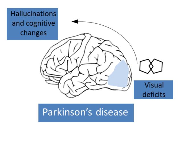
Patients with Parkinson's disease have a number of specific visual disturbances. These include changes in colour vision and contrast sensitivity and difficulties with complex visual tasks such as mental rotation and emotion recognition. We review changes in visual function at each stage of visual processing from retinal deficits, including contrast sensitivity and colour vision deficits to higher cortical processing impairments such as object and motion processing and neglect. We consider changes in visual function in patients with common Parkinson's disease-associated genetic mutations including GBA and LRRK2 . We discuss the association between visual deficits and clinical features of Parkinson's disease such as rapid eye movement sleep behavioural disorder and the postural instability and gait disorder phenotype. We review the link between abnormal visual function and visual hallucinations, considering current models for mechanisms of visual hallucinations. Finally, we discuss the role of visuo-perceptual testing as a biomarker of disease and predictor of dementia in Parkinson's disease.
Overview
The claims and statistics of this paper haven’t been checked yet.
Run the check — free, no account needed. It reads the open-access text and checks the sources it cites.
Integrity signals
Registry
Cites
Statistics
Figures
Verification
Claims the paper makes
The claims and statistics of this paper haven’t been checked yet.
Run the check — free, no account needed. It reads the open-access text and checks the sources it cites.
Self-check
Percentages, sums and reported differences re-computed from the paper’s own numbers.
The self-check needs the paper’s full text.
Citations
Cites
The precuneus: a review of its functional anatomy and behavioural correlates
Cavanna, A. E., & Trimble, M. R. (2006) · Brain · doi:10.1093/brain/awl004 · Cited by 5,128 · cited 2×
“Finally, in a structure from motion task, where volunteers detect an object that is formed from the contrast of moving dots, patients with Parkinson’s disease show higher thresholds for correctly identifying objects (Uc et al., 2005).”
BackgroundApplication of fourier analysis to the visibility of gratings
Campbell, F. W., & Robson, J. G. (1968) · The Journal of Physiology · doi:10.1113/jphysiol.1968.sp008574 · Cited by 3,019 · cited 3×
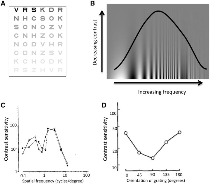
“This is more suggestive of a cortical rather than retinal or geniculate site of disruption as cortical neurons show stronger selectivity for different spatial frequencies than neurons in the retina or thalamus (Campbell and Robson, 1968; De Valois et al., 1982; Regan, 1982).”
BackgroundStages in the development of Parkinson’s disease-related pathology
Braak, H., Ghebremedhin, E., Rüb, U., Bratzke, H., & Del Tredici, K. (2004) · Cell and Tissue Research · doi:10.1007/s00441-004-0956-9 · Cited by 2,689 · cited 1×
“One theory of disease progression in Parkinson’s disease has suggested that the cerebral cortex is not damaged until the later stages of the disease (Braak et al., 2004).”
BackgroundThe Sydney multicenter study of Parkinson's disease: The inevitability of dementia at 20 years
Hely, M. A., Reid, W. G. J., Adena, M. A., Halliday, G. M., & Morris, J. G. L. (2008) · Movement Disorders · doi:10.1002/mds.21956 · Cited by 2,246 · cited 1×
“Visual hallucinations are also common in Parkinson’s disease, with a reported prevalence as high as 74% after 20 years of disease (Fenelon et al., 2000; Hely et al., 2008).”
BackgroundA direct demonstration of functional specialization in human visual cortex
Zeki, S., Watson, J., Lueck, C., Friston, K., Kennard, C., & Frackowiak, R. (1991) · The Journal of Neuroscience · doi:10.1523/jneurosci.11-03-00641.1991 · Cited by 1,716 · cited 2×
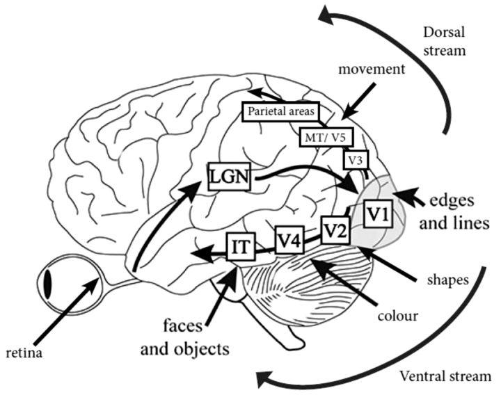
“IT = infero-temporal region; MT/V5 = motion processing region.”
BackgroundPharmacological Treatment of Parkinson Disease: A Review
Connolly, B. S., & Lang, A. E. (2014) · JAMA · doi:10.1001/jama.2014.3654 · Cited by 1,498 · cited 2×
“General advice is to consider visuo-perceptual ability and if there is any doubt, to refer the patient for a formal driving assessment via the national driving authority (Crizzle et al., 2012).”
BackgroundSpatial frequency selectivity of cells in macaque visual cortex
De Valois, R. L., Albrecht, D. G., & Thorell, L. G. (1982) · Vision Research · doi:10.1016/0042-6989(82)90113-4 · Cited by 1,489 · cited 2×
“This is more suggestive of a cortical rather than retinal or geniculate site of disruption as cortical neurons show stronger selectivity for different spatial frequencies than neurons in the retina or thalamus (Campbell and Robson, 1968; De Valois et al., 1982; Regan, 1982).”
Functional analysis of human MT and related visual cortical areas using magnetic resonance imaging
Tootell, R., Reppas, J., Kwong, K., Malach, R., Born, R., Brady, T., Rosen, B., & Belliveau, J. (1995) · The Journal of Neuroscience · doi:10.1523/jneurosci.15-04-03215.1995 · Cited by 1,415 · cited 2×
“IT = infero-temporal region; MT/V5 = motion processing region.”
BackgroundMazzulli, J. R., Xu, Y.-H., Sun, Y., Knight, A. L., McLean, P. J., Caldwell, G. A., Sidransky, E., Grabowski, G. A., & Krainc, D. (2011) · Cell · doi:10.1016/j.cell.2011.06.001 · Cited by 1,341 · cited 1×
“Both the GBA and SNCA genes are linked with the lysosomal pathway (Mazzulli et al., 2011), suggesting the visual deficits associated with these genes may share a pathophysiological mechanism, with lysosomal dysfunction associated with cortical visual dysfunction.”
BackgroundTHE DESIGN OF A NEW LETTER CHART FOR MEASURING CONTRAST SENSITIVITY
Pelli, D. G., Robson, J. G., & Wilkins, A. J. (1988) · Cited by 1,308
Research works printed but not linked: 3
- Ogden, 1990, Deficits on visuospatiaol tests involving forward planning in high-functioning Parkinsonians, Neuropsychiatry Neuropsychol Behav Neurol, 3, 125
- 10.1037/0033-295X.89.4.407 · doi:10.1037/0033-295x.89.4.407
- 10.1097/00006324-199404000-00002 · doi:10.1097/00006324-199404000-00002
Cited by
Abnormal intrinsic brain functional network dynamics in Parkinson’s disease
Kim, J., Criaud, M., Cho, S. S., Díez-Cirarda, M., Mihaescu, A., Coakeley, S., Ghadery, C., Valli, M., Jacobs, M. F., Houle, S., et al. (2017) · Brain · doi:10.1093/brain/awx233 · Cited by 369
“Visual function is also a major complex sensory domain affected by Parkinson’s disease (Mosimann et al., 2004; Weil et al., 2016).”
BackgroundDiederich, N. J., Goetz, C. G., & Stebbins, G. T. (2005) · Movement Disorders · doi:10.1002/mds.20308 · Cited by 290
Ko, F., Muthy, Z. A., Gallacher, J., Sudlow, C., Rees, G., Yang, Q., Keane, P. A., Petzold, A., Khaw, P. T., Reisman, C., et al. (2018) · JAMA Neurology · doi:10.1001/jamaneurol.2018.1578 · Cited by 241
Levodopa unresponsive symptoms in Parkinson disease: L-Dopa Unresponsive Symptoms in PD
Sethi, K. (2008) · Movement Disorders · doi:10.1002/mds.22049 · Cited by 175
Phosphorylated α‐synuclein in the retina is a biomarker of Parkinson's disease pathology severity
Ortuño‐Lizarán, I., Beach, T. G., Serrano, G. E., Walker, D. G., Adler, C. H., & Cuenca, N. (2018) · Movement Disorders · doi:10.1002/mds.27392 · Cited by 167
“Visual dysfunction and retinal changes in PD have been widely reported.(5,6) Patients suffering from PD have functional visual alterations such as reduced electroretinogram (ERG) responses and prolonged latency in visual evoked potentials.”
Background
Metadata
- Authors
- Weil, R. S., Schrag, A. E., Warren, J. D., Crutch, S. J., Lees, A. J., & Morris, H. R.
- Affiliations
- Royal Free London NHS Foundation Trust (GB)
- National Hospital for Neurology and Neurosurgery (GB)
- UCL Queen Square Institute of Neurology (GB)
- Corresponding author
- Weil, Rimona S.
- ORCID
- Venue
- Brain
- Publisher
- Oxford University Press (OUP)
- Published
- 2016-07-13
- Volume
- 139
- Issue
- 11
- Pages
- 2827-2843
- ISSN
- 0006-8950 (ISSN-L)
- 1460-2156
- PMC
- PMC5091042
- OpenAlex
- W2474900144
- Cited by
- 481
- References
- 232
- Citations by year
- 2026: 41 · 2025: 55 · 2024: 54 · 2023: 48 · 2022: 52 · 2021: 55 · 2020: 66 · 2019: 37 · 2018: 34 · 2017: 28 · 2016: 2
- FWCI
- 4.67
- Type
- journal article
- Language
- en
- Open access
- green
- www.ncbi.nlm.nih.gov
- Licence
- cc-by
- Locations
- Brain (publishedVersion)
- PubMed Central (publishedVersion, cc-by)
- PubMed (publishedVersion)
- UCL Discovery (University College London) (submittedVersion)
- HighWire Press Open Archive (submittedVersion)
- Funders
- National Institute for Health and Care Research
- Parkinson's UK (K-1501, J‐0804)
- Academy of Medical Sciences (AMS-SGCL13-Weil)
- Engineering and Physical Sciences Research Council (EP/M006093/1)
- Economic and Social Research Council (ES/L001810/1)
- Topics
- Health Sciences › Medicine › Clinical Neurology › Parkinson's Disease Mechanisms and Treatments
- Life Sciences › Neuroscience › Cognitive Neuroscience › Visual perception and processing mechanisms
- Life Sciences › Neuroscience › Cognitive Neuroscience › Hallucinations in medical conditions
- Keywords
- Parkinson's disease, visual dysfunction, contrast sensitivity, mental rotation, emotion recognition, visual neglect, motion processing, object processing, GBA mutations, LRRK2 mutations, REM sleep behavior disorder, postural instability, gait disturbance, visual hallucinations, biomarkers, dementia prediction
- MeSH terms
- Leucine-Rich Repeat Serine-Threonine Protein Kinase-2, beta-Glucosidase, Humans, Parkinson Disease, Vision Disorders, Visual Pathways, Visual Perception
About these checks
Registry, retraction, Cites and statistics results are computed, not judged, with one exception: a printed reference that no index lists is read once by a model before it counts as a citation error. Claims are extracted by a model and kept only when their quote appears verbatim in the text; each cited source is then checked, with a model’s help, for whether it says what the claim relies on. Those verdicts can be wrong — read the claim’s quote and the cited source before relying on one.
Statistics and arithmetic are recomputed, not judged.
Confidence bands are shown only for fields calibrated on our annotated test set. None is calibrated yet, so no band is shown.
What counts as a claim ›How we measure confidence ›How we check this ›
Are you an author, or do you see a problem with this page? Request a review
How to cite
Cite CoChat for the checks on this page, and the paper for its own findings.
- This page
- CoChat. (2026). Citation check: Visual dysfunction in Parkinson’s disease. https://app.cochat.ai/papers/10.1093/brain/aww175
- The paper
- Weil, R. S., Schrag, A. E., Warren, J. D., Crutch, S. J., Lees, A. J., & Morris, H. R. (2016). Visual dysfunction in Parkinson’s disease. Brain. https://doi.org/10.1093/brain/aww175
Data sources
Bibliographic data: Semantic Scholar, OpenAlex, Crossref.
Contains information from the Semantic Scholar Academic Graph Datasets, made available under the ODC Attribution License (ODC-BY 1.0).
Semantic Scholar’s data is described in: Kinney, R., et al. (2023). The Semantic Scholar Open Data Platform. arXiv:2301.10140.
